
Introduction
The onset of Legg-Calve´-Perthes disease (LCP), avas-
cular necrosis of the femoral head, frequently remains
clinically undetectable for months before presentation.
Plain films are not sensitive enough for the diagnosis of
early LCP and the diagnostic accuracy of standard mag-
netic resonance (MR) imaging remains controversial
because changes in bone marrow signal are delayed
and/or non-specific [1–4]. Changes in perfusion of the
femoral head provide one of the earliest indications of
LCP. The ability of bone scintigraphy to visualize bone
perfusion, based on the mechanisms of radionuclide lo-
calization, provides, in current practice, a highly sensi-
tive and specific means of early recognition of ischemia
[5]. Recently, findings in animal models [6, 7] and human
studies [8–11] have demonstrated the ability of contrast-
enhanced MR imaging to depict bone perfusion with in-
creased spatial and contrast resolution. To our knowl-
edge, MR perfusion studies have not yet been carried
out in children with early LCP. The purpose of this pre-
liminary study was to determine whether the simple tech-
nique of dynamic gadolinium-enhanced subtraction MR
imaging, available on standard units, can detect de-
creased femoral perfusion in these patients.
Materials and methods
MR evaluation
MR imaging was performed using a 0.5-T system and head coil
(Gyrex, Elscint, Haifa, Israel). Imaging parameters included: field
of view 27
×
27 cm, three to five sections with a 1-mm gap, matrix
size 192
×
256 and one to three signal acquisitions. The following
sequences were obtained: sagittal spin-echo, T1-weighted (TR
500/TE 20), and coronal spin-echo, proton-density and T2-weigh-
ted (2000/20/80). A dynamic T1-weighted spin-echo sequence
(200/20; acquisition time 1 min) was obtained at five levels in the
coronal plane.
Administration of a bolus of 0.1 mmol gadolinium tetraaza-
cyclododecanetetraacetic acid (Dota; Dotarem, Guerbet, Aulnay,
Guy Sebag
Hubert Ducou Le Pointe
Isabelle Klein
Djamila Maiza
Kevan Mazda
Henri Bensahel
Max Hassan
Dynamic gadolinium-enhanced
subtraction MR imaging – a simple
technique for the early diagnosis
of Legg-Calve´-Perthes disease:
preliminary results
Received: 12 April 1996
Accepted: 25 September 1996
G. Sebag (
)
)
⋅
I. Klein
⋅
D. Maiza
⋅
M. Hassan
Department of Pediatric Radiology,
Robert Debre Hospital, 48,
boulevard Serurier,
F-75935 Paris Cedex 19, France
H. Ducou Le Pointe
Department of Pediatric Radiology,
Trousseau Hospital, Paris, France
K. Mazda
⋅
H. Bensahel
Department of Pediatric Orthopedics,
Robert Debre Hospital, Paris, France
Abstract Purpose.
To determine
whether the simple technique of dy-
namic gadolinium-enhanced sub-
traction MR imaging, which is
available on standard MR units, can
detect ischemia of the femoral head
in children with early Legg-Calve´-
Perthes disease (LCP).
Materials and methods. Bone perfu-
sion of eight hips in four patients
(mean age 7.5 years) was studied
using dynamic gadolinium-en-
hanced subtraction MR imaging at
the onset of proven LCP (with initial
negative plain films). Enhancement
of subtracted images was compared
with that on standard MR images
and with bone scintigraphy find-
ings.
Results. Subtraction MR imaging
depicted ischemia as a widespread
absence of enhancement and was in
good agreement with bone scintig-
raphy. The subtraction technique
improved the sensitivity and the
specificity of MR imaging in two
children. Furthermore, subtraction
MR imaging allowed recognition of
the pattern of early reperfusion.
Conclusion. Our preliminary results
indicate that dynamic gadolinium-
enhanced subtraction MRI is a sim-
ple and promising means of early
recognition of ischemia in LCP.
Pediatr Radiol (1997) 27: 216–220
Springer-Verlag 1997

France) per kg body weight was injected by hand in less than 15 s,
followed by a 10-ml saline flush via an angiographic catheter in an
antecubital vein (22, 24 G). Images were acquired at the same five
levels as before, every minute for 5 min after the beginning of the
injection. The 1-, 2-, 3-, 4- and 5-min postenhancement images
were then subtracted from the precontrast images. Subtraction
was performed on a pixel-by-pixel basis, using the subtraction
function available as standard software on our console [12]. The fi-
nal images are the result of subtracting absolute pixel values and
are not affected by the window width and settings of the original
images [12].
Bone scintigraphy
Patients were injected with technetium-99m-labelled hydroxymeth-
ylene diphosphonate (7 mBq/kg). Three hours later, scintigrams of
the hips were acquired using a ds 7 gamma camera (Sopha Medical
Vision) fitted with a pinhole collimator (diameter of aperture
3 mm); 100 000 counts/image were obtained. Magnified frontal
views of the hips were obtained with pinhole collimation at the
bone phase.
Patients
Four patients with early LCP (three boys, one girl, mean age
7.5 years, range 5–9 years) were included in the study. Three pa-
tients had a recent history of hip pain and/or limping (duration 3–
30 days). One patient was asymptomatic, and the onset of right
LCP was detected during follow-up imaging for left LCP. None of
these patients had radiographic evidence of LCP. The delay be-
tween MR imaging and bone scintigraphy did not exceed 2 days
(average 1 day). The diagnosis of LCP was confirmed by the clini-
cal course and the appearance of typical radiographic signs on fol-
low-up plain films.
Results
In normal contralateral hips (n = 3), gadolinium injec-
tion resulted in an early, rapid (from 0 to 2 min) and in-
tense enhancement of the femoral head, the femoral
neck and the acetabulum which decreased slowly from
2 to 5 min. Enhancement was most intense at the pe-
riphery of the femoral head and along the femoral phy-
sis, resulting in a rim-like enhancement of the femoral
head and a linear enhancement of the physis (Fig. 1).
Since the marrow in the femoral head is fatty, dynamic
gadolinium-enhanced T1-weighted images without sub-
traction are not able to depict bone perfusion and is-
chemia (Fig. 1). In two hips, subtraction MR imaging
and scintigraphy demonstrated total whole-head avas-
cularity with no signal abnormality on static MR imag-
ing in one case (Fig. 1), whereas, in the second case,
static MR imaging showed a mild, diffuse, nonspecific
change in epiphyseal marrow signal (decreased T1 sig-
nal and increased T2 signal). Therefore, the dynamic ga-
dolinium-enhanced subtraction technique as an adjunct
increased both the sensitivity and specificity of MR im-
aging in the early detection of LCP (Fig. 1) in the case
of these two patients.
In another two hips, subtraction MR imaging and
scintigraphy demonstrated extensive avascularity asso-
ciated with signs of reperfusion, appearing as a lateral
column of increased enhancement and activity. In these
two cases, static MR imaging demonstrated a focal area
of necrosis involving the anterior half of the femoral
head (decreased T1 and T2 signal; Fig. 2). In one of
these two latter cases, subtraction MR imaging depicted
a medial column of increased enhancement without me-
dial isotopic uptake (Fig. 2). In summary, subtraction
MR imaging results were in good agreement with those
of bone scintigraphy, except to some extent in this last
case.
Discussion
The role of MR imaging in the diagnosis and manage-
ment of children with LCP is still controversial. Experi-
mental
and
clinical
investigations
have
clearly
demonstrated that MR imaging is very sensitive and al-
lows early detection of the disease when plain films still
appear normal [1, 13]. However, dead bone may not
show signal abnormalities on standard MR images, be-
cause at this stage of early marrow necrosis, the fat tis-
sue is “mummified” and preserves a fat-like signal
intensity, as Van de Berg et al. demonstrated [2]. Studies
in the literature comparing MR imaging and bone scin-
tigraphy findings indicate an equivalent [14], higher
[15, 16] or lower sensitivity [3] of MR imaging for LCP;
the number of series of LCP in its early stage that com-
pare MR imaging with scintigraphy is only limited. The
specificity of MR imaging has also been questioned, be-
cause the signal changes of LCP can be mimicked by
other conditions [3, 4]. The gold standard for detecting
early signs of LCP is scintigraphy with pinhole collima-
tion, because this radionuclide method allows a direct
study of bone marrow perfusion [17, 18] and a more spe-
cific identification of the reperfusion process than MR
imaging [5]. Contrast-enhanced subtraction MR imag-
ing, which has previously been applied to the study of
benign and malignant musculoskeletal tumors [13, 19,
20], can also depict tissue vascularization and perfusion
directly [11]. Verstraete et al. have clearly demon-
strated, using histologic correlation, that the pattern of
enhancement is directly related to the local blood pool
and the tissue perfusion [20]. In our experience, the
time of maximal marrow enhancement was approxi-
mately 2 min, and imaging at this early vascular phase
allowed the optimal depiction of ischemia [21]. In con-
trast, conventional T1-weighted postenhancement im-
ages, usually obtained during the late vascular phase
(between 4 and 9 min after injection) are less informa-
tive, since the contrast agent is already more diffusely
217
distributed in the tissues [11, 13]. Furthermore, subtrac-
tion techniques are essential to detect the presence or
absence of enhancement within the bright, fatty epiphy-
seal marrow. Postenhancement T1-weighted sequences
without subtraction are not appropriate (Fig. 1).
Although early diagnosis of LCP may not be of great
significance for treatment and outcome, it may be im-
portant in excluding other diseases requiring more
aggressive investigation methods and in instituting ap-
propriate therapy. In our limited series, the dynamic ga-
218
a
b
c
d
e
f
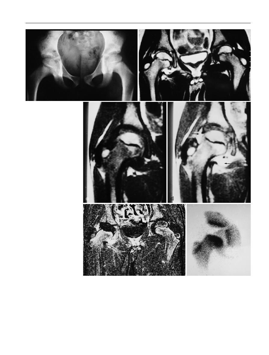
Fig. 1 a–f
Right-sided Legg-
Calve´-Perthes disease (LCP) in
an 8-year-old boy: a a normal
plain film; b a coronal T2-
weighted spin-echo image
showing left hip fluid effusion
(arrow) without any abnormal-
ity of the femoral heads; c a
coronal T1-weighted spin-echo
image showing no abnormality
of the right hip; d a coronal ga-
dolinium-enhanced T1-weight-
ed spin-echo image showing
synovial enhancement (arrows)
and effusion; e a 2-min gado-
linium-enhanced subtracted
image showing absence of en-
hancement in the entire femo-
ral head of the right hip
(arrows) and normal enhance-
ment of the left hip; f bone
scintigraphy: avascularity of the
whole femoral head
dolinium-enhanced subtraction technique improved the
sensitivity and specificity of MR imaging in the early
diagnosis of the disease in the case of two children. Fur-
ther studies will be needed to confirm this finding.
Of great importance is the fact that subtraction MR
imaging depicted the reperfusion pattern, which may
be directly related to prognosis [5, 17, 18, 22]. In our se-
ries, laterally increased enhancement was seen in two
cases which correlated well with the scintigraphic pat-
tern of “lateral column” uptake. It has been suggested
that this pattern is associated with a good prognosis for
the outcome of LCP [5]. In one case, medially increased
enhancement on MR was associated with an absence of
radionuclide uptake. The reason for this discrepancy is
unclear, but localization of bone-avid radiopharmaceu-
ticals depends on both local bone perfusion and metab-
olism and our data suggest that subtraction MR
imaging may depict reperfusion of metabolically inac-
tive bone. We are presently conducting further investi-
gations in this field.
In summary, contrast-enhanced subtraction MR im-
aging is a simple, non-ionizing technique available on
standard MR units which directly reflects blood per-
fusion and, we believe, allows early detection of epi-
physeal ischemia and revascularization processes. We
conclude from our limited series that it greatly improves
diagnostic accuracy and that these preliminary results
will provide a basis for the evaluation and comparison
of this technique with bone scintigraphy.
Acknowledgement
The authors would like to express their appre-
ciation of Christine Beslier’s help in preparing this manuscript.
219
a
b
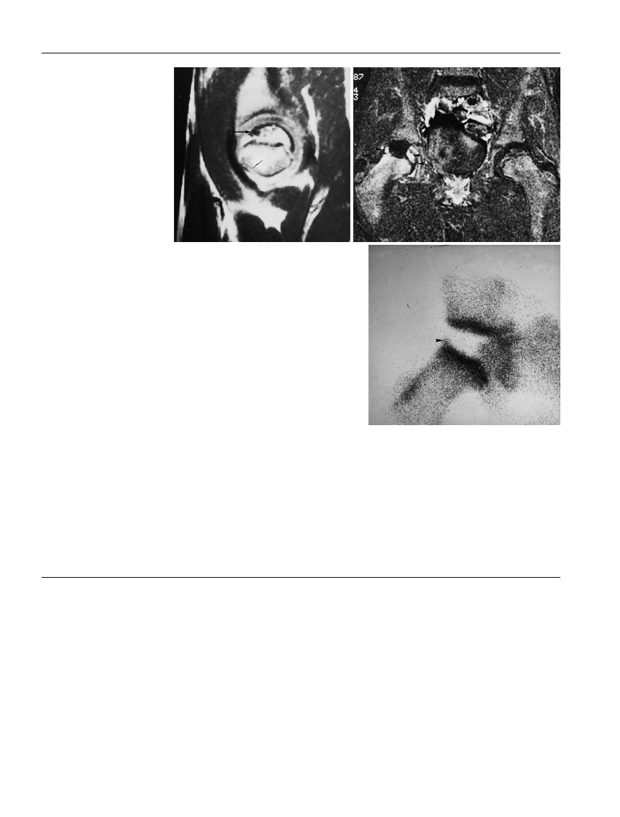
Fig. 2 a–c
Right-sided LCP in a
7-year-old boy: a a sagittal T1-
weighted image showing ante-
rior necrosis with decreased
signal (arrow); b a coronal 2-
min gadolinium-enhanced sub-
tracted image showing central
absence of enhancement and
reperfusion with lateral and
medial columns of increased
enhancement (arrowheads);
c
a bone scintigram showing
widespread absence of uptake
and reperfusion with a lateral
column pattern (arrowhead)
c
References
1. Brody AS, Strong M, Babikian G, et al
(1991) Avascular necrosis: early MR
imaging and histological findings in a
canine model. AJR 157: 341–345
2. Van de Berg B, Malghem J, Labaisse
MA, Noel H, Maldague B (1992) Avas-
cular necrosis of the hip: comparison of
contrast-enhanced and nonenhanced
MR imaging with histologic correlation.
Radiology 182: 445–450
3. Elsig JP, Exner GU, Von Schulthess
GK, Weitzel M (1989) False negative
magnetic resonance imaging in early
stage of Legg-Calve´-Perthes disease.
J Pediatr Orthop 9: 231–235
4. Norman TP, Singer WS, Bartal E (1989)
Hip pain in three children accompanied
by transient abnormal findings on MR
images. Radiology 171: 147–149
5. Conway JJ (1993) A scintigraphic clas-
sification of Legg-Calve´-Perthes dis-
ease. Semin Nucl Med 23: 274–295
6. Cova M, Young SK, Tsukamoto HT,
et al (1991) Bone marrow perfusion
evaluated with gadolinium-enhanced
dynamic fast MR imaging in a dog
model. Radiology 179: 535–539
7. Jaramillo D, Villegas-Medina OL, Doty
DK, et al (1995) Gadolinium-enhanced
MR imaging demonstrates abduction-
caused hip ischemia and its reversal in
piglets. Pediatr Radiol 25: 578–587

220
8. Saifuddin A, Bann K, Ridgway JP, et al
(1994) Bone marrow blood supply in
gadolinium-enhanced magnetic reso-
nance imaging. Skeletal Radiol 23: 455–
457
9. Bonnerot V, Charpentier A, Frouin F,
Kalifa C, Vanel D, Di Paola R (1992)
Factor analysis of dynamic magnetic
resonance imaging in predicting the re-
sponse of osteosarcoma to chemother-
apy. Invest Radiol 27: 847–855
10. Lang P, Mauz M, Scho¨rner W, et al
(1993) Acute fracture of the femoral
neck: assessment of femoral head per-
fusion with gadopentate dimeglumine-
enhanced MR imaging. AJR 160: 335–
341
11. Bluemke DA, Petri M, Zerhouni EA
(1995) Femoral head perfusion and
composition: MR imaging and spectro-
scopic evaluation of patients with sys-
temic lupus erythematosus and at risk
for avascular necrosis. Radiology 197:
433–438
12. Hanna LH, Langston JW, Gronemeyer
SA, Fletcher BD (1990) Subtraction
technique for contrast-enhanced MR
images of musculo-skeletal tumors.
Magn Reson Imaging 8: 213–215
13. Ranner G, Ebner F, Fotter R, et al
(1989) Magnetic resonance imaging in
children with acute hip pain. Pediatr
Radiol 20: 67–71
14. Scoles PV, Yoo YS, Makley JT, et al
(1984) Nuclear magnetic resonance im-
aging in Legg-Calve´-Perthes disease.
J Bone Joint Surg 666: 1357–1363
15. Pinto MR, Peterson HA, Barquist TH
(1989) Magnetic resonance imaging in
early diagnosis of Legg-Calve´-Perthes
disease. J Pediatr Orthop 9: 19–22
16. Toby EB, Koman LA, Bechtold RE
(1985) Magnetic resonance imaging of
pediatric hip disease. J Pediatr Orthop
5: 665–671
17. Bensahel H, Bok B (1979) Apport de la
scintigraphie a` la pathologie non infec-
tieuse de la hanche de l’enfant. Arch Fr
Pediatr 36: 1040–1045
18. Cavailloles F, Bok B, Bensahel H (1982)
Bone scintigraphy in the diagnosis and
follow up of Perthes’ disease. Eur J
Nucl Med 7: 327–330
19. De Baere T, Vanel D, Shapeero LG,
et al (1992) Osteosarcoma after chemo-
therapy: evaluation with contrast mate-
rial-enhanced subtraction MR imaging.
Radiology 185: 587-592
20. Verstraete KL, Dierick A, De Deene Y,
et al (1994) First-pass images of muscu-
loskeletal lesions: a new and useful di-
agnostic application of dynamic
contrast-enhanced MRI. Magn Reson
Imaging 12: 687–702
21. Theron J (1980) Angiography in Legg-
Calve´-Perthes disease. Radiology 135:
81–92
22. Ducou Le Pointe H, Haddad S, Silber-
man B, Filipe G, Monroc M, Montagne
JP (1994) Legg-Calve´-Perthes disease:
staging by MRI using gadolinium. Ped-
iatr Radiol 24: 88–91
Wyszukiwarka
Podobne podstrony:
Femoral head vascularisation in Legg Calvé Perthes disease comparison of dynamic gadolinium enhanced
Best Available Techniques for the Surface Treatment of metals and plastics
Aspects of the development of casting and froging techniques from the copper age of Eastern Central
Jagota, Dani 1982 A New Calorimetric Technique for the Estimation of Vitamin C Using Folin Phenol
Rapid Preconcentration Enrichment Techniques for the Analysi
Al Mann No Man Within Advanced Techniques for the Mentalist
CEREBRAL VENTICULAR ASYMMETRY IN SCHIZOPHRENIA A HIGH RESOLUTION 3D MR IMAGING STUDY
Pomiar lepkości dynamicznej i kinematycznej metodą swobodnego opadania ciała, SGGW Technika Rolnicza
CEREBRAL VENTICULAR ASYMMETRY IN SCHIZOPHRENIA A HIGH RESOLUTION 3D MR IMAGING STUDY
MR IMAGING OF THE NEONATAL BRAIN AT 3 TESLA
Test 3 notes from 'Techniques for Clasroom Interaction' by Donn Byrne Longman
A Digital Control Technique for a single phase PWM inverter
Gronostajski,podstawy i techniki wytwarzania II,Metody diagnostyki maszyn
Techniques for controlled drinking
19 Non verbal and vernal techniques for keeping discipline in the classroom
A Simple Circuit For Driving Microcontroller Friendly Pwm Generator 91085A
Data and memory optimization techniques for embedded systems
LEAPS Trading Strategies Powerful Techniques for Options Trading Success with Marty Kearney
więcej podobnych podstron