
10.1128/CDLI.7.5.719-723.2000.
2000, 7(5):719. DOI:
Clin. Diagn. Lab. Immunol.
Jean M. Bidlack
on Cells from the Immune System
Detection and Function of Opioid Receptors
http://cvi.asm.org/content/7/5/719
Updated information and services can be found at:
These include:
REFERENCES
http://cvi.asm.org/content/7/5/719#ref-list-1
at:
This article cites 64 articles, 26 of which can be accessed free
CONTENT ALERTS
articles cite this article),
Receive: RSS Feeds, eTOCs, free email alerts (when new
http://journals.asm.org/site/misc/reprints.xhtml
Information about commercial reprint orders:
http://journals.asm.org/site/subscriptions/
To subscribe to to another ASM Journal go to:
http://cvi.asm.org/
Downloaded from
http://cvi.asm.org/
Downloaded from

C
LINICAL AND
D
IAGNOSTIC
L
ABORATORY
I
MMUNOLOGY
,
1071-412X/00/$04.00
⫹0
Sept. 2000, p. 719–723
Vol. 7, No. 5
Copyright © 2000, American Society for Microbiology. All Rights Reserved.
MINIREVIEW
Detection and Function of Opioid Receptors on
Cells from the Immune System
JEAN M. BIDLACK*
Department of Pharmacology and Physiology, School of Medicine and Dentistry,
University of Rochester, Rochester, New York 14642
INTRODUCTION
Opioid alkaloids and peptides, such as morphine and the
endogenous opioid peptides, including
-endorphin and the
dynorphin peptides, modulate the function of lymphocytes and
other cells involved in host defense and immunity. In recent
years, investigations from several laboratories have indicated
that opioids can operate as cytokines, the principal communi-
cation signals of the immune system. All of the major proper-
ties of cytokines are shared by opioids, i.e., production by
immune cells with paracrine, autocrine, and endocrine sites of
action, functional redundancy, pleiotropy, and effects that are
both dose and time dependent (45). If opioids have a direct
effect on immune function, they must act through opioid re-
ceptors expressed on immune cells. Evidence indicates that
opioid receptors expressed by immune cells are often the same
as or similar to neuronal-type opioid receptors, particularly
-
and
␦-opioid receptors. Studies also point to the existence of
novel opioid receptors or binding sites on lymphocytes that are
selective for morphine. Opioids and their receptors appear to
function in an autocrine or paracrine manner. For example,
opioid peptides generated from immune-derived proenkepha-
lin A may act in a manner similar to that for cytokines, capable
of regulating many functions of both granulocytes and mono-
nuclear cells. The immunomodulatory effects of
-endorphin
have been shown to depend on both naloxone-sensitive and
naloxone-insensitive receptors, suggesting both brain-type and
non-neuronal-type opioid receptors on immune cells. Mea-
surements of the mRNAs that encode the neuronal types of
opioid receptor have detected rather low levels of receptor
mRNA in immune cells. Likewise, the use of radiolabeled
binding assays has not been successful in detecting opioid re-
ceptors on mixed populations of lymphocytes, probably be-
cause the receptor is expressed at a low density on a restricted
subpopulation of lymphocytes. Further identification and char-
acterization of receptors and signal tranduction pathways that
account for some of the unique properties of opioid binding
and immunomodulation represent major research challenges.
FUNCTIONAL EVIDENCE FOR PRESENCE OF
-OPIOID RECEPTORS ON LYMPHOCYTES
opioids modulate both cellular and humoral immune re-
sponses. The endogenous
-opioid-selective peptide dynorphin
has been shown to increase macrophage superoxide produc-
tion (53), modulate macrophage oxidative burst (61), enhance
macrophage tumoricidal activity (25, 29), and increase the level
of production of the cytokine interleukin-1 (IL-1) from bone
marrow macrophages (1). In the macrophage cell line P388D
1
,
the
-opioid-selective agonist U50,488 inhibited the synthesis
of IL-1 and tumor necrosis factor alpha (TNF-
␣) (4). U50,488
failed to modulate IL-6 production in these cells. Both T cells
and macrophages are targets for
-opioid agonists for produc-
tion of inhibition of T-cell-mediated antibody production (27).
These studies suggest that
-opioid receptors on T cells and
macrophages are involved in maintenance of the homeosta-
sis of the cells. Overstimulation of the
-opioid receptors on
T cells and macrophages by exogenous opioids or endoge-
nous opioid peptides may alter the levels of many cytokines.
Changes in cytokine levels may lead to the suppression of
antibody production (47, 60).
With cocultures of human fetal brain cells and a chronically
human immunodeficiency virus type 1 (HIV-1)-infected pro-
monocytic line, U1, the endogenous
-opioid peptide dynor-
phin A(1-13) and the
-opioid alkaloid U50,488 promoted
HIV-1 expression (14). Pretreatment with the
-opioid-se-
lective antagonist nor-binaltorphimine (nor-BNI) complete-
ly blocked this enhancement. The stimulation of HIV-1 expres-
sion was largely blocked by antibodies to the cytokines TNF-
␣
and IL-6 but not by IL-10. In addition, dynorphin stimulated
TNF-
␣ and IL-6 expression in the brain cell cultures at both
the mRNA and the protein levels, suggesting that
-opioid
agonists enhanced HIV-1 expression by increasing the levels of
TNF-
␣ and IL-6. In contrast to the chronically infected U1
cells, U50,488, dynorphin A(1-13), and dynorphin A(1-17) in-
hibited HIV-1 expression in acutely infected human microglial
cell cultures (16). This inhibition was blocked by the
-opioid-
selective antagonist nor-BNI. Collectively, these studies strong-
ly suggest the presence of
-opioid receptors on T cells, mac-
rophages, and microglia.
EVIDENCE FROM BINDING AND MOLECULAR
STUDIES FOR PRESENCE OF
-OPIOID RECEPTORS
ON CELLS FROM IMMUNE SYSTEM
Demonstration of radioligand binding of
opioids to a
mixed population of lymphocytes has been difficult, probably
due to the low density of opioid receptors on lymphocytes and
the fact that only small subpopulations of lymphocytes may
express the receptor. Consequently, cell lines have been used
to characterize the presence of
-opioid receptors on immune
cells. Fiorica and Spector (23) identified (
⫺)-[
3
H]bremazocine
binding sites on the EL-4 thymoma cell line; however, this
binding site was not stereoselective, and U50,488 concentra-
tions of greater than 1
M were needed to observe complete
* Mailing address: Department of Pharmacology and Physiology,
P.O. Box 711, School of Medicine and Dentistry, University of Roch-
ester, 601 Elmwood Ave., Rochester, NY 14642-8711. Phone: (716)
275-5600. Fax: (716) 273-2652. E-mail: Jean_Bidlack@urmc.rochester
.edu.
719
http://cvi.asm.org/
Downloaded from
inhibition of binding. The macrophage cell line P388D
1
ex-
pressed binding sites for the
-opioid agonist [
3
H]U69,593, but
neither the endogenous opioid peptide dynorphin nor the
antagonist naltrexone completely displaced binding (12).
However, the mouse R1.1 thymoma cell line, derived from a
thymoma from a C58/J mouse, expressed a single site with
high-affinity binding for [
3
H]naloxone and [
3
H]U69,593 (8).
The order of potency of competing ligands, including dynor-
phin peptides, was consistent with the presence of a
-opioid
receptor. This binding site was further characterized with (
⫺)-
[
3
H]bremazocine, which also bound with a high affinity to a
single binding site on R1.1 membranes (33). Competition
experiments showed that the site was stereoselective and
displayed a binding profile consistent with that of the brain
1
-opioid receptor described by Clark et al. (20), particular-
ly because the site had a high affinity for binding for both
U50,488 and
␣-neo-endorphin. In addition, (⫺)-[
3
H]bremazo-
cine binding to R1.1 membranes was potently inhibited by both
mono- and divalent cations (33), similar to results reported for
-opioid binding to brain membranes (44). The nucleotides
GTP and GDP and the nonhydrolyzable analog guanylyl-5
⬘-
imidodiphosphate further reduced the level of (
⫺)-[
3
H]brema-
zocine binding in the presence of NaCl, whereas other nucle-
otides were ineffective (33). That study suggested that the
-opioid binding site on R1.1 membranes was coupled to a G
protein, as has been reported for brain
-opioid receptors (41).
The R1.G1 and the R1EGO cell lines, two derivative cell lines
obtained from the mouse R1.1 thymoma, express the
-opioid
receptor at densities that are three- and sixfold greater than
the density at which it is expressed by the parent R1.1 cell line,
respectively, (34). These three thymoma cell lines were nega-
tively coupled to adenylyl cyclase through a pertussis toxin-
sensitive G protein (34). By using the R1.1 and related cell
lines, radioligand binding and second messenger studies have
demonstrated that a cell derived from the immune system can
express a brain-type
-opioid binding site.
The
-opioid receptor is a member of the family of seven
transmembrane receptors that are coupled to G proteins. A
partial
-opioid receptor amino acid sequence was deduced
from cDNA sequences from human and monkey lymphocytes
(18). Recently, the full-length nucleotide sequence for the
-opioid receptor expressed on the R1.1 thymoma cell line was
reported (5, 6). The nucleotide sequence shares 99.8% se-
quence homology and the deduced amino acid sequence shares
100% sequence homology with the reported murine brain
-opioid receptor (66). Another mRNA population obtained
from the R1.1 cells possesses a 30-bp insertion 15 bp upstream
of the initiation codon. This 30-bp insertion is also present in
the cDNA of the rat brain
-opioid receptor (38). These results
suggest that multiple
-opioid receptor mRNA species are
present in the R1.1 cell line. Splice variants of the
-opioid
receptor may exist on cells from the immune system and may
provide a source for protein heterogeneity. The R1.1 cell line
is negative for the cell surface phenotypic markers CD4 and
CD8, characteristics of thymocytes in one of the early stages of
differentiation (6). By cell fractionation techniques, CD4
⫺
/
CD8
⫺
thymocytes were isolated, and analysis by reverse tran-
scription-PCR showed that these primary immature thymo-
cytes also expressed mRNA for the
-opioid receptor (6). The
full-length sequence for the
-opioid receptor has also been
detected on human fetal microglia, the resident macrophages
of the brain (16). There was 100% identity between microglial
cell cDNA and the human brain
-opioid receptor gene (67).
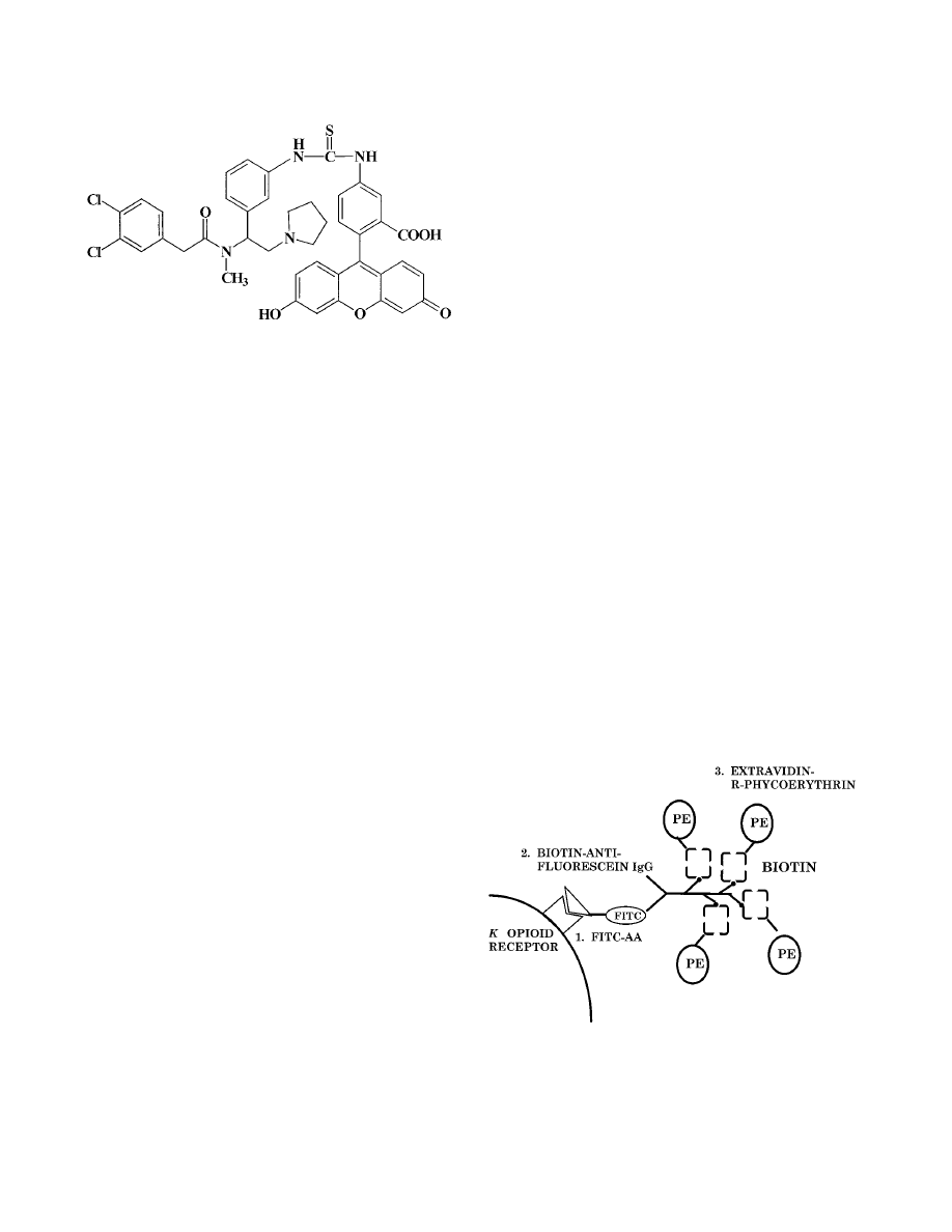
Human microglia were also shown to express the
-opioid
receptor protein, as detected by flow cytometry with the fluo-
rescent opioid fluorescein isothiocyanate (FITC)–acrylacet-
amide 2-(3,4-dichlorophenyl-N-methyl-N-[1-3-(aminophenyl)-
2-(1-pyrrolidinyl)ethyl]acetamide (AA) (Fig. 1) (16). These
studies demonstrate that cDNA for the brain-type
-opioid
receptor is present on cells from the immune system and that
these cells express the
-opioid receptor protein. Thus, a clear
molecular basis for the effects of opioid alkaloids and peptides
that bind to
-opioid receptors has been established.
In order to detect the expression of
-opioid receptors on a
mixed lymphocyte population, such as thymocytes and spleno-
cytes, an indirect immunofluorescence method that is more
sensitive than radioligand binding assays was developed in this
laboratory (35). By using a fluorescein-conjugated arylacet-
amide, a high-affinity
-opioid agonist shown in Fig. 1, fol-
lowed by amplification of FITC-AA binding with biotinylated
antifluorescein immunoglobulin G and extravidin-R–phyco-
erythrin (Fig. 2), specific labeling of
-opioid receptors on
thymocytes from C57BL/6ByJ mice was detected by flow cy-
tometry (31, 35). The
-opioid selective antagonist nor-BNI
inhibited greater than 50% of the phycoerythrin fluorescence,
FIG. 1. Structure of FITC-AA. FITC was condensed with the arylacetamide
2-(3,4-dichlorophenyl)-N-methyl-N-[1-(3-aminophenyl)-2-(1-pyrrolidinyl)ethy-
l]acetamide as previously described (35). FITC-AA was a
-opioid-selective
ligand used to label the
-opioid receptor on lymphocytes.
FIG. 2. Amplification procedure used to detect
-opioid receptors on cells
from the immune system. Unfixed mouse thymocytes (31, 35), splenocytes (32),
peritoneal macrophages (32), or human microglia (16) were incubated with
FITC-AA followed by centrifugal washes to remove unbound FITC-AA. Biotin-
ylated antifluorescein was then added, followed by washing of the cells. Finally,
extravidin-R–phycoerythrin was added. Phycoerythrin fluorescence was mea-
sured by flow cytometry (35). This procedure amplified the signal by having
multiple phycoerythrin molecules for each FITC-AA molecule that bound to the
-opioid receptor. IgG, immunoglobulin G; PE, phycoerythrin.
720
MINIREVIEW
C
LIN
. D
IAGN
. L
AB
. I
MMUNOL
.
http://cvi.asm.org/
Downloaded from

while
- and ␦-opioid-selective antagonists did not inhibit
the labeling. Double-labeling experiments with fluorescent
antibodies directed against the cell surface markers CD4 (T
helper) and CD8 (T cytotoxic) and labeling of the
-opioid
receptor were performed with thymocytes from 6- to 8-week-
old C57BL/6ByJ mice. Greater than 80% of the thymocytes
were positive for both CD4 and CD8 cell surface markers (31).
The
-opioid receptor was expressed on greater than 60% of
the CD4
⫹
/CD8
⫹
thymocytes. That study demonstrated that
the majority of mouse thymocytes express the
-opioid recep-
tor, but at a density that is too low to be detected by radio-
ligand binding. These findings are consistent with those ob-
tained with the R1.1 thymoma cell line, which expresses the
-opioid receptor (8).
To address whether mature lymphocytes expressed the
-opioid receptor, unfixed primary splenocytes from 6- to 8-
week-old C57BL/6ByJ male mice were incubated with the flu-
orescein-containing
-opioid-selective ligand FITC-AA, as de-
scribed above for the labeling of thymocytes. Amplification of
FITC-AA binding to the
-opioid receptor was attained by
adding a biotin-conjugated antifluorescein antibody, followed
by the addition of extravidin-R–phycoerythrin. As mentioned
above, greater than 60% of immature thymocytes (CD4
⫹
/
CD8
⫹
) demonstrated specific
-opioid receptor labeling. How-
ever, less than 25% of either T-helper or T-cytotoxic splenic
lymphocytes expressed the
-opioid receptor (32). Likewise,
only 16% of all splenic B lymphocytes expressed the
-opioid
receptor (32). These findings demonstrate a decrease in
-opi-
oid receptor expression upon maturation of mouse lympho-
cytes. However, recent studies have shown that mitogen acti-
vation of splenocytes increased
-opioid receptor expression
on both CD4
⫹
and CD8
⫹
cells, suggesting that the
-opioid
receptor may modulate the functions of activated T cells (7).
Interestingly, resident peritoneal macrophages showed a great-
er magnitude of specific receptor labeling compared to either
thymocytes or splenocytes, and approximately 50% of the rest-
ing macrophages expressed the
-opioid receptor (32). Also,
human microglia, the brain’s macrophages, possess a high lev-
el of
-opioid receptors, which have been shown to modu-
late HIV-1 expression in microglia (16). Taken together,
these findings demonstrate the diversity in the expression of
the
-opioid receptor on immune cells at various stages of
differentiation, with preferential expression demonstrated by
thymocytes, resident peritoneal macrophages, and microglia.
The detection of high levels of opioid receptors on peritoneal
macrophages and microglia correlates with the modulation of
TNF-
␣ and IL-1 production by opioids (4).
FUNCTIONAL EVIDENCE FOR PRESENCE OF
⌬-OPIOID RECEPTORS ON LYMPHOCYTES
Methionine-enkephalin stimulated chemotaxis in human
peripheral blood mononuclear and polymorphonuclear leu-
kocytes (24, 63). Studies of human T-lymphocyte chemotaxis
have shown that both leucine-enkephalin and methionine-
enkephalin and the enkephalin analogs [
D
-Ala
2
,
D
-Leu
5
]en-
kephalin and [
D
-Pen
2
,
D
-Pen
5
]enkephalin stimulated chemo-
taxis (30). The stimulation of chemotaxis was concentration
dependent and was inhibited by the opioid antagonist nalox-
one. Stefano et al. (58) observed that [
D
-Ala
2
,
D
-Met
5
]en-
kephalinamide stimulated immunocytes obtained from he-
molymphs of the mollusc Mytilas edulis such that the cellular
area was increased and the immunocytes clustered, with a peak
effect achieved with the opioid peptide at 10 pM. These effects
were blocked by naloxone. Similar effects were observed with
other
␦-selective opioid peptides, but the effects were not con-
centration dependent (59).
The expression of proenkephalin A mRNA by concanavalin
A-stimulated thymocytes was modulated in a biphasic manner
by the
␦-opioid agonist deltorphin I (40). Deltorphin I concen-
trations between 10
⫺13
and 10
⫺11
M increased the level of pro-
enkephalin A mRNA expression, while concentrations of 10
⫺9
to 10
⫺7
M inhibited proenkephalin A mRNA expression. The
␦-opioid antagonists naltrindole and naltriben blocked both
the enhancing and inhibiting effects of deltorphin I, suggesting
the direct involvement of
␦-opioid receptors. IL-2 secretion
from CD4
⫹
cells was also suppressed by
␦-opioid agonists (52).
Both the endogenous enkephalin-like agonists produced by
thymic T cells (39, 40) and the addition of
␦-opioid selective
peptides have been shown to exert complex effects on T-cell pro-
liferation. Deltorphin at picomolar concentrations enhanced
concanavalin A-stimulated splenocyte proliferation, an effect
blocked by the
␦-opioid selective antagonist naltrindole (11,
46). In contrast, three enkephalin analogs, including deltor-
phin, inhibited the proliferation of highly purified CD4
⫹
and
CD8
⫹
murine T cells that were activated by cross-linking the
T-cell receptor complex with anti-CD3-
ε
(52). This effect was
blocked by the
␦-opioid-selective antagonist naltrindole. In
order to observe inhibition of T-cell proliferation, it was nec-
essary to pretreat the purified lymphocytes with the
␦-opioid
peptides before the activation of the cells with anti-CD3-
ε
. In
summary, these investigations suggest that murine T cells ex-
press the
␦-opioid receptor and that activation of these recep-
tors may enhance or inhibit T-cell proliferation, depending on
the conditions, such as conditions in which purified cells versus
accessory cells are present.
EVIDENCE FROM BINDING AND MOLECULAR
STUDIES FOR PRESENCE OF
⌬-OPIOID RECEPTORS
ON CELLS FROM IMMUNE SYSTEM
As with the
-opioid receptor, the observation of classical
brain-type
␦ opioid binding of a
3
H-labeled
␦-selective opioid
ligand to a mixed population of lymphocytes has not been
achieved. [
3
H]deltorphin binding to a single high-affinity bind-
ing site on membranes from human peripheral blood poly-
morphonuclear leukocytes has been reported (59). This high-
affinity binding site for
␦ opioids also had a high affinity for
the
-opioid-selective peptide [
D
-Ala
2
, (Me)Phe
4
, Gly-(ol)
5
]
enkephalin. This result brings into question whether the [
3
H]
deltorphin binding site was the classical brain-type
␦-opioid re-
ceptor binding site or a unique binding site.
Simian peripheral blood mononuclear cells express the
␦-
opioid receptor mRNA identical to the
␦-opioid receptor mRNA
expressed by brain cells (17). Another laboratory has reported
that
␦-opioid receptor transcripts were undetectable in human
peripheral blood lymphocyte and monocyte populations after
PCR amplification but were found at low levels in human T-
cell, B-cell, and monocyte cell lines (26). In addition,
␦-opioid
receptor transcripts were found in murine splenocytes and on
some B- and T-cell lines. Human peripheral blood lymphocytes
and several human lymphoid cell lines expressed
␦-opioid re-
ceptor transcripts that were nearly identical to the known se-
quence from the human brain (65). Sharp et al. (55) have
reported that the sequence of a PCR transcript amplified from
enriched mouse splenic and lymph node T cells had 98% iden-
tity with the mouse brain
␦-opioid receptor (21).
V
OL
. 7, 2000
MINIREVIEW
721
http://cvi.asm.org/
Downloaded from

FUNCTIONAL EVIDENCE FOR PRESENCE OF
-OPIOID RECEPTORS ON LYMPHOCYTES
Many studies have used the prototypic ligand morphine to
study the effect of this clinically relevant opiate on immune
function. While being a
-opioid-preferring ligand, morphine
is not selective for the
-opioid receptor. Morphine increased
the rate of mortality among infected mice (15, 62). Also, mor-
phine inhibited the cytolytic activity of natural killer cells and
mitogen-stimulated proliferation (2, 3, 22, 64; Y. Shavit, F. C.
Martin, L. H. Angarita, R. P. Gale, and J. C. Liebeskind, Soc.
Neurosci. Abstr. 12:339, 1986). Morphine was shown to affect
the brain-immune axis by modulating an IL-1
-dependent path-
way (13). After chronic exposure in vivo, morphine attenuated
lymphocyte proliferation (9), natural killer cell cytotoxicity (37;
Shavit et al., Soc. Neurosci. Abstr. 12:339, 1986), antibody and
serum hemolysin formation (28), and the phagocytic properties
of peripheral mononuclear leukocytes (62). Morphine is known
to activate the hypothalamic-pituitary-adrenal axis and release
glucocorticoid, which is immunosuppressive (10). Therefore,
not knowing if an effect is centrally mediated or peripherally
mediated, or both, has complicated studies of chronic mor-
phine administration.
EVIDENCE FROM BINDING AND MOLECULAR
STUDIES FOR PRESENCE OF
-OPIOID RECEPTORS
ON CELLS FROM IMMUNE SYSTEM
Binding studies with lymphocytes suggest that morphine may
bind to a site that is not the classical brain
-opioid receptor
(42, 50, 57). Morphine receptors expressed on resting thymo-
cytes have a low affinity for morphine, with a K
d
value of
approximately 100 nM (48, 50). IL-1 activation of thymocytes
increased the level of [
3
H]morphine binding to the thymocytes.
The existence of a low-affinity, naloxone-insensitive morphine
binding site designated
3
on human peripheral blood macro-
phages has been reported by Makman et al. (43). Two mor-
phine binding sites have been observed on the murine macro-
phage/monocyte cell line Bac 1.2F5 (50).
Sedqi et al. (51) were the first to report the existence of
mRNA for the
-opioid receptor on rat peritoneal macro-
phages. Chuang et al. (19) reported the presence of mRNA for
the
-opioid receptor in human T- and B-cell lines, CD4
⫹
T
cells, monocytes, macrophages, and granulocytes. In addition,
transcripts have been found in simian peripheral blood mono-
nuclear cells and granulocytes (19). Collectively, these investi-
gations demonstrate that mRNAs for the
-, -, and ␦-opioid
receptors are expressed on cells from the immune system.
CONCLUSION
By understanding the synthesis of opioid peptides by lym-
phocytes and the localization of the multiple opioid receptors
on lymphocytes, the mechanisms involved in opioid-mediated
regulation of immunocompetence will be determined. Al-
though the roles of opiates and opioids in the physiological and
pathological functions of the immune system are only begin-
ning to be unraveled, multiple lines of evidence indicate that
the opioid receptors expressed by immune cells are often the
same or identical to the neuronal opioid receptors. Further
identification and characterization of the receptors and the
signal transduction pathways that account for some of the
unique properties of opioid binding and immunomodulation
represent major research challenges that lie ahead (56). Elu-
cidation of mechanisms such as these may provide unique
therapeutic opportunities through the application of opioid
immunopharmacology.
ACKNOWLEDGMENTS
This work was supported by grants K05-DA00360 and DA04355
from the National Institute on Drug Abuse.
REFERENCES
1. Apte, R. N., S. K. Durum, and J. J. Oppenheim. 1990. Opioids modulate
interleukin-1 production and secretion by bone-marrow macrophages. Im-
munol. Lett. 24:141–148.
2. Bayer, B. M., S. Daussin, M. Hernandez, and L. Irvin. 1990. Morphine
inhibition of lymphocyte activity is mediated by an opioid dependent mech-
anism. Neuropharmacology 29:369–374.
3. Bayer, B. M., M. R. Gastonguay, and M. C. Hernandez. 1992. Distinction
between the in vitro and in vivo inhibitory effects of morphine on lymphocyte
proliferation based on agonist selectivity and naltrexone reversibility. Immu-
nopharmacology 23:117–124.
4. Belkowski, S. M., C. Alicea, T. K. Eisenstein, M. W. Adler, and T. J. Rogers.
1995. Inhibition of interleukin-1 and tumor necrosis factor-
␣ synthesis fol-
lowing treatment of macrophages with the kappa opioid agonist U50,488H.
J. Pharmacol. Exp. Ther. 273:1491–1496.
5. Belkowski, S. M., J. M. Zhu, L.-Y. Liu-Chen, T. K. Eisenstein, M. W. Adler,
and T. J. Rogers.
1995. Sequence of kappa-opioid receptor cDNA in the
R1.1 thymoma cell line. J. Neuroimmunol. 62:113–117.
6. Belkowski, S. M., J. Zhu, L.-Y. Liu-Chen, T. K. Eisenstein, M. W. Adler, and
T. J. Rogers.
1995. Detection of kappa-opioid receptor mRNA in immature
T-cells. Adv. Exp. Med. Biol. 373:11–16.
7. Bidlack, J. M., and M. K. Abraham. Mitogen-induced activation of mouse T
cells increases kappa opioid receptor expression. Adv. Exp. Med. Biol., in
press.
8. Bidlack, J. M., L. D. Saripalli, and D. M. P. Lawrence. 1992.
-Opioid
binding sites on a murine lymphoma cell line. Eur. J. Pharmacol. 227:257–
265.
9. Bryant, H. U., E. W. Bernton, and J. W. Holaday. 1987. Immunosuppressive
effects of chronic morphine treatment in mice. Life Sci. 41:1731–1738.
10. Bryant, H. U., E. W. Bernton, J. R. Kenner, and J. W. Holaday. 1991. Role
of adrenal cortical activation in the immunosuppressive effects of chronic
morphine treatment. Endocrinology 128:3253–3258.
11. Caroleo, M. C., M. Arbitrio, D. Melchiorri, and G. Nistico. 1994. A reap-
praisal of the role of the various opioid receptor subtypes in cell-mediated
immunity. Neuroimmunomodulation 1:141–147.
12. Carr, D. J. J., B. R. DeCosta, C.-H. Kim, A. E. Jacobson, V. Guarcello, K. C.
Rice, and J. E. Blalock.
1989. Opioid receptors on cells of the immune
system: evidence for
␦- and -classes. J. Endocrinol. 122:161–168.
13. Chang, S. L., R. L. Moldow, S. D. House, and J. E. Zadina. 1996. Morphine
affects the brain-immune axis by modulating an interleukin-1 beta dependent
pathway. Adv. Exp. Med. Biol. 402:35–42.
14. Chao, C. C., G. Gekker, S. Hu, W. S. Sheng, P. S. Portoghese, and P. K.
Peterson.
1995. Upregulation of HIV-1 expression in co-cultures of chroni-
cally infected promonocytes and human brain cells by dynorphin. Biochem.
Pharmacol. 50:715–722.
15. Chao, C. C., B. M. Sharp, C. Pomeroy, G. A. Filice, and P. K. Peterson. 1990.
Lethality of morphine in mice infected with Toxoplasma gondii. J. Pharma-
col. Exp. Ther. 252:605–609.
16. Chao, C. C., G. Gekker, S. Hu, W. S. Sheng, D.-F. Bu, S. Archer, J. M.
Bidlack, and P. K. Peterson.
1996. Kappa opioid receptors in human micro-
glia downregulate human immunodeficiency virus-1 expression. Proc. Natl.
Acad. Sci. USA 93:8051–8056.
17. Chuang, L. F., T. K. Chuang, K. F. Killam, Jr., A. J. Chuang, H. Kung, L. Yu,
and R. Y. Chuang.
1994. Delta opioid receptor gene expression in lympho-
cytes. Biochem. Biophys. Res. Commun. 202:1291–1299.
18. Chuang, L. F., T. K. Chuang, K. F. Killam, Jr., Q. Qui, X. R. Wang, J. J. Lin,
H. F. Kung, W. Sheng, C. Chao, L. Yu, and R. Y. Chuang.
1995. Expression
of kappa opioid receptors in human and monkey lymphocytes. Biochem.
Biophys. Res. Commun. 209:1003–1010.
19. Chuang, T. K., K. F. Killam, Jr., L. F. Chuang, H. F. Kung, W. S. Sheng,
C. C. Chao, L. Yu, and R. Y. Chuang.
1995. Mu opioid receptor gene
expression in immune cells. Biochem. Biophys. Res. Commun. 216:922–930.
20. Clark, J. A., L. Liu, M. Price, B. Hersh, M. Edelson, and G. W. Pasternak.
1989. Kappa opioid receptor multiplicity: evidence for two U50,488-sensitive
kappa 1 subtypes and a novel kappa 3 subtype. J. Pharmacol. Exp. Ther.
251:
461–468.
21. Evans, C. J., D. E. Keith, Jr., H. Morrison, K. Magendzo, and R. H. Ed-
wards.
1992. Cloning of a delta opioid receptor by functional expression.
Science 258:1952–1955.
22. Fecho, K., L. A. Dykstra, and D. T. Lysle. 1993. Evidence for
-adrenergic
receptor involvement in the immunomodulatory effects of morphine. J. Phar-
macol. Exp. Ther. 265:1079–1087.
23. Fiorica, E., and S. Spector. 1988. Opioid binding site in EL-4 thymoma cell
line. Life Sci. 42:199–206.
24. Foris, G., G. A. Medgyesi, J. T. Nagy, and Z. Varga. 1987. Concentration-
dependent effect of met-enkephalin on human polymorphonuclear leuko-
cytes. Ann. N. Y. Acad. Sci. 496:151–157.
722
MINIREVIEW
C
LIN
. D
IAGN
. L
AB
. I
MMUNOL
.
http://cvi.asm.org/
Downloaded from

25. Foster, J. S., and R. N. Moore. 1987. Dynorphin and related opioid peptides
enhance tumoricidal activity mediated by murine peritoneal macrophages.
J. Leukoc. Biol. 42:171–174.
26. Gaveriaux, C., J. Peluso, F. Simonin, J. LaForet, and B. Kieffer. 1995.
Identification of kappa- and delta-opioid receptor transcripts in immune
cells. FEBS Lett. 369:272–276.
27. Guan, L., R. Townsend, T. K. Eisenstein, M. W. Adler, and T. J. Rogers.
1994. Both T-cells and macrophages are target of
-opioid-induced immu-
nosuppression. Brain Behav. Immun. 8:229–240.
28. Gungor, M., E. Genc, H. Sogduyu, L. Eroglu, and H. Koyuncuoglu. 1980.
Effect of chronic administration of morphine on primary immune response
in mice. Experientia 36:1309–1310.
29. Hagi, K., K. Uno, K. Inaba, and S. Muramatsu. 1994. Augmenting effect of
opioid peptides on murine macrophage activation. J. Neuroimmunol. 50:71–
76.
30. Heagy, W., M. Laurance, E. Cohen, and R. Finberg. 1990. Neurohormones
regulate T-cell function. J. Exp. Med. 171:1625–1633.
31. Ignatowski, T. A., and J. M. Bidlack. 1998. Detection of kappa opioid
receptors on mouse thymocyte phenotypic subpopulations as assessed by
flow cytometry. J. Pharmacol. Exp. Ther. 284:298–306.
32. Ignatowski, T. A., and J. M. Bidlack. 1999. Differential
-opioid receptor
expression on mouse lymphocytes at varying stages of maturation and on
mouse macrophages after selective elicitation. J. Pharmacol. Exp. Ther.
290:
863–870.
33. Lawrence, D. M. P., and J. M. Bidlack. 1992. Kappa opioid binding sites on
the R1.1 murine lymphoma cell line: sensitivity to cations and guanine
nucleotides. J. Neuroimmunol. 41:223–230.
34. Lawrence, D. M. P., D. B. Joseph, and J. M. Bidlack. 1995. Kappa opioid
receptors expressed on three related thymoma cell lines. Differences in
receptor-effector coupling. Biochem. Pharmacol. 49:81–89.
35. Lawrence, D. M. P., W. El-Hamouly, S. Archer, J. F. Leary, and J. M.
Bidlack.
1995. Identification of
opioid receptors in the immune system by
indirect immunofluorescence. Proc. Natl. Acad. Sci. USA 92:1062–1066.
36. Lawrence, D. M. P., I. Hutchinson, A. Seyed-Mozaffari, S. Archer, and J. M.
Bidlack.
1997. Fluorescent staining of kappa opioid receptors in the immune
system using naltrexamine derivatives and phycoerythrin. J. Immunol. Meth-
ods 201:173–181.
37. Lefkowiz, S. S., and C. Y. Chiang. 1975. Effects of certain abused drugs on
hemolysin forming cells. Life Sci. 17:1763–1768.
38. Li, S., J. Zhu, C. Chen, Y.-W. Chen, J. K. DeRiel, B. Ashby, and L.-Y.
Liu-Chen.
1993. Molecular cloning and expression of a rat kappa opioid
receptor. Biochem. J. 295:629–633.
39. Linner, K. M., H. S. Beyer, and B. M. Sharp. 1991. Induction of the mes-
senger ribonucleic acid for proenkephalin A in cultured murine CD4-posi-
tive thymocytes. Endocrinology 128:717–724.
40. Linner, K. M., H. E. Quist, and B. M. Sharp. 1995. Met-enkephalin-con-
taining peptides encoded by proenkephalin A mRNA expressed in activated
murine thymocytes inhibit thymocyte proliferation. J. Immunol. 154:5049–
5060.
41. Mack, K. J., M. F. Lee, and J. A. Weyhenmeyer. 1985. Effects of guanyl
nucleotides and ions on kappa opioid binding. Brain Res. Bull. 14:301–306.
42. Madden, J. J., W. L. Whaley, and D. Ketelsen. 1998. Opiate binding sites in
the cellular immune system: expression and regulation. J. Neuroimmunol.
83:
57–62.
43. Makman, M. H., B. Dvorkin, and G. B. Stefano. 1995. Murine macrophage
cell lines contain
3-opiate receptors. Eur. J. Pharmacol. 273:5–6.
44. Paterson, S. J., L. E. Robson, and H. W. Kosterlitz. 1986. Control by cations
of opioid binding in guinea pig brain membranes. Proc. Natl. Acad. Sci. USA
83:
6216–6220.
45. Peterson, P. K., T. W. Molitor, and C. C. Chao. 1998. The opioid-cytokine
connection. J. Neuroimmunol. 83:63–69.
46. Portoghese, P. S., M. Sultana, and A. E. Takemori. 1988. Naltrindole, a
highly selective and potent non-peptide delta opioid receptor antagonist.
Eur. J. Pharmacol. 146:185–186.
47. Radulovic, J., C. Miljevic, D. Djergovic, V. Vujic, J. Antic, S. Von Hsrstein,
and B. D. Jankovic.
1995. Opioid receptor-mediated suppression of humoral
immune response in vivo and in vitro: involvement of
opioid receptors.
J. Neuroimmunol. 57:55–62.
48. Roy, S., B. L. Ge, S. Ramakrishan, N. M. Lee, and H. H. Loh. 1991. [
3
H]-
morphine binding to thymocytes is enhanced by IL-1 stimulation. FEBS Lett.
287:
93–96.
49. Roy, S., S. Ramakrishnan, H. H. Loh, and N. M. Lee. 1991. Chronic mor-
phine treatment selectively suppresses macrophage colony formation in bone
marrow. Eur. J. Pharmacol. 195:359–363.
50. Roy, S., M. Sedqi, S. Ramakrishnan, R. A. Barke, and H. H. Loh. 1996.
Differential effects of opioids on the proliferation of a macrophage cell line.
Cell. Immunol. 169:271–277.
51. Sedqi, M., S. Roy, S. Ramakrishnan, R. Elde, and H. H. Loh. 1995. Com-
plementary DNA cloning of a
-opioid receptor from rat peritoneal macro-
phages. Biochem. Biophys. Res. Commun. 209:563–574.
52. Shahabi, N. A., and B. M. Sharp. 1995. Antiproliferative effects of delta
opioids on highly purified CD4(
⫹) and CD8(⫹) murine T cells. J. Pharma-
col. Exp. Ther. 273:1105–1113.
53. Sharp, B. M., W. F. Keane, H. J. Suh, G. Gekker, D. Tsukayama, and P. K.
Peterson.
1985. Opioid peptides rapidly stimulate superoxide production by
human polymorphonuclear leukocytes and macrophages. Endocrinology
117:
793–795.
54. Sharp, B. M., N. A. Shahabi, W. Heagy, K. McAllen, M. Bell, C. Huntoon,
and D. J. McKeane.
1996. Dual signal transduction through delta opioid
receptors in a transfected human T-cell line. Proc. Natl. Acad. Sci. USA 93:
8294–8299.
55. Sharp, B. M., N. Shahabi, D. McKean, M. D. Li, and K. McAllen. 1997.
Detection of basal levels and induction of delta opioid receptor mRNA in
murine splenocytes. J. Neuroimmunol. 78:198–202.
56. Sharp, B. M., S. Roy, and J. M. Bidlack. 1998. Evidence for opioid receptors
on cells involved in host defense and the immune system. J. Neuroimmunol.
83:
45–56.
57. Sibinga, N. E. S., and A. Goldstein. 1988. Opioid peptides and opioid re-
ceptors in cells of the immune system. Annu. Rev. Immunol. 6:219–249.
58. Stefano, G. B., P. Cadet, and B. Scharrer. 1989. Stimulatory effects of opioid
neuropeptides on locomotory activity and conformational changes in inver-
tebrate and human immunocytes: evidence for a subtype of delta receptor.
Proc. Natl. Acad. Sci. USA 86:6307–6311.
59. Stefano, G. B., P. Melchiorri, L. Negri, T. K. Hughes, Jr., and B. Scharrer.
1992. [
D
-Ala
2
]deltorphin I binding and pharmacological evidence for a spe-
cial subtype of delta opioid receptor on human and invertebrate immune
cells. Proc. Natl. Acad. Sci. USA 89:9316–9320.
60. Taub, D. D., T. K. Eisenstein, E. B. Geller, M. W. Adler, and T. J. Rogers.
1991. Immunomodulatory activity of
- and -selective opioid agonists. Proc.
Natl. Acad. Sci. USA 88:360–364.
61. Tosk, J. M., J. R. Grim, K. M. Kinback, E. J. Sale, L. P. Bozetti, and A. D.
Will.
1993. Modulation of chemiluminescence in a murine macrophage cell
line by neuroendocrine hormones. Int. J. Immunopharmacol. 15:615–620.
62. Tubaro, E., G. Borelli, C. Croce, G. Cavallo, and C. Santiangeli. 1983. Effect
of morphine on resistance to infection. J. Infect. Dis. 148:656–666.
63. VanEpps, D. E., and L. Saland. 1984.
-Endorphin and met-enkephalin
stimulate human peripheral blood mononuclear cell chemotaxis. J. Immunol.
132:
3046–3053.
64. Weber, R. J., and A. Pert. 1989. The periaqueductal gray matter mediates
opiate-induced immunosuppression. Science 245:188–190.
65. Wick, M. J., S. R. Minnerath, S. Roy, S. Ramakrishnan, and H. H. Loh. 1995.
Expression of alternate forms of brain opioid orphan receptor mRNA in
activated human peripheral blood lymphocytes and lymphocytic cell lines.
Mol. Brain Res. 32:342–347.
66. Yasuda, K., K. Raynor, H. Kong, C. D. Breder, J. Takeda, T. Reisine, and
G. I. Bell.
1993. Cloning and functional comparison of
and ␦ opioid
receptors from mouse brain. Proc. Natl. Acad. Sci. USA 90:6736–6740.
67. Zhu, J., C. Chen, J. C. Xue, S. Kunapuli, J. K. DeRiel, and L.-Y. Liu-Chen.
1995. Cloning of a human kappa opioid receptor from the brain. Life Sci.
56:
201–207.
V
OL
. 7, 2000
MINIREVIEW
723
http://cvi.asm.org/
Downloaded from
Wyszukiwarka
Podobne podstrony:
Advances in the Detection and Diag of Oral Precancerous, Cancerous Lesions [jnl article] J Kalmar (
Why Do People Hurt Themselves New Insights into the Nature and Functions of Self Injury
Revealing the Form and Function of Self Injurious Thoughts and Behaviours A Real Time Ecological As
Multiscale Modeling and Simulation of Worm Effects on the Internet Routing Infrastructure
Generic Detection and Classification of Polymorphic Malware Using Neural Pattern Recognition
Static detection and identification of X86 malicious executables A multidisciplinary approach
Formation and growth of calcium phosphate on the surface of
6 6 Detection and Identification of Drugs; Summary
Intraindividual stability in the organization and patterning of behavior Incorporating psychological
Effects of kinesio taping on proprioception at the ankle
Intraindividual stability in the organization and patterning of behavior Incorporating psychological
The effects of plant flavonoids on mammalian cells implication for inflammation, heart disease, and
53 755 765 Effect of Microstructural Homogenity on Mechanical and Thermal Fatique
Possible Effects of Strategy Instruction on L1 and L2 Reading
32 425 436 Ifluence of Vacuum HT on Microstructure and Mechanical Properties of HSS
więcej podobnych podstron