Collagens—structure, function, and biosynthesis
K. Gelse
a
, E. Po¨schl
b
, T. Aigner
a,
*
a
Cartilage Research, Department of Pathology, University of Erlangen-Nu¨rnberg, Krankenhausstr. 8-10, D-91054 Erlangen, Germany
b
Department of Experimental Medicine I, University of Erlangen-Nu¨rnberg, 91054 Erlangen, Germany
Received 20 January 2003; accepted 26 August 2003
Abstract
The extracellular matrix represents a complex alloy of variable members of diverse protein families defining structural
integrity and various physiological functions. The most abundant family is the collagens with more than 20 different collagen
types identified so far. Collagens are centrally involved in the formation of fibrillar and microfibrillar networks of the
extracellular matrix, basement membranes as well as other structures of the extracellular matrix. This review focuses on the
distribution and function of various collagen types in different tissues. It introduces their basic structural subunits and points
out major steps in the biosynthesis and supramolecular processing of fibrillar collagens as prototypical members of this protein
family. A final outlook indicates the importance of different collagen types not only for the understanding of collagen-related
diseases, but also as a basis for the therapeutical use of members of this protein family discussed in other chapters of this
issue.
D 2003 Elsevier B.V. All rights reserved.
Keywords: Collagen; Extracellular matrix; Fibrillogenesis; Connective tissue
Contents
1. Collagens—general introduction . . . . . . . . . . . . . . . . . . . . . . . . . . . . . . . . . . . . . . . . . . . . .
1532
2. Collagens—the basic structural module. . . . . . . . . . . . . . . . . . . . . . . . . . . . . . . . . . . . . . . . . .
1532
3. Distribution, structure, and function of different collagen types . . . . . . . . . . . . . . . . . . . . . . . . . . . . . . .
1535
3.1. Collagen types I, II, III, V and XI—the fibril-forming collagens . . . . . . . . . . . . . . . . . . . . . . . . . . .
1535
3.2. Collagen types IX, XII, and XIV—The FACIT collagens . . . . . . . . . . . . . . . . . . . . . . . . . . . . . . .
1537
3.3. Collagen type VI—a microfibrillar collagen . . . . . . . . . . . . . . . . . . . . . . . . . . . . . . . . . . . . .
1538
3.4. Collagen types X and VIII—short chain collagens . . . . . . . . . . . . . . . . . . . . . . . . . . . . . . . . . .
1538
3.5. Collagen type IV—the collagen of basement membranes . . . . . . . . . . . . . . . . . . . . . . . . . . . . . . .
1538
4. Biosynthesis of collagens . . . . . . . . . . . . . . . . . . . . . . . . . . . . . . . . . . . . . . . . . . . . . . . .
1540
4.1. Transcription and translation . . . . . . . . . . . . . . . . . . . . . . . . . . . . . . . . . . . . . . . . . . . .
1540
4.2. Posttranslational modifications of collagen . . . . . . . . . . . . . . . . . . . . . . . . . . . . . . . . . . . . .
1540
4.3. Secretion of collagens . . . . . . . . . . . . . . . . . . . . . . . . . . . . . . . . . . . . . . . . . . . . . . .
1541
4.4. Extracellular processing and modification . . . . . . . . . . . . . . . . . . . . . . . . . . . . . . . . . . . . . .
1541
0169-409X/$ - see front matter
D 2003 Elsevier B.V. All rights reserved.
doi:10.1016/j.addr.2003.08.002
* Corresponding author. Tel.: +49-9131-8522857; fax: +49-9131-8524745.
E-mail address: thomas.aigner@patho.imed.uni-erlangen.de (T. Aigner).
www.elsevier.com/locate/addr
Advanced Drug Delivery Reviews 55 (2003) 1531 – 1546

5. Functions of collagens beyond biomechanics . . . . . . . . . . . . . . . . . . . . . . . . . . . . . . . . . . . . . .
1542
6. Perspectives . . . . . . . . . . . . . . . . . . . . . . . . . . . . . . . . . . . . . . . . . . . . . . . . . . . . .
1542
Acknowledgements . . . . . . . . . . . . . . . . . . . . . . . . . . . . . . . . . . . . . . . . . . . . . . . . . . . .
1543
References . . . . . . . . . . . . . . . . . . . . . . . . . . . . . . . . . . . . . . . . . . . . . . . . . . . . . . . .
1543
1. Collagens—general introduction
The extracellular matrix of connective tissues rep-
resents a complex alloy of variable members of
diverse protein families defining structural integrity
and various physiological functions. The supramolec-
ular arrangement of fibrillar elements, microfibrillar
networks as well as soluble proteins, glycoproteins
and a wide range of other molecules define the
biophysical characteristics. Composition and structure
vary considerably among different types of connective
tissues. Tissue-specific expression and synthesis of
structural proteins and glycoprotein components result
in the unique functional and biological characteristics
at distinct locations.
The primary function of extracellular matrix is to
endow tissues with their specific mechanical and
biochemical properties. Resident cells are responsible
for its synthesis and maintenance, but the extracellular
matrix, in turn, has also an impact on cellular func-
tions. Cell – matrix interactions mediated by specific
cell receptors and cell binding epitopes on many
matrix molecules do not only play a dominant role
in cell attachment and migration, but also regulate or
promote cellular differentiation and gene expression
levels. The pericellular matrix provides a special
physiological microenvironment for the cells protect-
ing them from detrimental mechanical influences and
also mediating mechanically induced signal transmis-
sion. An additional influence of the extracellular
matrix on morphogenesis and cellular metabolism
can be ascribed to the storage and release of growth
factors which is modulated by their binding to specific
matrix components
The most abundant proteins in the extracellular
matrix are members of the collagen family. Colla-
gens were once considered to be a group of proteins
with a characteristic molecular structure with their
fibrillar structures contributing to the extracellular
scaffolding. Thus, collagens are the major structural
element of all connective tissues and are also found
in the interstitial tissue of virtually all parenchymal
organs, where they contribute to the stability of
tissues and organs and maintain their structural
integrity. However, in the last decade, the knowledge
increased and the collagen family expanded dramat-
ically
. All members are characterized by
containing domains with repetitions of the proline-
rich tripeptide Gly-X-Y involved in the formation of
trimeric collagen triplehelices. The functions of this
heterogeneous family are not confined to provide
structural components of the fibrillar backbone of the
extracellular matrix, but a great variety of additional
functional roles are defined by additional protein
domains.
The knowledge about the molecular structure,
biosynthesis, assembly and turnover of collagens is
important to understand embryonic and fetal develop-
mental processes as well as pathological processes
linked with many human diseases. The exploration of
expression and function of the different collagen types
also contributes to a better understanding of diseases
which are based on molecular defects of collagen
genes such as chondrodysplasias, osteogenesis imper-
fecta, Alport syndrome, Ehler’s Danlos Syndrome, or
epidermolysis bullosa
. Additionally, collagen
degradation and disturbed metabolism are important
in the course of osteoarthritis and osteoporosis. A
profound knowledge of the properties of the different
types of collagens may also be beneficial in thera-
peutical aspects. Due to their binding capacity, they
could serve as delivery systems for drugs, growth
factors or cells and the network-forming capacity and
anchoring function of certain collagen types could
contribute to the formation of scaffolds promoting
tissue repair or regeneration
2. Collagens—the basic structural module
The name ‘‘collagen’’ is used as a generic term for
proteins forming a characteristic triple helix of three
polypeptide chains and all members of the collagen
family form these supramolecular structures in the
K. Gelse et al. / Advanced Drug Delivery Reviews 55 (2003) 1531–1546
1532

Table 1
Table showing the various collagen types as they belong to the major collagen families
Type
Molecular composition
Genes (genomic localization) Tissue distribution
Fibril-forming collagens
I
[a1(I)]
2
a2(I)
COL1A1 (17q21.31 – q22)
bone, dermis, tendon, ligaments, cornea
COL1A2 (7q22.1)
II
[a1(II)]
3
COL2A1 (12q13.11 – q13.2)
cartilage, vitreous body, nucleus pulposus
III
[a1(III)]
3
COL3A1 (2q31)
skin, vessel wall, reticular fibres of most tissues (lungs, liver, spleen, etc.)
V
a1(V),a2(V),a3(V)
COL5A1 (9q34.2 – q34.3)
lung, cornea, bone, fetal membranes; together with type I collagen
COL5A2 (2q31)
COL5A3 (19p13.2)
XI
a1(XI)a2(XI)a3(XI)
COL11A1 (1p21)
cartilage, vitreous body
COL11A2 (6p21.3)
COL11A3 = COL2A1
Basement membrane collagens
IV
[a1(IV)]
2
a2(IV); a1 – a6 COL4A1 (13q34)
basement membranes
COL4A2 (13q34)
COL4A3 (2q36 – q37)
COL4A4 (2q36 – q37)
COL4A5 (Xq22.3)
COL4A6 (Xp22.3)
Microfibrillar collagen
VI
a1(VI),a2(VI),a3(VI)
COL6A1 (21q22.3)
widespread: dermis, cartilage, placenta, lungs, vessel wall,
COL6A2 (21q22.3)
intervertebral disc
COL6A3 (2q37)
Anchoring fibrils
VII
[a1(VII)]
3
COL7A1 (3p21.3)
skin, dermal – epidermal junctions; oral mucosa, cervix,
Hexagonal network-forming collagens
VIII
[a1(VIII)]
2
a2(VIII)
COL8A1 (3q12 – q13.1)
endothelial cells, Descemet’s membrane
COL8A2 (1p34.3 – p32.3)
X
[a3(X)]
3
COL10A1 (6q21 – q22.3)
hypertrophic cartilage
FACIT collagens
IX
a1(IX)a2(IX)a3(IX)
COL9A1 (6q13)
cartilage, vitreous humor, cornea
COL9A2 (1p33 – p32.2)
XII
[a1(XII)]
3
COL12A1 (6q12 – q13)
perichondrium, ligaments, tendon
XIV
[a1(XIV)]
3
COL9A1 (8q23)
dermis, tendon, vessel wall, placenta, lungs, liver
XIX
[a1(XIX)]
3
COL19A1 (6q12 – q14)
human rhabdomyosarcoma
XX
[a1(XX)]
3
corneal epithelium, embryonic skin, sternal cartilage, tendon
XXI
[a1(XXI)]
3
COL21A1 (6p12.3 – 11.2)
blood vessel wall
Transmembrane collagens
XIII
[a1(XIII)]
3
COL13A1 (10q22)
epidermis, hair follicle, endomysium, intestine, chondrocytes, lungs, liver
XVII
[a1(XVII)]
3
COL17A1 (10q24.3)
dermal – epidermal junctions
Multiplexins
XV
[a1(XV)]
3
COL15A1 (9q21 – q22)
fibroblasts, smooth muscle cells, kidney, pancreas,
XVI
[a1(XVI)]
3
COL16A1 (1p34)
fibroblasts, amnion, keratinocytes
XVIII [a1(XVIII)]
3
COL18A1 (21q22.3)
lungs, liver
Given are the molecular composition, the genomic localization of the different chains as well as the basic tissue distribution.
K. Gelse et al. / Advanced Drug Delivery Reviews 55 (2003) 1531–1546
1533
extracellular matrix although their size, function and
tissue distribution vary considerably. So far, 26 ge-
netically distinct collagen types have been described
Based on their structure and supramolecular orga-
nization, they can be grouped into fibril-forming
collagens, fibril-associated collagens (FACIT), net-
work-forming collagens, anchoring fibrils, transmem-
brane collagens, basement membrane collagens and
others with unique functions (see
The different collagen types are characterized by
considerable complexity and diversity in their struc-
ture, their splice variants, the presence of additional,
non-helical domains, their assembly and their func-
tion. The most abundant and widespread family of
collagens with about 90% of the total collagen is
represented by the fibril-forming collagens. Types I
and V collagen fibrils contribute to the structural
backbone of bone and types II and XI collagens
predominantly contribute to the fibrillar matrix of
articular cartilage. Their torsional stability and tensile
strength lead to the stability and integrity of these
tissues
. Type IV collagens with a more
flexible triple helix assemble into meshworks restrict-
ed to basement membranes. The microfibrillar type VI
collagen is highly disulfide cross-linked and contrib-
utes to a network of beaded filaments interwoven with
other collagen fibrils
. Fibril-associated collagens
with interrupted triplehelices (FACIT) such as types
IX, XII, and XIV collagens associate as single mol-
ecules with large collagen fibrils and presumably play
a role in regulating the diameter of collagen fibrils
. Types VIII and X collagens form hexagonal
networks while others (XIII and XVII) even span cell
membranes
Despite the rather high structural diversity among
the different collagen types, all members of the
collagen family have one characteristic feature: a
right-handed triple helix composed of three a-chains
. These might be formed by three
identical chains (homotrimers) as in collagens II, III,
VII, VIII, X, and others or by two or more different
chains (heterotrimers) as in collagen types I, IV, V, VI,
IX, and XI. Each of the three a-chains within the
molecule forms an extended left-handed helix with a
pitch of 18 amino acids per turn
. The three
chains, staggered by one residue relative to each other,
are supercoiled around a central axis in a right-handed
manner to form the triple helix
. A structural
prerequisite for the assembly into a triple helix is a
glycine residue, the smallest amino acid, in every third
position of the polypeptide chains resulting in a (Gly-
X-Y)
n
repeat structure which characterizes the ‘‘col-
lagenous’’ domains of all collagens. The a-chains
assemble around a central axis in a way that all
glycine residues are positioned in the center of the
triple helix, while the more bulky side chains of the
other amino acids occupy the outer positions. This
allows a close packaging along the central axis of the
molecule. The X and Y position is often occupied by
proline and hydroxyproline. Depending on the colla-
gen type, specific proline and lysine residues are
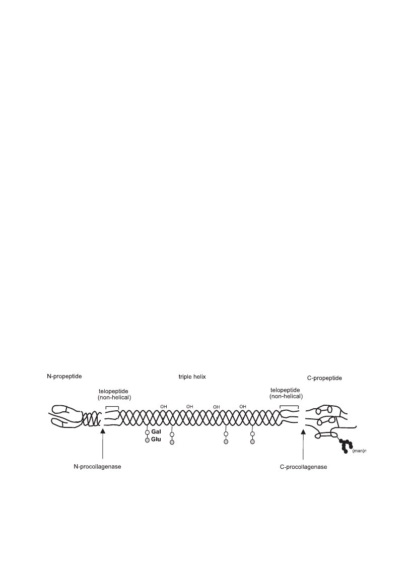
Fig. 1. Molecular structure of fibrillar collagens with the various subdomains as well as the cleavage sites for N- and C-procollagenases (shown
is the type I collagen molecule). Whereas they are arranged in tendon in a parallel manner they show a rather network-like supramolecular
arrangement in articular cartilage.
K. Gelse et al. / Advanced Drug Delivery Reviews 55 (2003) 1531–1546
1534

modified by post-translational enzymatic hydroxyl-
ation. The content of 4-hydroxyproline is essential
for the formation of intramolecular hydrogen bonds
and contributes to the stability of the triple helical
conformation. Some of the hydroxylysines are further
modified by glycosylation. The length of the triple
helical part varies considerably between different
collagen types. The helix-forming (Gly-X-Y) repeat
is the predominating motif in fibril-forming collagens
(I, II, III) resulting in triple helical domains of 300 nm
in length which corresponds to about 1000 amino
acids
. In other collagen types, these collagenous
domains are much shorter or contain non-triple helical
interruptions. Thus, collagen VI or X contains triple
helices with about 200 or 460 amino acids, respec-
tively
. Although the triple helix is a key feature of
all collagens and represents the major part in fibril-
forming collagens, non-collagenous domains flanking
the central helical part are also important structural
components
. Thus, the C-propeptide is
thought to play a fundamental role in the initiation
of triple helix formation, whereas the N-propeptide is
thought to be involved in the regulation of primary
fibril diameters
. The short non-helical telopeptides
of the processed collagen monomers (see
) are
involved in the covalent cross-linking of the collagen
molecules as well as linking to other molecular
structures of the surrounding matrix
FACIT collagens are characterized by several
non-collagenous domains interrupting the triple he-
lices, which may function as hinge regions
. In
other collagens like collagens IV, VI, VII, VIII or
X, non-collagenous domains are involved in net-
work formation and aggregation. In contrast to the
highly conserved structure of the triple helix, non-
collagenous domains are characterized by a more
structural and functional diversity among different
collagen families and types. Interruptions of the
triple helical structure may cause intramolecular
flexibility and allow specific proteolytic cleavage.
Native triple helices are characterized by their
resistance to proteases such as pepsin, trypsin or
chymotrypsin
and can only be degraded by
different types of specific collagenases. Collagenase
A (MMP-1)
, the interstitial collagenase, is
expressed by a large variety of cells and is thought
to be centrally involved in tissue remodeling, e.g.
during wound healing. MMP-8 (collagenase B) is
largely specific for neutrophil granulocytes
and,
thus, thought to be mainly involved in tissue
destruction during acute inflammatory processes.
MMP-13 (collagenase C)
is expressed by
hypertrophic chondrocytes as well as osteoblasts
and osteoclasts
and therefore most likely plays
an important role in cartilage and bone remodeling.
Many other matrix metalloproteinases are able to
cleave the denatured collagen (‘‘gelatin’’). The de-
tailed analysis of the interplay of MMPs as well as
specific inhibitors will describe the reactivities in
vivo as well as potential pharmaceutical options for
intervention
3. Distribution, structure, and function of different
collagen types
3.1. Collagen types I, II, III, V and XI—the fibril-
forming collagens
The classical fibril-forming collagens include col-
lagen types I, II, III, V, and XI. These collagens are
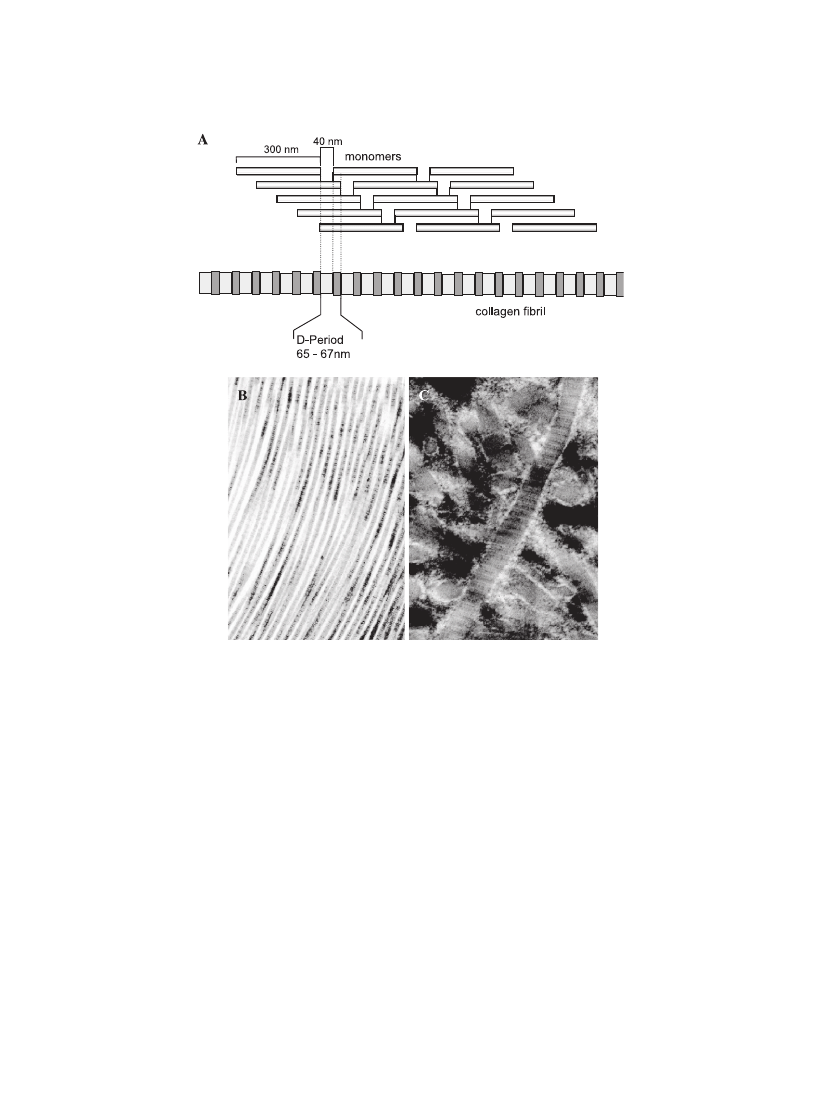
characterized by their ability to assemble into highly
orientated supramolecular aggregates with a charac-
teristic suprastructure, the typical quarter-staggered
fibril-array with diameters between 25 and 400 nm
. In the electron microscope, the fibrils are
defined by a characteristic banding pattern with a
periodicity of about 70 nm (called the D-period) based
on a staggered arrangement of individual collagen
monomers
Type I collagen is the most abundant and best
studied collagen. It forms more than 90% of the
organic mass of bone and is the major collagen of
tendons, skin, ligaments, cornea, and many intersti-
tial connective tissues with the exception of very few
tissues such as hyaline cartilage, brain, and vitreous
body. The collagen type I triple helix is usually
formed as a heterotrimer by two identical a1(I)-
chains and one a2(I)-chain. The triple helical fibres
are, in vivo, mostly incorporated into composite
containing either type III collagen (in skin and
reticular fibres)
or type V collagen (in bone,
tendon, cornea)
. In most organs and notably in
tendons and fascia, type I collagen provides tensile
stiffness and in bone, it defines considerable biome-
chanical properties concerning load bearing, tensile
K. Gelse et al. / Advanced Drug Delivery Reviews 55 (2003) 1531–1546
1535
strength, and torsional stiffness in particular after
calcification.
The fibril-forming type II collagen is the charac-
teristic and predominant component of hyaline carti-
lage. It is, however, not specifically restricted to
cartilage where it accounts for about 80% of the
total collagen content since it is also found in the
vitreous body, the corneal epithelium, the notochord,
the nucleus pulposus of intervertebral discs, and
embryonic epithelial – mesenchymal transitions
The triple helix of type II collagen is composed of
three a1(II)-chains forming a homotrimeric molecule
similar in size and biomechanical properties to that
of type I collagen
. Collagen fibrils in cartilage
represent heterofibrils containing in addition to the
dominant collagen II, also types XI and IX collagens
which are supposed to limit the fibril diameter to
about 15 – 50 nm
as well as other non-collage-
nous proteins. Compared to type I collagen, type II
collagen chains show a higher content of hydroxy-
lysine as well as glucosyl and galactosyl residues
which mediate the interaction with proteoglycans,
another typical component of the highly hydrated
matrix of hyaline cartilage
. Alternative splicing
of the type II collagen pre-mRNA results in two
forms of the a1(II)-chains. In the splice variant IIB,
Fig. 2. (A) Schematic representation of the supramolecular assembly of the collagen fibrils in the characteristic quarter-staggered form. The
monomers are 300-nm long and 40-nm gaps separate consecutive monomers causing the characteristic appearance of the collagen type I fibrils
on the ultrastructural level. (B + C) Collagen type I (B) and II (C) fibrils as they are arranged in normal tendon (B) and articular cartilage (C).
Whereas they are arranged in tendon in a parallel manner, they show a rather network-like supramolecular arrangement in articular cartilage.
K. Gelse et al. / Advanced Drug Delivery Reviews 55 (2003) 1531–1546
1536

the dominant form in mature cartilage, the second
exon coding for a globular cystein-rich domain in the
N-terminal propeptide is excluded, whereas it is
retained in the IIA variant, the embryonic form found
in prechondrogenic mesenchyme
, osteo-
phytes
, perichondrium, vertebrae
and
chondrogenic tumors
. The switch from IIA to
IIB suggests a role during developmental processes
and the IIB variant represents a characteristic marker
for mature cartilage
Type III collagen is a homotrimer of three a1(III)-
chains and is widely distributed in collagen I contain-
ing tissues with the exception of bone
. It is an
important component of reticular fibres in the inter-
stitial tissue of the lungs, liver, dermis, spleen, and
vessels. This homotrimeric molecule also often con-
tributes to mixed fibrils with type I collagen and is
also abundant in elastic tissues
Types V and XI collagens are formed as hetero-
trimers of three different a-chains (a1, a2, a3). It is
remarkable that the a3-chain of type XI collagen is
encoded by the same gene as the a1-chain of type II
collagen and only the extent of glycosylation and
hydroxylation differs from a1(II)
. Although it is
finally not sorted out, a combination between differ-
ent types V and XI chains appears to exist in various
tissues
. Thus, types V and XI collagens form
a subfamily within fibril-forming collagens, though
they share similar biochemical properties and func-
tions with other members of this family. As men-
tioned before, type V collagen typically forms
heterofibrils with types I and III collagens and
contributes to the organic bone matrix, corneal stro-
ma and the interstitial matrix of muscles, liver, lungs,
and placenta
. Type XI collagen codistributes
largely in articular cartilage with type II collagen
. The large amino-terminal non-collagenous
domains of types V and XI collagens are processed
only partially after secretion and their incorporation
into the heterofibrils is thought to control their
assembly, growth, and diameter
. Since their
triple helical domains are immunologically masked
in tissues, they are thought to be located central in
the fibrils rather than on their surface
. Thus,
type V collagen may function as a core structure of
the fibrils with types I and III collagens polymerizing
around this central axis. Analogous to this model,
type XI collagen is supposed to form the core of
collagen II heterofibrils
. A high content of
tyrosine-sulfate in the N-terminal domains of
a1(V)- and a2(V)-chains, with 40% of the residues
being O-sulfated, supports a strong interaction with
the more basic triple helical part and is likely to
stabilize the fibrillar complex
3.2. Collagen types IX, XII, and XIV—The FACIT
collagens
The collagen types IX, XII, XIV, XVI, XIX, and
XX belong to the so-called Fibril-Associated Colla-
gens with Interrupted Triple helices (FACIT colla-
gens). The structures of these collagens are
characterized by ‘‘collagenous domains’’ interrupted
by short non-helical domains and the trimeric mole-
cules are associated with the surfaces of various
fibrils.
Collagen type IX codistributes with type II colla-
gen in cartilage and the vitreous body
. The
heterotrimeric molecule consists of three different a-
chains (a1(IX), a2(IX), and a3(IX)) forming three
triple helical segments flanked by four globular
domains (NC1 – NC4)
. Type IX collagen mole-
cules are located periodically along the surface of type
II collagen fibrils in antiparallel direction
. This
interaction is stabilized by covalent lysine-derived
cross-links to the N-telopeptide of type II collagen.
A hinge region in the NC3 domain provides flexibility
in the molecule and allows the large and highly
cationic globular N-terminal domain to reach out from
the fibril where it presumably interacts with proteo-
glycans or other matrix components
. A chon-
droitin-sulfate side chain is covalently linked to a
serine residue of the a2(IX)-chain in the NC3 domain
and the size may vary between tissues
. It might
be involved in the linkage of various collagen fibres
as well as their interaction with molecules of the
extracellular matrix. Additionally, collagen type XVI
is found in hyaline cartilage and skin
and is
associated with a subset of the collagen ‘‘type II
fibers’’ (Graessel, personal communication).
Types XII and type XIV collagens are similar in
structure and share sequence homologies to type IX
collagen. Both molecules associate or colocalize with
type I collagen in skin, perichondrium, periosteum,
tendons, lung, liver, placenta, and vessel walls
The function of these collagens, as well as of collagen
K. Gelse et al. / Advanced Drug Delivery Reviews 55 (2003) 1531–1546
1537

types XIX
and XX
, within the tissue is still
poorly understood.
3.3. Collagen type VI—a microfibrillar collagen
Type VI collagen is an heterotrimer of three differ-
ent a-chains (a1, a2, a3) with short triple helical
domains and rather extended globular termini
. This is in particular true for the a3-chain
which is nearly as twice as long as the other chains
due to a large N- and C-terminal globular domains.
However, these extended domains are subject not only
to alternative splicing, but also to extensive posttrans-
lational processing, both within and outside the cell
. The primary fibrils assemble already inside
the cell to antiparallel, overlapping dimers, which then
align in a parallel manner to form tetramers. Following
secretion into the extracellular matrix, type VI collagen
tetramers aggregate to filaments and form an indepen-
dent microfibrillar network in virtually all connective
tissues, except bone
. Type VI collagen
fibrils appear on the ultrastructural level as fine fila-
ments, microfibrils or segments with faint crossband-
ing of 110-nm periodicity
, although not all
fine filaments represent type VI collagen
3.4. Collagen types X and VIII—short chain collagens
Types X and VIII collagens are structurally related
short-chain collagens. Type X collagen is a charac-
teristic component of hypertrophic cartilage in the
fetal and juvenile growth plate, in ribs and vertebrae
. It is a homotrimeric collagen with a large C-
terminal and a short N-terminal domain and experi-
ments in vitro are indicative for its assembly to
hexagonal networks
. The function of type X
collagen is not completely resolved. A role in endo-
chondral ossification and matrix calcification is dis-
cussed. Thus, type X collagen is thought to be
involved in the calcification process in the lower
hypertrophic zone
, a possibility supported
by the restricted expression of type X collagen in the
calcified zone of adult articular cartilage
and
its prevalence in the calcified chick egg shell
. In
fetal cartilage, type X collagen has been localized in
fine filaments as well as associated with type II
fibrils.
. Mutations of the COL10A1 gene are
causative for the disease Schmid type metaphyseal
chondrodysplasia (SMCD) impeding endochondral
ossification in the metaphyseal growth plate. This
leads to growth deficiency and skeletal deformities
with short limbs
Type VIII collagen is very homologous to type X
collagen in structure but shows a distinct distribution
and may therefore have different functions
. This
network-forming collagen is produced by endothelial
cells and assembles in hexagonal lattices, e.g. in the
Descemet’s membrane in the cornea
3.5. Collagen type IV—the collagen of basement
membranes
Type IV collagen is the most important structural
component of basement membranes integrating lam-
inins, nidogens and other components into the
visible two-dimensional stable supramolecular ag-
gregate. The structure of type IV collagen is
characterized by three domains: the N-terminal 7S
domain, a C-terminal globular domain (NC1), and
the central triple helical part with short interruptions
of the Gly-X-Y repeats resulting in a flexible triple
helix. Six subunit chains have been identified
yet, a1(IV) – a6(IV), associating into three distinct
heterotrimeric molecules. The predominant form is
represented by a1(IV)
2
a2(IV) heterotrimers forming
the essential network in most embryonic and adult
basement membranes. Specific dimeric interactions
of the C-terminal NC1 domains, cross-linking
of four 7S domains as well as interactions of the
triple helical domains, are fundamental for the
stable network of collagen IV
. The isoforms
a3(IV) – a6(IV) show restricted, tissue-specific ex-
pression patterns and are forming either an inde-
pendent homotypic network of a3(IV)a4(IV)a6(IV)
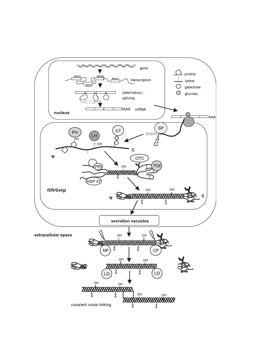
Fig. 3. Schematic representation of collagen synthesis starting form the nuclear transcription of the collagen genes, mRNA processing,
ribosomal protein synthesis (translation) and post-translational modifications, secretion and the final steps of fibril formation. (SP: signal
peptidase; GT: hydroxylysyl galactosyltransferase and galactosylhydroxylysyl glucosyltransferase; LH: lysyl hydroxylase; PH: prolyl
hydroxylase; OTC: oligosaccharyl transferase complex; PDI: protein disulphide isomerase; PPI: peptidyl-prolyl cis-trans-isomerase; NP:
procollagen N-proteinase; CP: procollagen C-proteinase; LO: lysyl oxidase; HSP47: heat shock protein 47, colligin1).
K. Gelse et al. / Advanced Drug Delivery Reviews 55 (2003) 1531–1546
1538
K. Gelse et al. / Advanced Drug Delivery Reviews 55 (2003) 1531–1546
1539

heterotrimers (kidney, lung) or a composite network
of a5(IV)
2
a6(IV)/a1(IV)
2
a2(IV) molecules
Mutations of the major isoform a1(IV)
2
a2(IV) are
assumed to be embryonic lethal, but defects of the
a5(IV), as well as a3(IV) or a4(IV)-chains are
causative for various forms of Alport syndrome
due to the importance of the a3a4a6 heterotrimer
for stability and function of glomerular and alveolar
basement membranes
4. Biosynthesis of collagens
The biosynthesis of collagens starting with gene
transcription of the genes within the nucleus to the
aggregation of collagen heterotrimers into large fibrils
is a complex multistep process
. Since most of
our knowledge of these mechanisms is based on fibril-
forming collagens, this discussion will mostly focus
on type I collagen. It is likely that the basic mecha-
nisms of triple helix formation and processing will
also apply for other collagen types.
4.1. Transcription and translation
The regulation of the transcriptional activities of
collagens depends largely on the cell type, but may
also be controlled by numerous growth factors and
cytokines (for review, see
). Thus, bone
formation is stimulated, at least in the adult, by
members of the TGF-h-superfamily as well as the
insulin-like-growth factors. In other tissues, fibro-
blast-growth-factors and many other agents are even
more important. To discuss this in more detail is
beyond the scope of this review and needs to be
evaluated for all collagens and tissues separately.
Most collagen genes revealed a complex exon –
intron pattern, ranging from 3 to 117 exons, with the
mRNAs of fibrillar collagens encoded by more than
50 exons. Therefore, in many cases, different mRNA
species could be detected, caused by multiple tran-
scription initiation sites, alternative splicing of exons
or combination of both. For example, in the cornea
and the vitreous body, a shorter form of type IX
collagen mRNA is generated by an additional start
site between exons 6 and 7
. Alternative splicing
has been reported for many collagen types and was
first described for type II collagen. A longer form of
type II collagen (COL2A) is expressed by chondro-
progenitor cells and varies from a shorter form
(COL2B) where exon 2 is excluded
and which
is the main gene product of mature articular chon-
drocytes. More recently, more than 17 different tran-
scripts have been reported for type XIII collagen
and alternative splicing also generates heterogeneous
transcription products for collagens VI, XI, XII
85]
. In addition to splicing, the pre-mRNA undergoes
capping at the 5Vend and polyadenylation at the 3Vend
and the mature mRNA is transported to the cytoplasm
and translated at the rough endoplasmatic reticulum.
Ribosome-bound mRNA is translated into prepro-
collagen molecules which protrude into the lumen of
the rough endoplasmatic reticulum with the help of a
signal recognition domain recognized by the cor-
responding receptors.
4.2. Posttranslational modifications of collagen
After removal of the signal peptide by a signal
peptidase
, the procollagen molecules undergo
multiple steps of post-translational modifications. Hy-
droxylation of proline and lysine residues catalyzed
by prolyl 3-hydroxylase, prolyl 4-hydroxylase, and
lysyl hydroxylase, respectively. All three enzymes
require ferrous ions, 2-oxoglutarate, molecular oxy-
gen, and ascorbate as cofactors. In fibril-forming
collagens, approximately 50% of the proline residues
contain a hydroxylgroup at position 4 and the extent
of prolyl-hydroxylation is species-dependent. The
organisms living at lower environmental temperatures
show a lower extent of hydroxylation
. The
presence of 4-hydroxyproline is essential for intramo-
lecular hydrogen bonds and thus contributes to the
thermal stability of the triple helical domain, and
therefore also to the integrity of the monomer and
collagen fibril. The function of 3-hydroxyproline is
not known
. The extent of lysine hydroxylation
also varies between tissues and collagen types
Hydroxylysine residues are able to form stable inter-
molecular cross-linking of collagen molecules in
fibrils and additionally represent sites for the attach-
ment of carbohydrates. Glucosyl- and galactosyl-
residues are transferred to the hydroxyl groups of
hydroxylysine; this is catalyzed by the enzymes
hydroxylysyl galactosyltransferase and galactosylhy-
droxylysyl-glucosyltransferase, respectively
K. Gelse et al. / Advanced Drug Delivery Reviews 55 (2003) 1531–1546
1540

The C-propeptides have an essential function in the
assembly of the three a-chains into trimeric collagen
monomers. The globular structure of the C-propepti-
des is stabilized by intrachain disulphide bonds and a
N-linked carbohydrate group is added by the oligo-
saccharyl transferase complex. The formation to triple
helices is preceded by the alignment of the C-terminal
domains of three a-chains and initiates the formation
of the triple helix progressing to the N-terminus. The
efficient formation and folding of the procollagen
chains depends on the presence of further enzymes
like PPI (peptidyl-prolyl cis-trans-isomerase)
and
collagen-specific chaperones like HSP47
. The
importance of these activities was substantiated by
the pharmacological influence of cyclosporine A, an
inhibitor of PPI-activity on the triple-helix formation
in vitro
as well as the fatal consequences seen
with a knock-out model of murine HSP47
. Addi-
tionally, the enzyme protein disulphide isomerase PDI,
identical with the h-subunit of prolyl 4-hydroxylase
, is involved in the formation of intra- and inter-
chain disulphide bonds in procollagen molecules
4.3. Secretion of collagens
After processing and procollagen assembly, the
triple-helical molecules are packaged within the Golgi
compartment into secretory vesicles and released into
the extracellular space. Following the secretion, the
procollagen trimers are processed depending on the
collagen type. The C-propeptides and N-propeptides
are cleaved off by two specific proteases, the procol-
lagen N-proteinase and the procollagen C-proteinase.
Both proteins belong to a family of Zn
2 +
-dependent
metalloproteinases
and the binding to the cell
membrane and internalization of the released N- and
C-propeptides was seen in studies of collagen-synthe-
sizing fibroblasts
. Therefore, a feedback mecha-
nism for the control of expression was discussed
suggesting a collagen-type specific modulating effect
of the propeptides on the collagen synthesis by
inhibiting chain initiation
. However, due to the
lack of further studies, the mechanism and their
physiological relevance remain unclear. Another study
showed that the C-propeptide of type I collagen is
internalized by fibroblasts and becomes localized
within the nucleus
. A potential effect on tran-
scription was discussed, but again, the potential
mechanisms of regulation remained largely unre-
solved
4.4. Extracellular processing and modification
The collagen fibril assembly is a complex process
and the current understanding is largely based on in
vitro experiments. The fibril-forming collagens I, II,
III, V, XI spontaneously aggregate after processing of
procollagens into ordered fibrillar structures in vitro, a
process which has been compared to crystallization
with initial nucleation and subsequent organized ag-
gregation
. The ability for the ‘‘self-assembly’’
is encoded in the structure of the collagens and several
models describe the mechanism for the periodic fibril-
lar assembly. Hydrophobic and electrostatic interac-
tions of collagen monomers are involved in the
quarter-staggered arrangement of collagen monomers,
which may aggregate into five-stranded fibrils and
subsequently into larger fibrils
The formed fibrils can be orientated differently in
distinct types of tissues. In tendons, the type I collagen
fibrils align parallel to each other and form bundles or
fibres, whereas in the skin, the orientation is more
randomly with the formation of a complex network of
interlaced fibrils
. Furthermore, the fibril forma-
tion is influenced by the propeptides of procollagen
molecules. Thus, the cleavage of the C-propeptides of
type I collagen is an essential step for regulating fibril
formation, but the function of the N-terminal propep-
tides in this process is still not fully understood and
may differ between collagen types. It has been sug-
gested that they may regulate the diameter of the
forming fibrils and their removal from type I procolla-
gen influences the regular fibril morphology
The molecular arrangement into fibrils is addition-
ally stabilized by the formation of covalent cross-links
which finally contribute to the mechanical resilience
of collagen fibrils. The hydroxylation state of telopep-
tide lysine residues is crucial in defining collagen
cross-links. Lysine hydroxylation within the telopep-
tides is catalyzed by an enzyme system different from
the lysyl hydroxylase responsible for helical residues.
The extent of hydroxylation in the telopeptides varies
between different tissues with complete hydroxylation
of lysine residues in cartilage, but no detectable
hydroxylation of telopeptide lysine in the skin
The copper-dependent enzyme lysyl oxidase catalyzes
K. Gelse et al. / Advanced Drug Delivery Reviews 55 (2003) 1531–1546
1541

the formation of aldehydes from lysine and hydrox-
ylysine residues in the telopeptides. Subsequent spon-
taneous reactions result in the formation of intermediate
cross-links. Lysine-derived telopeptide aldehydes in-
teract with adjacent lysine residues from adjacent
molecules to form Schiff base (aldimin) cross-links,
whereas the presence of hydroxylysine-derived telo-
peptide aldehydes allows to form more stable ketoi-
mine bonds. During maturation of the tissue, the
reducible intermediate cross-links (aldimines and
ketoimines) are converted to non-reducible mature
products: The Schiff bases are converted to non-
reducible histidin adducts while the ketoimines react
either with hydroxylysine aldehyde or a second ketoi-
mine to form pyridinium cross-links. Alternatively,
pyrrolic cross-links are formed in case of ketoimines
reacting with lysyl aldehyde components
. Pyridi-
nium compounds and pyrroles result in a cross-link
between three collagen molecules. Most cross-links
have been shown to be located at the overlap position
connecting the N- or C-telopeptides with specific
residues within the helical part of adjacent molecules
These intermolecular cross-links are a prerequisite
for the physical and mechanical properties of collagen
fibrils and a stable network formation.
5. Functions of collagens beyond biomechanics
As discussed earlier, collagens serve within the
body to a large extent for the maintenance of the
structural integrity of tissues and organs. This is true
for all parenchymal organs where they represent the
major component of the ‘‘interstitial’’ matrix as well
as the basement membranes. This is even more
obvious for all ‘‘connective’’ tissues and in particular
bone and cartilage where collagens provide the major
functional backbone of the structures. Besides this, the
formation of a defined pericellular microenvironment
is important for the cellular integrity, as seen with
collagen VI in articular cartilage, but presumably also
in bone (own unpublished observation). Besides the
biomechanical aspects, however, collagens are also
involved in a plethora of additional functions. Specific
receptors mediate the interaction with collagens, like
integrins, discoidin-domain receptors, glycoprotein VI
or specialized proteoglycan receptors
Signaling by these receptors defines adhesion, differ-
entiation, growth, cellular reactivities as well as the
survival of cells in multiple ways.
Collagens contribute to the entrapment, local stor-
age and delivery of growth factors and cytokines and
therefore play important roles during organ develop-
ment, wound healing and tissue repair
. Col-
lagen type I has been shown to bind decorin, and
thereby, it might block indirectly TGF-h-action within
the tissue
. Collagens also bind a number of other
growth factors and cytokines. Thus, IGF-I and -II are
bound to the collagenous matrix of bone and, there-
fore, bone represents a major reservoir of these growth
factors within the body
. In bone, degradation of
the collagen network by osteoclasts during bone
remodeling is thought to release matrix-bound IGFs
and, thus, to induce new bone formation via stimula-
tion of osteoblastic activity in a paracrine manner.
Similar effects may be active in articular cartilage and
could be due to anabolic activation of chondrocytes
via release of bound growth factors after cartilage
matrix degradation. Type IIA collagen was recently
shown to be able to bind TGFh and BMP-2
Thus, collagens are very likely to be relevant for
certain cellular reactions. This potential of collagens
to bind growth factors and cytokines qualifies these
molecules also as transport vehicles for therapeutic
factor delivery (for review, see other chapters of this
issue).
Recently, it became evident that collagens are
involved in more subtle and sophisticated functions
than just the architecture of extracellular matrices.
Non-collagenous fragments of collagens IV, XV and
XVIII have been shown to influence angiogenesis and
tumorigenesis and their biological functions may not
only be limited to these processes, but also influence
various cellular reactivities
. Therefore,
these fragments (matricryptins) attracted great interest
for potential pharmaceutical uses.
6. Perspectives
Collagens are the most abundant group of organic
macro-molecules in an organism. First, collagens
serve important mechanical functions within the body,
particularly in connective tissues. Thus, in bone,
tendon, fascia, articular cartilage, etc., fibrillar colla-
K. Gelse et al. / Advanced Drug Delivery Reviews 55 (2003) 1531–1546
1542

gens are providing most of the biomechanical prop-
erties essential for the functioning of these organ
systems. Second, collagens also exert important func-
tions in the cellular microenvironment and are in-
volved in the storage and release of cellular mediators,
such as growth factors. All aspects mentioned above
define collagens as interesting targets as well as tools
of pharmacological intervention. A proper collagen
matrix in terms of its composition and supramolecular
organization is the target of any repair process of
connective tissue whether occurring naturally, like
during fracture healing or following treatment of bone
non-unions after trauma, tumor-surgery or of cartilage
defects (for review, see Aigner and Sto¨ve, this issue).
Finally, it should be considered that some additional
features of collagens, such as biodegradability, low
immunogenicity and the possibilities for large-scale
isolation make them interesting compounds for a
widespread industrial use in medicine, cosmetics or
food industry.
Acknowledgements
This work was supported by the Ministry of
Science and Technology (grant 01GG9824).
References
[1] Y. Yamaguchi, D.M. Mann, E. Ruoslathi, Negative regula-
tion of transforming growth factor-h by the proteoglycan
decorin, Nature 346 (1990) 281 – 284.
[2] D. Schuppan, M. Schmid, R. Somasundaram, R. Ackermann,
M. Ruehl, T. Nakamura, E.O. Riecken, Collagens in the liver
extracellular matrix bind hepatocyte growth factor, Gastro-
enterology 114 (1998) 139 – 152.
[3] J.F. Bateman, S.R. Lamande, J.A.M. Ramshaw, Collagen
superfamily, in: W.D. Comper (Ed.), Extracellular Matrix,
Harwood Academic Press, Melbourne, 1996, pp. 22 – 67.
[4] K. von der Mark, Structure, biosynthesis and gene regula-
tion of collagens in cartilage and bone, Dynamics of Bone
and Cartilage Metabolism, Academic Press, Orlando, 1999,
pp. 3 – 29.
[5] S.R. Frenkel, B. Toolan, D. Menche, M.I. Pitman, J.M. Pa-
chence, Chondrocyte transplantation using a collagen bilayer
matrix for cartilage repair, J. Bone Jt. Surg. 79-B (1997)
831 – 836.
[6] S. Wakitani, T. Kimura, A. Hirooka, T. Ochi, M. Yoneda,
N. Yasui, H. Owaki, K. Ono, Repair of rabbit articular
surfaces with allograft chodnrocytes embedded in colla-
gen gel, J. Bone Jt. Surg. 71-B (1989) 74 – 80.
[7] K. Ku¨hn, The collagen family-variations in the molecular and
supermolecular structure, Rheumatology 10 (1986) 29 – 69.
[8] R. Mayne, R.G. Brewton, New members of the collagen
superfamily, Curr. Opin. Cell Biol. 5 (1993) 883 – 890.
[9] M. van der Rest, R. Garrone, Collagen family of proteins,
FASEB J. 5 (1991) 2814 – 2823.
[10] J. Myllyharju, K.I. Kivirikko, Collagens and collagen-related
diseases, Ann. Med. 33 (2001) 7 – 21.
[11] K. Sato, K. Yomogida, T. Wada, T. Yorihuzi, Y. Nishimune,
N. Hosokawa, K. Nagata, Type XXVI collagen, a new mem-
ber of the collagen family, is specifically expressed in the
testis and ovary, J. Biol. Chem. 277 (2002) 37678 – 37684.
[12] D.E. Birk, J.M. Fitch, J.P. Babiarz, T.F. Linsenmayer, Colla-
gen type I and type V are present in the same fibril in the
avian corneal stroma, J. Cell Biol. 106 (1988) 999 – 1008.
[13] R. Mayne, Cartilage collagens—what is their function, and
are they involved in articular disease? Arthritis Rheum. 32-3
(1989) 241 – 246.
[14] H. von der Mark, M. Aumailley, G. Wick, R. Fleischmajer, R.
Timpl, Immunochemistry, genuine size and tissue localization
of collagen VI, Eur. J. Biochem. 142 (1984) 493 – 502.
[15] R. van der Rest, R. Mayne, Regulation of matrix accumula-
tion, in: R. Mayne, R. Burgeson (Eds.), Structure and Func-
tion of Collagen Types, Academic Press, Orlando, 1987.
[16] K.A. Piez, Molecular and aggregate structure of the colla-
gens, in: K.A. Pietz, H. Reddi (Eds.), Extracellular Matrix
Biology, Elsevier, Amsterdam, 1984, pp. 1 – 39.
[17] H. Hofmann, P.P. Fietzek, K. Kuhn, The role of polar and
hydrophobic interactions for the molecular packing of type I
collagen: a three-dimensional evaluation of the amino acid
sequence, J. Mol. Biol. 125 (1978) 137 – 165.
[18] R.D. Fraser, T.P. MacRae, E. Suzuki, Chain conformation in
the collagen molecule, J. Mol. Biol. 129 (1979) 463 – 481.
[19] L.M. Shaw, B.R. Olsen, FACIT collagens: diverse molecular
bridges in extracellular matrices, Trends Biochem. Sci. 16
(1991) 191 – 194.
[20] P. Bruckner, D.J. Prockop, Proteolytic enzymes as probes for
the triple – delical conformation of procollagen, Anal. Bio-
chem. 110 (1981) 360 – 368.
[21] G.I. Goldberg, S.M. Wilhelm, A. Kronberger, E.A. Bauer,
G.A. Grant, A.Z. Eisen, Human fibroblast collagenase, J. Biol.
Chem. 261 – 14 (1986) 6600 – 6605.
[22] K.A. Hasty, T.F. Pourmotabbed, G.I. Goldberg, J.P.
Thompson, D.G. Spinelly, R.M. Stevens, C.L. Mainardi,
Human neutrophil collagenase, J. Biol. Chem. 265 (1990)
11421 – 11424.
[23] J.M. Freije, I. Diez-Itza, M. Balbin, L.M. Sanchez, R. Blasco,
J. Tolivia, C. Lopez-Otin, Molecular cloning and expression
of collagenase-3, a novel human matrix metalloproteinase
produced by breast carcinomas, J. Biol. Chem. 269 (1994)
16766 – 16773.
[24] N. Johansson, U. Saarialho-Kere, K. Airola, R. Herva,
L. Nissinen, J. Westermarck, E. Vuorio, J. Heino, V.-M. Ka¨-
ha¨ri, Collagenase-3 (MMP-13) is expressed by hypertrophic
chondrocytes, periosteal cells, and osteoblasts during human
fetal bone development, Dev. Dyn. 208 (1997) 387 – 397.
[25] G. Giannelli, S. Antonaci, Gelatinases and their inhibitors in
K. Gelse et al. / Advanced Drug Delivery Reviews 55 (2003) 1531–1546
1543

tumor metastasis: from biological research to medical appli-
cations, Histol. Histopathol. 17 (2002) 339 – 345.
[26] C.M. Overall, C. Lopez-Otin, Strategies for MMP inhibition
in cancer: innovations for the post-trial era, Nat. Rev., Cancer
2 (2002) 657 – 672.
[27] K. Brand, Cancer gene therapy with tissue inhibitors of metal-
loproteinases (TIMPs), Curr. Gene Ther. 2 (2002) 255 – 271.
[28] D.J. Hulmes, A. Miller, Molecular packing in collagen, Nature
293 (1981) 234 – 239.
[29] R. Fleischmajer, E.D. MacDonald, J.S. Perlish, R.E. Bur-
geson, L.W. Fisher, Dermal collagen fibrils are hybrids
of type I and type III collagen molecules, J. Struct.
Biol. 105 (1990) 162 – 169.
[30] C. Niyibizi, D.R. Eyre, Bone type V collagen: chain compo-
sition and location of a trypsin cleavage site, Connect. Tissue
Res. 20 (1989) 247 – 250.
[31] P. Bruckner, M. van der Rest, Structure and function
of cartilage collagens, Microsc. Res. Tech. 28 (1994)
378 – 384.
[32] M. Mendler, S.G. Eich-Bender, L. Vaughan, K.H. Win-
terhalter, P. Bruckner, Cartilage contains mixed fibrils of
collagen types II, IX and XI, J. Cell Biol. 108 (1989)
191 – 197.
[33] M.C. Ryan, L.J. Sandell, Differential expression of a cys-
teine-rich domain in the amino- terminal propeptide of type
II (cartilage) procollagen by alternative splicing of mRNA,
J. Biol. Chem. 265 (1990) 10334 – 10339.
[34] L.J. Sandell, N.P. Morris, J.R. Robbins, M.B. Goldring, Al-
ternatively spliced type II procollagen mRNAs define dis-
tinct populations of cells during vertebral development:
differential expression of the amino-propeptide, J. Cell Biol.
114 (1991) 1307 – 1319.
[35] J.R. Matyas, L.J. Sandell, M.E. Adams, Gene expression of
type II collagens in chondro-osteophytes in experimental
osteoarthritis, Osteoarthr. Cartil. 5 (1997) 99 – 105.
[36] K. Gelse, S. So¨der, W. Eger, T. Diemtar, T. Aigner, Osteo-
phyte development—molecular characterization of differen-
tiation stages, Osteoarthr. Cartil. 11 (2003) 141 – 148.
[37] T. Aigner, S. Loos, S. Mu¨ller, L.J. Sandell, R. Perris, K.K.
Unni, T. Kirchner, Cell differentiation and matrix gene
expression in mesenchymal chondrosarcomas, Am. J. Pathol.
156 (2000) 1327 – 1335.
[38] J. Rossert, B. de Crombrugghe, Type I collagen: structure,
synthesis and regulation, in: J.P. Bilezkian, L.G. Raisz,
G.A. Rodan (Eds.), Principles in Bone Biology, Academic
Press, Orlando, 2002, pp. 189 – 210.
[39] K. von der Mark, Localization of collagen types in tissues,
Int. Rev. Connect. Tissue Res. 9 (1981) 265 – 324.
[40] M. Yamazaki, R.J. Majeska, H. Yoshioka, H. Moriya, T.A.
Einhorn, Spatial and Temporal expression of fibril-forming
minor collagen genes (types V and XI) during fracture heal-
ing, J. Orthop. Res. 15 (1997) 757 – 764.
[41] J.-P. Kleman, D.J. Hartmann, F. Ramirez, M. van der Rest,
The human rhabdomyosarcoma cell line A204 lays down a
highly insoluble matrix composed mainly of a1 type-XI and
a2 type-V collagen chains, Eur. J. Biochem. 210 (1992)
329 – 335.
[42] K.J. Bos, D.F. Holmes, K.E. Kadler, D. McLeod, N.P.
Morris, P.N. Bishop, Axial structure of the heterotypic colla-
gen fibrils of vitreous humour and cartilage, J. Mol. Biol. 306
(2001) 1011 – 1022.
[43] V.C. Lui, R.Y.C. Kong, J. Nicholls, A.N.Y. Cheung, K.S.E.
Cheah, The mRNAs for the three chains of h type XI are
widely distributed but not necessarily co-expressed: implica-
tions for homotrimeric and heterotypic collagen molecules,
Biochem. J. 311 (1995) 511 – 516.
[44] J.H. Fessler, N. Shigaki, L.I. Fessler, Biosynthesis and pro-
perties of procollagens V, Ann. N.Y. Acad. Sci. 460 (1985)
181 – 186.
[45] B. Petit, M.-C. Ronzie`re, D.J. Hartmann, D. Herbage, Ultra-
structural organization of type XI collagen in fetal bone epi-
physeal cartilage, Histochemistry 100 (1993) 231 – 239.
[46] L.I. Fessler, S. Brosh, S. Chapin, J.H. Fessler, Tyrosine sul-
fation in precursors of collagen V, J. Biol. Chem. 261 (1986)
5034 – 5040.
[47] M. van der Rest, R. Mayne, Y. Ninomiya, N.G. Seidah,
M. Chretien, B.R. Olsen, The structure of type IX col-
lagen, J. Biol. Chem. 260 (1985) 220 – 225.
[48] J.-J. Wu, D.R. Eyre, Structural analysis of cross-linking do-
mains in cartilage type XI collagen, J. Biol. Chem. 270
(1995) 18865 – 18870.
[49] M. van der Rest, R. Mayne, Type IX collagen proteoglycan
from cartilage is covalently cross-linked to type II collagen,
J. Biol. Chem. 263 (1988) 1615 – 1618.
[50] T. Yada, S. Suzuki, K. Kobayashi, M. Kobayashi, T. Hoshino,
K. Horie, K. Kimata, Occurrence in chick embryo vitreous
humor of a type IX collagen proteoglycan with an extraordi-
narily large chondroitin sulfate chain and short alpha 1 poly-
peptide, J. Biol. Chem. 265 (1990) 6992 – 6999.
[51] C.H. Lai, M.L. Chu, Tissue distribution and developmental
expression of type XVI collagen in the mouse, Tissue Cell 28
(1996) 155 – 164.
[52] J.C. Myers, D. Li, A. Bageris, V. Abraham, A.S. Dion, P.S.
Amenta, Biochemical and immunohistochemical character-
ization of human type XIX defines a novel class of basement
membrane zone collagens 14, Am. J. Pathol. 151 (1997)
1729 – 1740.
[53] M. Koch, J.E. Foley, R. Hahn, P. Zhou, R.E. Burgeson, D.R.
Gerecke, M.K. Gordon, Alpha 1(XX) collagen, a new mem-
ber of the collagen subfamily, fibril-associated collagens
with interrupted triple helices, J. Biol. Chem. 276 (2001)
23120 – 23126.
[54] D. Weil, M.-G. Mattei, E. Passage, N. Van Cong, D. Pribula-
Conway, K. Mann, R. Deutzmann, R. Timpl, M.-L. Chu,
Cloning and chromosomal localization of human genes en-
coding the three chains of type VI collagen, Am. J. Hum.
Genet. 42 (1988) 435 – 445.
[55] M.-L. Chu, K.-H. Mann, R. Deutzmann, D. Pribula-Conway,
C.C. Hsu-Chen, M.P. Bernard, R. Timpl, Charcterization of
three constituent chains of collagen type VI by peptide se-
quences and cDNA clones, Eur. J. Biochem. 168 (1987)
309 – 317.
[56] T. Aigner, L. Hambach, S. So¨der, U. Schlotzer-Schrehardt,
E. Poschl, The C5 domain of Col6A3 is cleaved off from
K. Gelse et al. / Advanced Drug Delivery Reviews 55 (2003) 1531–1546
1544

the Col6 fibrils immediately after secretion, Biochem. Bio-
phys. Res. Commun. 290 (2002) 743 – 748.
[57] R. Timpl, M.-L. Chu, Microfibrillar collagen type VI, in:
P.D. Yuchenco, D. Birk, R.P. Mecham (Eds.), Extracellular
Matrix Assembly and Structure, Academic Press, Orlando,
1994, pp. 207 – 242.
[58] D.R. Keene, E. Engvall, R. Glanville, Ultrastructure of type
VI collagen in human skin and cartilage suggests an anchor-
ing function for this filamenteous network, J. Cell Biol. 107
(1988) 1995 – 2006.
[59] J. Engel, H. Furthmayr, E. Obermatt, H. von der Mark, M.
Aumailley, R. Fleischmajer, R. Timpl, Structure and mac-
romolecular organization of type VI collagen, Ann. N.Y.
Acad Sci. 460 (1985) 25 – 37.
[60] C.A. Poole, S. Ayad, R.T. Gilbert, Chondrons from articular
cartilage—V. Immunohistochemical evaluation of type VI
collagen organisation in isolated chondrons by light, confocal
and electron microscopy, J. Cell. Sci. 103 (1992) 1101 – 1110.
[61] R.R. Bruns, Beaded filaments and long-spacing fibrils: re-
lation to type VI collagen, J. Ultrastruct. Res. 89 (1984)
136 – 146.
[62] R.R. Bruns, W. Press, E. Engvall, R. Timpl, J. Gross, Type
VI collagen in extracellular, 100-nm periodic filaments and
fibrils: identification by immunoelecton microscopy, J. Cell
Biol. 103 (1986) 393 – 404.
[63] M.-C. Ronzie`re, S. Ricard-blum, J. Tiollier, D.J. Hartmann,
R. Garrone, D. Herbage, Comparative analysis of collagens
solubilized from human foetal, and normal and osteoarthritic
adult articular cartilage, with emphasis on type VI collagen,
Biochim. Biophys. Acta 1038 (1990) 222 – 230.
[64] C.A. Poole, M.H. Flint, B.W. Beaumont, Morphology of the
pericellular capsule in articular cartilage revealed by hyalu-
ronidase digestion, J. Ultrastruct. Res. 91 (1985) 13 – 23.
[65] V.C. Duance, M. Shimokomaki, A.J. Bailey, Immunofluo-
rescence localization of type-M collagen in articular carti-
lage, Biosci. Rep. 2 (1982) 223 – 227.
[66] H.B. Evans, S. Ayad, M.Z. Abedin, S. Hopkins, K. Morgan,
K.W. Walton, J.B. Weiss, P.J.L. Holt, Localization of colla-
gen types and fibronectin in cartilage by immunofluores-
cence, Ann. Rheum. Dis. 42 (1983) 575 – 581.
[67] D.J. Hartmann, H. Magloire, S. Ricard-blum, A. Joffre, M.-L.
Couble, G. Ville, D. Herbage, Light and electron immunoper-
ixodase localization of mnor disulfide-bonded collagens in
fetal epiphyseal cartilage, Collagen Relat. Res. 3 (1983)
349 – 357.
[68] S. Ricard-blum, D.J. Hartmann, D. Herbage, C. Payen-
Meyran, G. Ville, Biochemical properties and immunolocal-
ization of minor collagens in foetal calf cartilage, FEBS Lett.
146 (1982) 343 – 347.
[69] T.M. Schmid, T.F. Linsenmayer, Type X collagen, in:
R. Mayne, R.E. Burgeson (Eds.), Structure and Func-
tion of Collagen Types, Academic Press, Orlando, 1987,
pp. 195 – 222.
[70] T. Kirsch, K. von der Mark, Ca
2 +
binding properties of type
X collagen, FEBS Lett. 294 (1992) 149 – 152.
[71] M. Alini, D.E. Carey, S. Hirata, M.D. Grynpas, I. Pidoux,
A.R. Poole, Cellular and matrix changes before and at the
time of calcification in the growth plate studied in vitro:
arrest of type X collagen synthesis and net loss of collagen
when calcification is initiated, J. Bone Miner. Res. 9 (1994)
1077 – 1087.
[72] A.P.L. Kwan, I.R. Dickson, A.J. Freemont, M.E. Grant,
Comparative studies of type X collagen expression in normal
and rachitic chicken epiphyseal cartilage, J. Cell Biol. 109
(1989) 1849 – 1856.
[73] J.M. Gannon, G. Walker, M. Fischer, R. Carpenter, R.C.
Thompson, T.R. Oegema, Localization of type X collagen
in canine growth plate and adult canine articular cartilage,
J. Orthop. Res. 9 (1991) 485 – 494.
[74] G.D. Walker, M. Fischer, J. Gannon, R.C. Thompson,
T.R. Oegema, Expression of type-X collagen in osteoar-
thritis, J. Orthop. Res. 13 (1995) 4 – 12.
[75] J.L. Arias, M.S. Fernandez, J.E. Dennis, A.I. Caplan, Colla-
gens of the chicken eggshell membranes, Connect. Tissue
Res. 26 (1991) 37 – 45.
[76] T.M. Schmid, T.F. Linsenmayer, Immunoelectron micro-
scopy of type X collagen: supramolecular forms within em-
bryonic chick cartilage, Dev. Biol. 138 (1990) 53 – 62.
[77] M.L. Warman, M. Abbott, S.S. Apte, T. Hefferon, I.
McIntosh, D.H. Cohn, J.T. Hecht, B.J. Olsen, C.A. Franco-
mano, A type X collagen mutation causes Schmid metaphy-
seal chondrodysplasia, Nat. Genet. 5 (1993) 79 – 82.
[78] N. Yamaguchi, R. Mayne, Y. Ninomiya, The alpha 1 (VIII)
collagen gene is homologous to the alpha 1 (X) collagen
gene and contains a large exon encoding the entire triple
helical and carboxyl-terminal non-triple helical domains of
the alpha 1 (VIII) polypeptide, J. Biol. Chem. 266 (1991)
4508 – 4513.
[79] H. Sawada, H. Konomi, K. Hirosawa, Characterization of the
collagen in the hexagonal lattice of Descemet’s membrane:
its relation to type VIII collagen, J. Cell Biol. 110 (1990)
219 – 227.
[80] B.G. Hudson, S.T. Reeders, K. Tryggvason, Type IV colla-
gen: structure, gene organization, and role in human dis-
eases. Molecular basis of Goodpasture and Alport syn-
dromes and diffuse leiomyomatosis, J. Biol. Chem. 268
(1993) 26033 – 26036.
[81] D.B. Borza, O. Bondar, Y. Ninomiya, Y. Sado, I. Naito, P.
Todd, B.G. Hudson, The NC1 domain of collagen IV en-
codes a novel network composed of the alpha 1, alpha 2,
alpha 5, and alpha 6 chains in smooth muscle basement mem-
branes, J. Biol. Chem. 276 (2001) 28532 – 28540.
[82] S. Peltonen, M. Rehn, T. Pihlajaniemi, Alternative splicing of
mouse alpha1(XIII) collagen RNAs results in at least 17
different transcripts, predicting alpha1(XIII) collagen chains
with length varying between 651 and 710 amino acid resi-
dues, DNA Cell Biol. 16 (1997) 227 – 234.
[83] B. Saitta, D.G. Stokes, H. Vissing, R. Timpl, M.-L. Chu,
Alternative splicing of the human a2(VI) collagen gene gen-
erates multiple mRNA transcripts which predict three protein
variants with distinct carboxyl termini, J. Biol. Chem. 265
(1990) 6473 – 6480.
[84] M. Moradi-Ameli, B. de Chassey, J. Farjanel, R.M. van der,
Different splice variants of cartilage alpha1(XI) collagen
K. Gelse et al. / Advanced Drug Delivery Reviews 55 (2003) 1531–1546
1545

chain undergo uniform amino-terminal processing, Matrix
Biol. 17 (1998) 393 – 396.
[85] M. Koch, B. Bohrmann, M. Matthison, C. Hagios, B. Trueb,
M. Chiquet, Large and small splice variants of collagen XII:
differential expression and ligand binding, J. Cell Biol. 130
(1995) 1005 – 1014.
[86] L. Cohen-Solal, J. Castanet, F.J. Meunier, M.J. Glimcher,
Proline hydroxylation of collagens synthesized at different
temperatures in vivo by two poikilothermic species, Comp.
Biochem. Physiol., B 83 (1986) 483 – 486.
[87] K.I. Kivirikko, L. Ryhanen, H. Anttinen, P. Bornstein, D.J.
Prockop, Further hydroxylation of lysyl residues in collagen
by protocollagen lysyl hydroxylase in vitro, Biochemistry 12
(1973) 4966 – 4971.
[88] K. Lang, F.X. Schmid, G. Fischer, Catalysis of protein fold-
ing by prolyl isomerase, Nature 329 (1987) 268 – 270.
[89] E.P. Clarke, G.A. Cates, E.H. Ball, B.D. Sanwal, A collagen-
binding protein in the endoplasmic reticulum of myoblasts
exhibits relationship with serine protease inhibitors, J. Biol.
Chem. 266 (1991) 17230 – 17235.
[90] H.P. Bachinger, N.P. Morris, J.M. Davis, Thermal stability
and folding of the collagen triple helix and the effects of
mutations in osteogenesis imperfecta on the triple helix of
type I collagen, Am. J. Med. Genet. 45 (1993) 152 – 162.
[91] B. Steinmann, P. Bruckner, A. Superti-Furga, Cyclosporin A
slows collagen triple-helix formation in vivo: indirect evi-
dence for a physiologic role of peptidyl-prolyl cis-trans-iso-
merase, J. Biol. Chem. 266 (1991) 1299 – 1303.
[92] N. Nagai, M. Hosokawa, S. Itohara, E. Adachi, T. Matsushita,
N. Hosokawa, K. Nagata, Embryonic lethality of molecular
chaperone hsp47 knockout mice is associated with defects in
collagen biosynthesis, J. Cell Biol. 150 (2000) 1499 – 1506.
[93] J. Koivu, R. Myllyla, T. Helaakoski, T. Pihlajaniemi, K.
Tasanen, K.I. Kivirikko, A single polypeptide acts both as the
beta subunit of prolyl 4-hydroxylase and as a protein disul-
fide-isomerase, J. Biol. Chem. 262 (1987) 6447 – 6449.
[94] T. Pihlajaniemi, T. Helaakoski, K. Tasanen, R. Myllyla, M.L.
Huhtala, J. Koivu, K.I. Kivirikko, Molecular cloning of the
beta-subunit of human prolyl 4-hydroxylase. This subunit
and protein disulphide isomerase are products of the same
gene, EMBO J. 6 (1987) 643 – 649.
[95] D.J. Prockop, A.L. Sieron, S.W. Li, Procollagen N-proteinase
and procollagen C-proteinase. Two unusual metalloprotei-
nases that are essential for procollagen processing probably
have important roles in development and cell signaling, Ma-
trix Biol. 16 (1998) 399 – 408.
[96] W. Schlumberger, M. Thie, H. Volmer, J. Rauterberg, H.
Robenek, Binding and uptake of Col 1(I), a peptide capable
of inhibiting collagen synthesis in fibroblasts, Eur. J. Cell
Biol. 46 (1988) 244 – 252.
[97] D. Ho¨rlein, J. McPherson, S. Han Gow, P. Bornstein, Regu-
lation of protein synthesis: translational control by procolla-
gen-derived fragments, Proc. Natl. Acad. Sci. U. S. A. 78-10
(1981) 6163 – 6167.
[98] C.H. Wu, C.M. Walton, G.Y. Wu, Propeptide-mediated reg-
ulation of procollagen synthesis in IMR-90 human lung
fibroblast cell cultures, J. Biol. Chem. 266-5 (1991)
2983 – 2987.
[99] A. Veis, K. Payne, Collagen fibrillogenesis, in: M.E. Nimni
(Ed.), Collagen. Biochemistry, CRC Press, Boca Raton,
1988, p. 113.
[100] D. Silver, J. Miller, R. Harrison, D.J. Prockop, Helical model
of nucleation and propagation to account for the growth of
type I collagen fibrils from symmetrical pointed tips: a spe-
cial example of self-assembly of rod-like monomers, Proc.
Natl. Acad. Sci. U. S. A. 89 (1992) 9860 – 9864.
[101] W.F. Vogel, Collagen-receptor signaling in health and dis-
ease, Eur. J. Dermatol. 11 (2001) 506 – 514.
[102] J.M. Levine, A. Nishiyama, The NG2 chondroitin sulfate
proteoglycan: a multifunctional proteoglycan associated
with immature cells, Perspect. Dev. Neurobiol. 3 (1996)
245 – 259.
[103] E.D. Hay, Extracellular matrix, J. Cell Biol. 91 (1981)
205s – 223s.
[104] C.M. Bautista, S. Mohan, D.J. Baylink, Insulin-like growth
factors I and II are present in the skeletal tissues of ten
vertebrates, Metabolism 39 (1990) 96 – 100.
[105] Y. Zhu, A. Oganesian, D.R. Keene, L.J. Sandell, Type IIA
procollagen containing the cysteine-rich amino propeptide is
deposited in the extracellular matrix of prechondrogenic tis-
sue and binds to TGF-h1 and BMP-2, J. Cell Biol. 144
(1998) 1069 – 1080.
[106] N. Ortega, Z. Werb, New functional roles for non-collage-
nous domains of basement membrane collagens, J. Cell Sci.
115 (2002) 4201 – 4214.
[107] G.E. Davis, K.J. Bayless, M.J. Davis, G.A. Meininger, Reg-
ulation of tissue injury responses by the exposure of matri-
cryptic sites within extracellular matrix molecules, Am. J.
Pathol. 156 (2000) 1489 – 1498.
[108] M.S. O’Reilly, T. Boehm, Y. Shing, N. Fukai, G. Vasios,
W.S. Lane, E. Flynn, J.R. Birkhead, B.R. Olsen, J. Folkman,
Endostatin: an endogenous inhibitor of angiogenesis and tu-
mor growth, Cell 88 (1997) 277 – 285.
K. Gelse et al. / Advanced Drug Delivery Reviews 55 (2003) 1531–1546
1546
Document Outline
- Collagens-structure, function, and biosynthesis
Wyszukiwarka
Podobne podstrony:
Building Collagen Molecules, Fibrils, and Suprafibrillar Structures
L 3 Complex functions and Polynomials
4 Plant Structure, Growth and Development, before ppt
Euler’s function and Euler’s Theorem
a relational perspective on turnover examining structural, attitudinal and behavioral predictors
Cell structure function
augocm h8 truck scanner function and model list
Derrida, Jacques Structure, Sign And Play In The Discourse Of The Human Sciences
L 3 Complex functions and Polynomials
4 Plant Structure, Growth and Development, before ppt
Euler’s function and Euler’s Theorem
Collagen stability, hydration and native state
06 The Korean Language Structure, Use and Context
He Clusters structural embeddedness and knowledge a sttructural embeddedness modle of clusters
więcej podobnych podstron