
Review
Outer membrane permeability and antibiotic resistance
Anne H. Delcour
Department of Biology and Biochemistry, University of Houston, 369 Science and Research Building II, Houston, TX 77204-5001, USA
a b s t r a c t
a r t i c l e i n f o
Article history:
Received 19 September 2008
Received in revised form 12 November 2008
Accepted 13 November 2008
Available online 27 November 2008
Keywords:
Outer membrane
Antibiotic
Porin
LPS
Resistance
To date most antibiotics are targeted at intracellular processes, and must be able to penetrate the bacterial
cell envelope. In particular, the outer membrane of gram-negative bacteria provides a formidable barrier that
must be overcome. There are essentially two pathways that antibiotics can take through the outer
membrane: a lipid-mediated pathway for hydrophobic antibiotics, and general diffusion porins for
hydrophilic antibiotics. The lipid and protein compositions of the outer membrane have a strong impact
on the sensitivity of bacteria to many types of antibiotics, and drug resistance involving modi
fications of
these macromolecules is common. This review will describe the molecular mechanisms for permeation of
antibiotics through the outer membrane, and the strategies that bacteria have deployed to resist antibiotics
by modi
fications of these pathways.
© 2008 Elsevier B.V. All rights reserved.
The outer membrane (OM) of gram-negative bacteria performs the
crucial role of providing an extra layer of protection to the organism
without compromising the exchange of material required for sustain-
ing life. In this dual capacity, the OM emerges as a sophisticated
macromolecular assembly, whose complexity has been unraveled only
in recent years. By combining a highly hydrophobic lipid bilayer with
pore-forming proteins of speci
fic size-exclusion properties, the OM
acts as a selective barrier. The permeability properties of this barrier,
therefore, have a major impact on the susceptibility of the micro-
organism to antibiotics, which, to date, are essentially targeted at
intracellular processes. Small hydrophilic drugs, such as
β-lactams,
use the pore-forming porins to gain access to the cell interior, while
macrolides and other hydrophobic drugs diffuse across the lipid
bilayer. The existence of drug-resistant strains in a large number of
bacterial species due to modi
fications in the lipid or protein
composition of the OM indeed highlights the importance of the OM
barrier in antibiotic sensitivity. This review will summarize the
properties of the OM lipid barrier and porin-mediated permeability,
and highlight the antibiotic resistance mechanisms that involve
modi
fications of these properties.
It is important to note that many of the alterations in outer
membrane permeability described below are often associated with
increased levels of antibiotic ef
flux. Even intrinsic antibiotic resistance
is likely to re
flect the synergistic action of the outer membrane acting
as a permeability barrier, and of the diverse and widely distributed
ef
flux pumps. The review below essentially focuses on the perme-
ability changes per se, as the roles of ef
flux pathways in antibiotic
resistance are treated by others. Whether changes in outer membrane
lipid or porin composition also mechanistically in
fluences the efflux
systems remains to be determined.
1. Organization of the OM
In most gram-negative bacteria, the OM is an asymmetric bilayer of
phospholipid and lipopolysaccharides (LPS), the latter exclusively
found in the outer lea
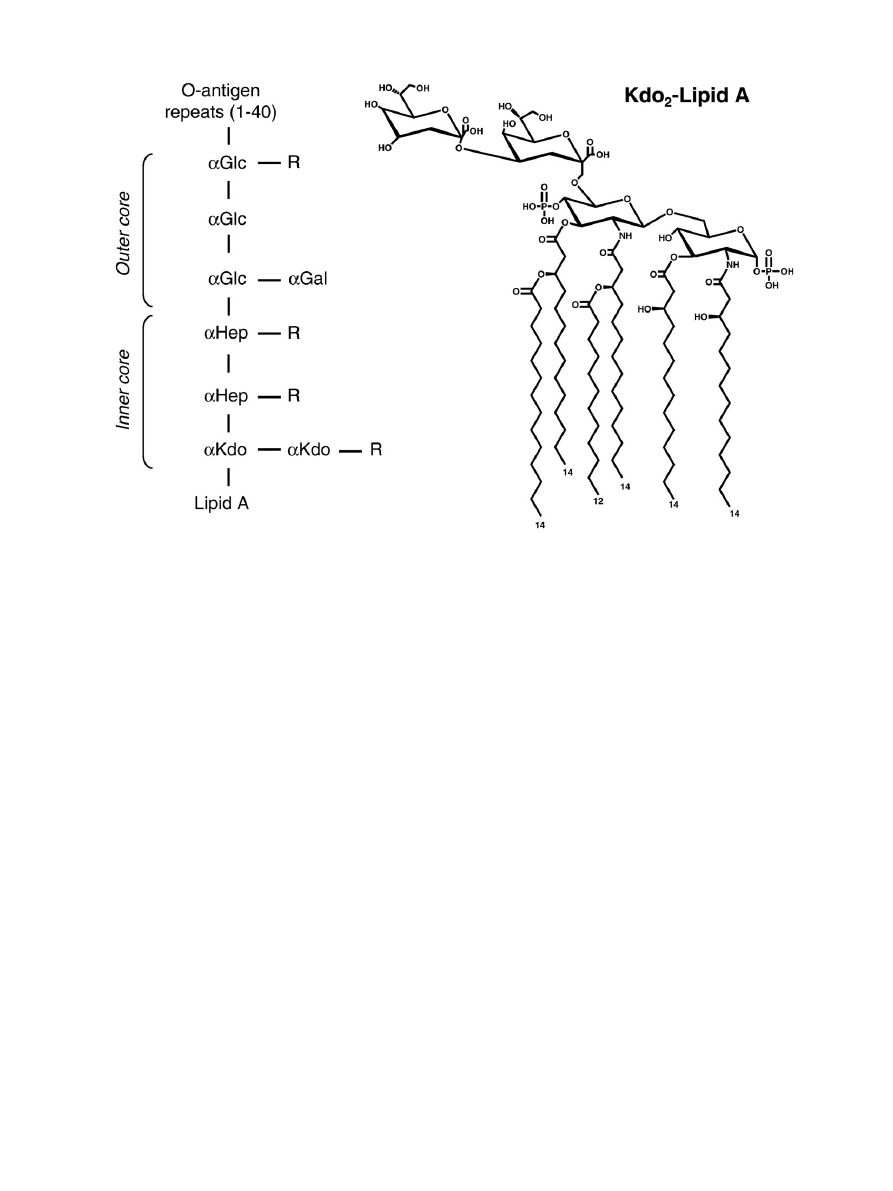
flet. A typical LPS molecule consists of three
parts (
): 1) lipid A, a glucosamine-based phospholipid, 2) a
relatively short core oligosaccharide, and 3) a distal polysaccharide
(O-antigen)
. Since part of the core oligosaccharide and the O-
antigen are not required for the growth of Escherichia coli, strains can
exhibit varying length of these structures. The phospholipid composi-
tion of the inner lea
flet of the OM is similar to that of the cytoplasmic
membrane, i.e. about 80% phosphatidylethanolamine, 15% phosphati-
dylglycerol and 5% cardiolipin
. In mutants with altered LPS structure,
phospholipids have also been detected in the outer lea
flet of the OM,
possibly due to consequent decrease in OM protein levels
.
A large number of different types of proteins reside in the OM.
Some of them are extremely abundant. For example, murein
lipoprotein (Lpp), OmpA and general diffusion porins are present
at
N10
5
copies per cell
. Lpp carries a fatty acid moiety that anchors
it into the OM, while about a third of the Lpp population is also
covalently attached to the peptidoglycan layer. Thus, Lpp is thought to
play a role in providing OM
–peptidoglycan interactions and in
maintaining OM integrity. Indeed, mutants lacking Lpp produce OM
vesicles and leak periplasmic enzymes
. Another abundant OM
protein is OmpA. The protein is believed to have a structural role and
the absence of OmpA and Lpp compromises the shape of the cell
.
Along with the Pseudomonas aeruginosa homolog OprF, OmpA has
pore-forming properties as well, but with extremely low permeation
Biochimica et Biophysica Acta 1794 (2009) 808
–816
⁎ Tel.: +1 713 7432684; fax: +1 713743 2636.
E-mail address:
1570-9639/$
– see front matter © 2008 Elsevier B.V. All rights reserved.
doi:
Contents lists available at
Biochimica et Biophysica Acta
j o u r n a l h o m e p a g e : w w w. e l s e v i e r. c o m / l o c a t e / b b a p a p
ef
ficiency. Recent experimental evidence suggests that these proteins
exhibit two different conformations, an abundant closed form that
exists as a monomeric 8-stranded
β-barrel with a C-terminal
periplasmic domain, and a rare oligomeric form, that comprises
large open
β-barrels similar to the general diffusion porin OmpF
Other than general diffusion porins, which will be described in
detail below, the OM also contains specialized protein channels and
receptors used for the uptake of speci
fic substrates (for example LamB
and BtuB for maltodextrins and vitamin B12 transport, respectively),
proteins involved in OM and surface appendages biogenesis (for
example, Omp85 for membrane protein insertion, and a large array of
translocators used in the assembly of adhesins, pili and
flagella),
translocons allowing release of secreted substrates (for example,
translocon of the Type II secretion system involved in toxin release),
various enzymes (such as the E. coli OmpT protease) and proteins
involved in LPS assembly. The reader is referred to recent reviews for
more information on these proteins
.
2. The OM lipid barrier
2.1. Molecular description
The asymmetric presence of LPS is a salient and unique feature of
the OM. LPS is composed of the hydrophobic, fatty acid chain bearing
lipid A, a core oligosaccharide and the O-antigen (
). The O
antigen is an immunogenic oligosaccharide of considerable variability
among gram-negative bacteria, consisting of 1 to 40 repeating units.
The core oligosaccharide is branched and contains 6 to 10 sugars in
addition to two Kdos (3-deoxy-
D
-manno-oct-2-ulosonic acid) linked
to lipid A. This core region is also heterogeneous due to the variable
presence and nature of additional substituents. Lipid A is a
glucosamine disaccharide, phosphorylated at the 1 and 4
′ positions,
and acetylated at the 2, 2
′, 3 and 3′ positions with 3-hydroxymyristic
acid. It differs from a typical phospholipid by having six saturated fatty
acid chains rather than two saturated or unsaturated chains. These
characteristics make the asymmetric OM bilayer much more hydro-
phobic than a typical phospholipid bilayer, due to strong lateral
interactions between LPS molecules and low
fluidity
. The
glucosamine backbone of lipid A and the core region bear multiple
anionic groups, and LPS is known to bind strongly divalent cations,
which compensates for the electrostatic repulsion between neighbor-
ing LPS molecules. Only the inner part of LPS, consisting of lipid A and
Kdo, is required to sustain growth in E. coli
. Thus, many mutants (R
or
“rough” mutants, due to colony appearance) exist with varying
length of core oligosaccharide, and have been classi
fied as Ra to Re
chemotypes
“Deep rough” mutants have the most truncated
core, and show high sensitivity to lipophylic agents such as detergents,
some antibiotics, bile salts, etc.
“Smooth” strains have an intact O-
antigen, of varying length, and are found among clinical isolates of
Enterobacteriaceae. Excellent descriptions of LPS structure and
biogenesis can be found in earlier reviews
2.2. Lipid-mediated antibiotic resistance
Hydrophobic antibiotics that appear to gain access to the cell
interior by permeating through the OM bilayer per se are aminoglyco-
sides (gentamycin, kanamycin), macrolides (erythromycin), rifamy-
cins, novobiocin, fusidic acid and cationic peptides
. Tetracylcine
and quinolones use both a lipid-mediated and a porin-mediated
pathway (see below). The core region of LPS plays a major role in
providing a barrier to hydrophobic antibiotics and other compounds,
and the strains which express full length LPS have an intrinsic
resistance to these. On the other hand, membrane permeabilizers,
such as Tris/EDTA, polymyxin B and polymyxin B nonapeptide
(PMBN), have the ability to increase the sensitivity of E. coli and Sal-
monella typhimurium to the hydrophobic antibiotics mentioned above
Fig. 1. Overall organization of LPS and structure of Kdo
2
-Lipid A. The left hand side shows the organization of LPS in 3 regions: Lipid A, core oligosaccharide (itself subdivided into
inner core and outer core), and O-antigen. Abbreviations are: Kdo, 3-deoxy-
D
-manno-oct-2-ulosonic acid; Hep,
L
-glycero-
D
-manno-heptose; Glc,
D
-glucose; Gal,
D
-galactose; R, a
variety of different substituents (see reference
for details). The right hand side shows the structure of Kdo
2
-Lipid A, the minimal entity required for E. coli growth.
809
A.H. Delcour / Biochimica et Biophysica Acta 1794 (2009) 808
–816

by tens- to hundreds fold, depending on the treatment and the
particular antibiotics
. The achieved sensitivities become similar to
those of deep rough mutants
. Treatment by Tris/EDTA leads to
massive release of LPS in the medium, and it is believed that the
reduced amount of LPS in the OM outer lea
flet is compensated by
glycerophospholipids, essentially creating patches of phospholipid
bilayer, which are much more permeable to lipophilic compounds
. A similar situation may also be found in deep rough mutants,
where there is a decrease in OM membrane protein incorporation,
leaving a void which is also
filled by phospholipids
The molecular mechanism for permeabilization by polymyxin B and
PMBN is thought to involve the competition for binding to LPS of these
cations with the divalent cations that normally cross-bridge neighbor-
ing LPS molecules. The displacement of these stabilizing interactions
leads to enhanced lateral diffusion of LPS. The resulting destabilization
of the LPS layer allows the penetration of polymyxin B into the
periplasm, providing essentially a
“self-promoted uptake pathway” for
polymyxin B to reach its target, the cytoplasmic membrane. Then, the
fatty acid tail on polymyxin B allows it to permeabilize the inner
membrane, thus leading to its antibacterial action. PMBN lacks the fatty
acid chain, and is less bactericidal, but the fact that it sensitizes cells to
hydrophobic antibiotics demonstrates that it retains OM permeabiliz-
ing properties
. To our knowledge there is no evidence that the
cationic peptides induce phospholipid patches.
Polymyxin-resistant mutants have been isolated in S. typhimurium
and E. coli
. The polymyxin-resistant mutants of S. typhimurium
bind only 25% of the amount of polymyxin bound by the parent strain,
and tolerate up to 100 times higher concentrations of polymyxin B
.
LPS isolated from these mutants also binds less polymyxin B
,
and contains 4 to 6 times more 4-aminoarabinose and also more
phosphoethanolamine
, due to esteri
fication of the lipid A
phosphates by these moieties. These substitutions effectively lower
the negative charge of the LPS molecule, and possibly decrease the
repulsion between adjacent LPS molecules
. The resulting more
closely packed LPS layer and decreased negative charge lead to a
reduced sensitivity of the mutants not only to polymyxin B, but also to
PMBN, EDTA and other cationic agents
. It was later found that the
addition of 4-aminoarabinose and phosphoethanolamine to the 1- and
4
′-phosphates of lipid A is operative in wildtype cells, creating a family
of variant LPS molecules in S. typhimurium
. It is now known that
the regulation of these modi
fications in wildtype and under antibiotic
stress is under the control of the two component system PmrA/PmrB,
itself regulated by the PhoP/PhoQ system
. Indeed the constitutive
expression of pmrA confers a polymyxin-resistant phenotype
and is associated with a larger amount of lipid A bearing 4-
aminoarabinose modi
fications than in wildtype cells
. In addition,
neither 4-aminoarabinose nor the ethanolamine substitutions occur in
a pmrA null mutant
, and genes controlled by the PmrAB system
are involved in the aminoarabinose modi
fication
The PhoP/PhoQ and PmrA/PmrB two-component systems play
important roles in the adaptation of S. typhimurium to cationic
antimicrobial peptides and survival inside macrophages
. This
adaptation is crucial for virulence as the bacteria need to be protected
from the host innate immune system, which comprises numerous
cationic peptides found at mucosal surfaces and in the phagosome.
PhoQ is a membrane-bound protein with a periplasmic sensor
domain, and a cytoplasmic kinase domain. It has been shown to be
directly activated by cationic peptides that are thought to bind the
acidic surface of the periplasmic domain of PhoQ
. The resulting
autophosphorylation of PhoQ and subsequent phosphotransfer to
PhoP lead to activation of PhoP, which itself negatively or positively
controls the expression of speci
fic genes, including the activation of
the pmrAB operon
. In addition, a PhoP activated gene, pagP, is also
required for resistance to a cationic antimicrobial peptide
. pagP
codes for a palmitate acyl transferase, which links an additional
palmitate to lipid A, creating a heptaacylated form
. The
palmitoylation of lipid A allows for increased hydrophobic interac-
tions between neighboring LPS molecules. Besides the addition of
aminoarabinose, phosphoethanolamine and palmitate, the remodel-
ing of LPS in response to antibiotic stress also includes the conversion
to 2-hydroxymyristic acid of the
“piggyback” fatty acid chain linked to
the hydroxymyristic acid at the 3
′-position, and the deacylation of the
3-hydroxymyristic acid at the 3-position
. Altogether, these
modi
fications lead to stabilization of the LPS leaflet and decreased
electrostatic interactions with cations, and have been shown to play
an important role in mediating resistance to lipophylic agents,
including cationic antimicrobial peptides
.
A pmr mutant of E. coli has also been shown to be somewhat
resistant to the aminoglycoside antibiotics gentamycin and kanamycin
. Like polymyxin B, aminoglycosides are thought to use a self-
promoted uptake pathway to penetrate the OM
. Indeed, they carry
three to six net positive charges, and bind to isolated LPS
. These
antibiotics increase the permeability of the OM to
fluorescent hydro-
phobic probes
, and thus can been considered as OM permeabilizers.
However, this effect is relatively weak, when one compares the ability of
the aminoglycoside streptomycin to sensitize S. typhimurium to the
hydrophobic antibiotic novobiocin to that of PMBN
.
3. Porin-mediated OM permeability
3.1. Structural and functional properties of general diffusion porins
3.1.1. Structure
Except for the capsular polysaccharide translocon Wza
, all OM
proteins crystallized to date are built on a
β-barrel structural motif.
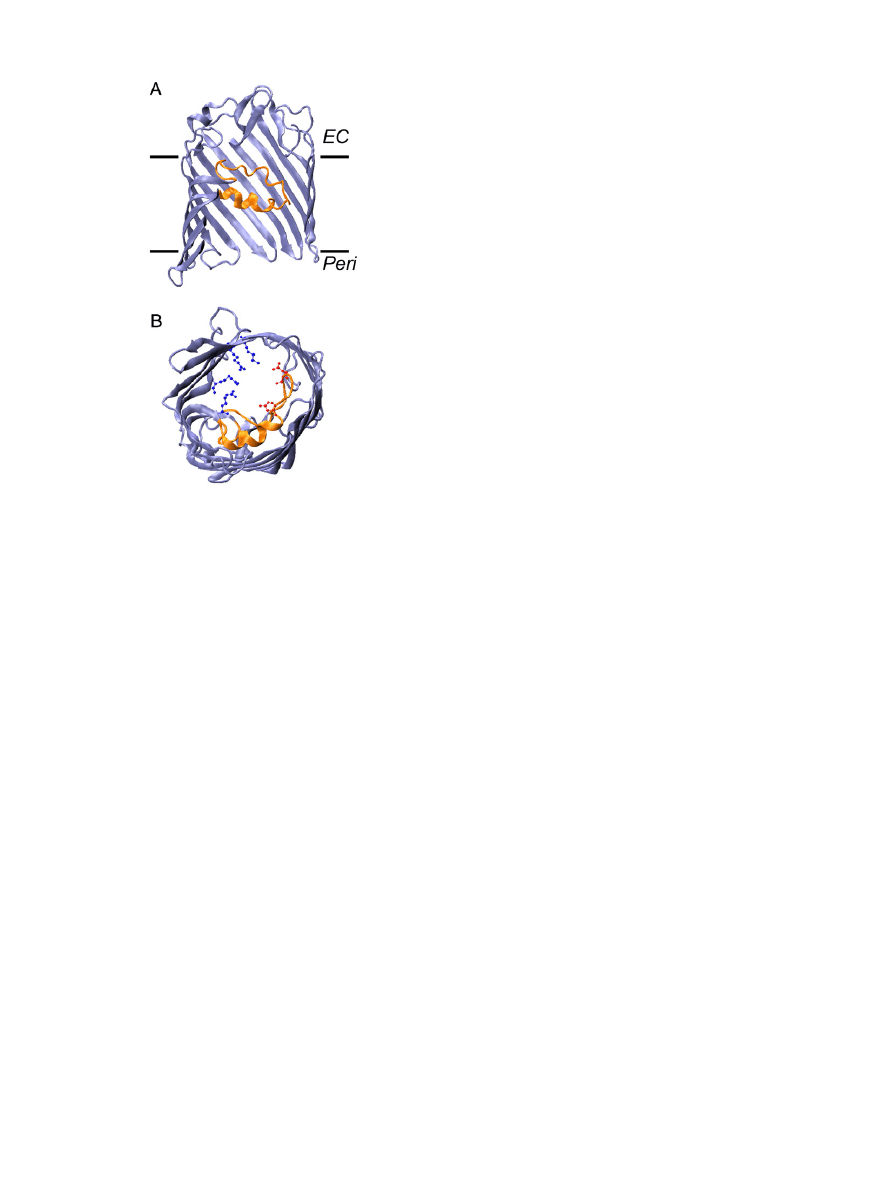
The E. coli general diffusion porins OmpF, PhoE and OmpC are trimers
of 16-stranded
β-barrels
(
). The large number and
con
figuration of the β-strands allow for the formation of a central
hydrophilic pore in each
β-barrel. The pore of some other OM
proteins, such as the enterobactin transporter FepA
or the adhesin
translocator FhaC
, is essentially obstructed by a globular plug
domain. But this is not the case for general diffusion porins. The pore
is, however, somewhat constricted by the inwardly folded extra-
cellular loop L3 (shown in orange in
). This loop, together with
the opposite barrel wall, form the so-called eyelet or constriction zone,
which determines the size exclusion limit and other permeation
properties of the barrel (see below). At this level, the pore size of
OmpF is 7 × 10 Å
. A conserved set of charged residues decorates
the eyelet: negatively charged residues (in red in
) are typically
found on the L3 loop itself, and positive charges (in blue in
often form a cluster on the opposite barrel wall. These residues have
been shown to play an important role in ionic movement and in ionic
selectivity (see below). The
β-strands are connected to each other by
short turns on the periplasmic side and long loops on the extracellular
side. This protruding extracellular domain provides a site for
interactions with speci
fic colicins and phages that use porins as
surface receptors
.
3.1.2. Pore properties and permeation
The functional properties of porins have been the subject of
investigation for over 30 years. Initial work established the size
exclusion cutoffs of porins by measuring the transport of various size
sugars using liposome swelling assays
. A value of about 600 Da
was determined for OmpF
, which implies that ions, amino acids,
and small sugars use general diffusion porins for gaining access to the
periplasm. Disaccharides, larger sugars and other molecules need to
use dedicated pathways for OM transport
. These early studies
established the molecular sieving properties of porins, and provided
an explanation for the high diffusion rates of these compounds
through the OM
.
The application of electrophysiology to the study of porins, along
with computational studies, has permitted a better understanding of
810
A.H. Delcour / Biochimica et Biophysica Acta 1794 (2009) 808
–816
porin permeation at the molecular level. The traditional electrophy-
siological approach is the study of porin-mediated ion currents in
planar lipid bilayers (also known as
“black lipid membranes” or
“BLM”). A lipid bilayer is formed over an aperture pierced through a
Te
flon film separating two chambers. Each chamber contains a
buffered ionic solution and an electrode used to measure electric
current due to the
flow of ions across the bilayer and to clamp the
transmembrane potential required to promote ion movement. Puri
fied
detergent-solubilized channel proteins or proteoliposomes are added
to one chamber (the so-called cis side), and spontaneously insert in the
bilayer over time. The sequential insertions of open channels in the
membrane lead to discrete current jumps due to ion movement
through the open channels. The conductance (i.e. the amount of
current per unit voltage) of a channel can be obtained from measuring
the size of these current jumps. In the case of porins, this would
represent the trimeric conductance, since porins typically purify and
insert in the bilayer as trimers. By manipulating the protein
concentration, it is possible to ensure that either many or only one
porin trimer inserts, and investigations can be performed on single
channels or on populations of channels. After insertion, the channel
activity can be studied in various conditions and membrane potentials.
The patch-clamp technique has also been applied to the study of
puri
fied porins reconstituted in artificial liposomes. Here a small patch
of liposome membrane is drawn at the tip of a 1
μM-diameter glass
pipette, and the current
flowing through this patch is recorded at a
fixed membrane potential. Because of the small area of membrane
under investigation, the patch clamp technique typically offers a
better signal-to-noise ratio than BLM. This technique permitted the
discovery that porins
flicker between multiple states, whose kinetics
and conductance can be affected in mutants and in the presence of
modulators (see below).
Studies performed by the Benz and the Rosenbusch groups in the
70s and 80s established some of the hallmark properties of the general
diffusion porins, such as high ionic current due to the relatively large
pore size, low ionic selectivity (although some porins show preference
for cations (OmpC) or anions (PhoE)), and high open probability, in
standard bilayer electrophysiology conditions of low voltage, neutral
pH and high ionic strength (but see below)
. Computational
modeling studies have suggested that the paths taken by anions and
cations are divergent at the eyelet, as cations are drawn close to the
negative charges of the L3 loop, and anions
flow near the positively
charged cluster of the opposite barrel wall
. This type of work
emphasizes the notion that the permeating ions interact with the wall
of the channel and that ion movement does not follow simple
diffusion. This was demonstrated experimentally by measuring the
conductance and selectivity of various general diffusion porins in
solutions of varying ionic strength or pH, and in variants with
mutations at speci
fic pore exposed residues
Bezrukov's group showed that the selectivity of OmpF for cations
relative to anions increases sharply in solutions of low ionic strength
. The channel reaches nearly ideal cation-selectivity in solutions
of
b100 mM KCl. Furthermore, at pHsb4, the channel reverses its
selectivity from preferring cations to preferring anions. The authors
combined these experimental observations with calculations of the
distribution of charged residues in the pore lumen and concluded that
electrostatic interactions exist between the permeating ions and the
charges of ionizable residues over the entire channel length
However, shifts in selectivity are detected upon mutations of single
residues. Substitution at the pore-exposed D113 residue in OmpF
and its homologs in OmpC
and the Vibrio cholerae porin OmpU
decreases cation-selectivity. Opposite effects are seen upon
charge removal at arginines of the constriction zone
The mutations also affect conductance, although there is no strict
correlation between an apparent increase in pore size due to removal
of a bulky side chain and increase conductance
. The results
highlight the notion that conductance is a re
flection not only of pore
size but also interaction of permeating ions with channels walls, and
strengthen the argument that the derivation of pore size from
conductance measurements should be avoided
. In addition, an
increase in conductance is not always a good predictor of an increased
permeation of larger substrates or antibiotic susceptibility, as was
shown for OmpF
and the V. cholerae porin OmpU
.
3.1.3. Functional modulation of porins
The fact that the activity of porins can be quickly modulated by
ligand binding and a variety of physico-chemical parameters is an
important
– but relatively unappreciated – aspect of outer membrane
permeability. Porins are thought of as permanently open pores, and
for years, the only documented mechanism to reduce outer mem-
brane permeability was through a lower porin expression due to
environmental factors or mutations. The knowledge of which
parameters lead to rapid closure of porins is important, since the
resulting tightening of the OM will decrease the ef
ficacy of penetra-
tion of antibiotics using the porin-mediated pathway. The
first rapid
modulation of porin function to be described was transmembrane
voltage
, but the signi
ficance of this phenomenon is often
dismissed because the OM is believed to be without a transmembrane
potential (but see below). Still, the voltage-dependent inactivation of
porins is a robust phenomenon, shown to differ among different
porins species, and affected by mutations at speci
fic pore residues
. The voltage sensitivity of porins is typically quanti
fied by
the so-called
“threshold” potential, i.e. the minimum membrane
potential at which porins start to close. When the membrane potential
is above this value, porin monomers close, often sequentially, in a
typical stepwise fashion. The protein appears to reach a deep
inactivated state, as it is reluctant to re-opening, even at lower
voltages, and hysteresis is observed when voltages are slowly ramped
Fig. 2. Structure of an OmpF monomer. (A) Side view of a single
β-barrel of the OmpF
trimer to highlight the location of the protein in the membrane bilayer (
“EC” refers to
the extracellular side, and
“Peri” refers to the periplasmic side); note that some of the
protein structure has been cut out of view in order to better visualize the constricting L3
loop (orange). (B) View of the OmpF monomer from the periplasmic side, highlighting
the con
figuration of the eyelet or constriction zone. Important residues of the eyelet are
acidic residues of the L3 loop (in red) and a cluster of basic amino acids of the opposite
barrel wall (in blue).
811
A.H. Delcour / Biochimica et Biophysica Acta 1794 (2009) 808
–816

up and down in bilayers containing many channels
. The threshold
potential is typically quite high (
∼150 mV for OmpF
and
∼200 mV
for OmpC
), but some porins are more voltage-sensitive (V.
cholerae OmpT has a threshold potential of
∼90 mV
). Nikaido
demonstrated that Donnan potentials established by accumulation of
periplasmic negatively charged membrane-derived oligosaccharides
(MDOs) are unable to decrease porin-mediated permeability to
β-
lactams
. However, it is possible that this negative result stems
from the asymmetric voltage dependence of porins
. OmpF might
close in vivo upon the opposite membrane potential (more positive on
the periplasmic side relative to the outside); this potential could be
established in vivo by a concentration gradient of potassium ions, for
example, if the absolute ionic strength of the periplasmic and external
solutions is relatively low (
b100 mM), i.e. in the range where OmpF
becomes a highly selective cation channel
. This still needs to be
demonstrated experimentally.
The voltage-dependent inactivation of porins demonstrated that
porins can exist in non-conducting, i.e. closed, forms, and set the stage
for the discovery of other possible modulators of porin function. In
particular, two other important forms of modulation lead to closing of
porins: acidic pH and binding of polyamines. Besides effects on
conductance and selectivity
, acid pH also promotes kinetic
changes in porins. Fast
flickering in the open channel noise drastically
increases, in particular at pHs
b4, and may be attributed to protona-
tion
–deprotonation events of key acidic residues in the pore
. In
addition, the channel increases its closing probability at acidic pH. In
OmpF, this is often seen as a sequential step-wise closing of
monomeric units after application of a transmembrane voltage. It is
similar to the effect of voltage, but occurs at much lower potentials
than at neutral pH. It is possible that it stems from an enhanced
voltage-sensitivity, as documented
. In OmpU, channels are
immediately stabilized in a closed con
figuration, and surprisingly,
individual closures of three monomers are not observed, but rather
closing events of an apparent single channel of increasingly larger size
as the pH is decreased
. In OmpF, extracellular loops L1, L7 and L8
have been implicated in the conformational changes that might lead
to acidic pH-induced channel closures
. Altogether, these loops
form a lid-type domain that might close up above the pore, as
suggested by atomic force microscopy of OmpF surface at low pH
.
E. coli OmpF and OmpC porins are inhibited by the polyamines
spermine, spermidine and cadaverine
. This is also true for the
V. cholerae porin OmpU (Delcour, unpublished). These linear, highly
charged, amine compounds are small enough to pass through porins,
and indeed, the kinetics of the modulation observed in patch clamp
experiments do not bear the hallmarks of open channel block. Rather, it
appears that the compounds bind to an internal pore-exposed site and
trigger channel closures. These effects are rather complex, with a greatly
increased
flickering activity to states of lower conductance than the
monomeric conductance (subconductance states)
, and prolonged
monomeric closures
. Mutagenesis work de
fined a binding site
involving the L3 loop acidic residues D113 and D121, and also Y294 for
the case of spermine
. We envisage a model whereby polyamines
would saddle over the L3 loop through ionic interactions involving the
amine groups, and cause a destabilization or a possible movement of the
L3 loop, leading to channel closure. Modulation of this kind by
polyamines has a marked impact on the overall outer membrane
permeability
.The porin closure induced by spermine might have
some important therapeutic consequences in treatment of infections of
tissues where spermine content is high, such as in the prostate.
Cadaverine is endogenously produced by E. coli and secreted in
conditions of acidic pH. By manipulating the Cad operon, we have
shown that the production and release of endogenous cadaverine
decreases outer membrane permeability
. The cadaverine-depen-
dent modulation of porin is part of adaptive response to a pH drop,
since a cadaverine-resistant porin mutant is outcompeted by wildtype
in acidic conditions
. These observations reinforce the notion that
the rapid modulation of porin function can provide cells with an
emergency mechanism to shut down OM permeability until slower
mechanisms involving regulation of porin expression are put in place.
Importantly, they suggest that the permeability of the OM to
antibiotics, for example, might be changing for cells in different
external conditions. Indeed polyamines were shown to inhibit the
flux
of cephaloridine through the porins OmpF and OmpC
.
3.2. Porin-mediated antibiotic permeability
The permeability of porins to
β-lactam antibiotics has been
demonstrated by various means. Evidence for a direct role of porins
in mediating the diffusion of
β-lactams was provided by purifying and
reconstituting porins into liposomes and using either a liposome
swelling assay
, or measuring the antibiotic degradation rate by an
entrapped
β-lactamase
. Measurement of antibiotic
flux in whole
cells was originally developed by Zimmermann and Rosselet
and
then extensively used by Nikaido's group to characterize the
permeability of cephaloridine and other cephalosporins in various
cells types (wildtype and porin mutants), by taking advantage of the
fast rate of cephalosporin degradation by periplasmic
β-lactamase
. Rates of the order of
∼10–50 10
− 5
cm/s were found for the
permeation of zwitterionic drugs through OmpF, but were much
reduced for anionic compounds.
A molecular explanation for these
findings has recently emerged
from a more detailed view of the interactions of the permeating drugs
with the porin channels, obtained from the combination of electro-
physiology and computational studies. Bezrukov et al. demonstrated
that ampicillin acts as a transient open channel blocker of the OmpF
porin in a pH dependent manner, with a maximum block in a pH range
where the ampicillin molecule is zwitterionic
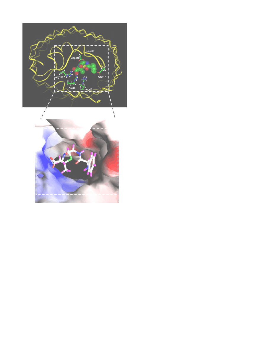
. Molecular dynamics
calculations explain this pH dependence, as they reveal that the drug
molecule perfectly occludes the pore in the zwitterionic form, as it
interacts simultaneously with negatively charged residues of L3 and
positively charged residues of the barrel wall (
). Such comple-
mentation between the charge distributions on the drug molecule and
the narrowest region of the OmpF pore has also been found for another
zwitterionic
β-lactam, amoxicillin
. On the contrary, poor interac-
tions were delineated for the di-anionic carbenicillin and the mono-
anionic
β-lactams azlocillin and piperacillin. This negligible binding
correlates with the poor diffusion rates measured from such compounds
from liposome swelling assays
. On the other hand, high diffusion
rates were obtained for ampicillin and amoxicillin. Thus, it appears that
interactions at the OmpF constriction zone facilitate the drug transloca-
tion, and that the nature and position of speci
fic charges on the
antibiotic molecule and on OmpF play a major role in these interactions.
Experimentally, site-directed mutations of many key charged
residues of the porin constriction zone affect
β-lactam flux and
sensitivity
. The involvement of speci
fic OmpF residues as
anchorage points for several cephalosporins has been suggested from
computational studies, as well
. Some mutations also involved
uncharged residues. For example, the diffusion of radiolabeled
cefepime was drastically decreased in the G119D and G119E mutants
. The X-ray structure of the G119D mutant OmpF shows that the
introduced aspartate residue protrudes in the eyelet and constricts the
diameter the pore
. Consequently, the channel conductance,
diffusion rate of various sugars and sensitivity to cephalosporins are
greatly reduced
. On the other hand, mutations at the R132
residues lead to improved growth on maltodextrins relative to
wildtype
and increased cefepime diffusion
, possibly due to
an increase in pore diameter
Carbapenems, such as imipenem and merpenem, are
β-lactam
antibiotics with a high resistance to the action of
β-lactamase. They
have been particularly effective against P. aeruginosa, an organism
which appears less susceptible to most antibiotics than Enterobacter-
iaceae, because of decreased OM permeability and an ef
ficient drug
812
A.H. Delcour / Biochimica et Biophysica Acta 1794 (2009) 808
–816
ef
flux system. The low OM permeability stems from the lack of general
diffusion porins, as P. aeruginosa acquires its nutrients through
dedicated speci
fic porins
. The isolation of an imipenem resistant
strain pointed to the role of the OprD porin (previously known as
protein D2) in the permeability to imipenem
, and indeed this
protein was later shown to allow the facilitated diffusion of
carbapenems and penems through the OM
. When the puri
fied
protein is reconstituted into arti
ficial bilayers, the formed channels
have a very low conductance, but can be blocked by imipenem,
indicating the presence of a speci
fic binding site
. Additional
studies demonstrated that, in fact, the OprD porin is used for the
uptake of basic amino acids and peptides, which share structural
similarity with the carbapenem molecule
.
Quinolones are believed to use a dual pathway for entry into
bacterial cells, because drug
flux and susceptibility are both sensitive to
the presence of porins (in particular OmpF) and to the manipulations
that disrupt the outer membrane LPS barrier
. The relative
contribution of the two pathways correlates with the hydrophobicity
and the protonation state of the quinolones, in the manners described
below. Hydrophobic quinolones are more effective in LPS mutants
There is a report that the quinolone
fleroxacin induces the same
perturbations of the OM as does gentamycin or EDTA, supporting the
contention that quinolones might act as chelating agents and use a
self-promoted pathway as aminoglycosides and cationic peptides do
. However, the sensitivity of cells to less hydrophobic quinolones,
such as nor
floxacin and ciprofloxacin and other drugs with similar
hydrophobicity coef
ficient of less than 0.1, was not much affected in
mutants in LPS structure
suggesting that they might use porin for
access through the OM. Indeed a reduced accumulation of radiolabeled
nor
floxacin was observed in E. coli strains lacking OmpF
Moreover, the
flux of norfloxacin in Enterobacter cloacae was inhibited
in the presence of spermine or cefepime, both known to use porins for
permeation through the OM, thus con
firming that norfloxacin diffuses
through the porin lumen
. Nikaido and Thanassi have proposed
that quinolones exist in an equilibrium of charged and uncharged
species depending on the solution pH
. For example, they
calculated that about 10% of nor
floxacin exists as an uncharged species
at pH 7.4, and this ratio is even higher (
∼40% at pH 6.5) for amifloxacin.
These authors have argued that the uncharged quinolone molecules
cross the OM through the lipid bilayer, while the negatively charged
molecules are likely to pass through porin channels as magnesium
chelates. Thus the relative contributions of the porin-mediated and
lipid-mediated pathways are likely to depend on the protonation
–
deprotonation states of the drug, which will themselves be in
fluenced
by external pH. In addition, the charged species is proposed to
accumulate in the periplasm due to the interior-negative Donnan
potential across the OM
. This accumulation leads to high
cytoplasmic levels as well, as the cytoplasm equilibrates very rapidly
with the periplasm, even for drugs with oil/water partition coef
ficient
less than 0.1. In porin-de
ficient mutants, quinolones still permeate
through the outer membrane bilayer itself in their uncharged form, but
do not accumulate in the periplasm because they are not sensitive to
the Donnan potential, thus leading to decreased cytoplasmic concen-
trations and ef
ficacy.
The uptake of tetracycline by E. coli cells was shown to be reduced
in a mutant lacking OmpF
, con
firming the suggestion that
tetracycline uses this pathway based on increased resistance in
mutants with decreased ompF expression
. This accumulation,
however, is not null in the absence of OmpF, and it positively
correlates with pH, i.e. there is less in
flux of tetracycline at lower pH
(pH 6.0) relative to neutral pH, or even 7.8
. Tetracycline has a pKa
of 7.7, and therefore exists mostly in a protonated form at pHs under
the pKa. In this uncharged form, tetracycline is believed to enter cells
by diffusion through the OM lipid barrier
. Thus, tetracycline, like
fluoroquinolones, uses both a porin- and a lipid-mediated pathway,
depending on its protonated status.
3.3. Porins and antibiotic resistance
As described above, porins provide a path through the OM to small
hydrophilic antibiotics, such as
β-lactams, as well as tetracycline,
chloramphenicol and
fluoroquinolones
. Any decrease in the ability
or rate of entry of these compounds can lead to resistance. There is
an abundance of reports of antibiotic resistance acquired through
loss or functional change of porins in a large number of organisms,
such as E. coli, P. aeruginosa, Neisseria gonorrhoeae, Enterobacter
aerogenes and Klebsiella pneumoniae (see references
for
reviews, and references therein). Although much of the mechanistic
studies described above have focused on OmpF because of its well
understood structural and functional properties relative to any other
major porins, many of the reports of changes in porin expression
Fig. 3. Docking of an ampicillin molecule at the constriction zone of an OmpF monomer.
The top panel shows the
fit of the ampicillin molecule within the pore, with the
carboxylate group attracted to the cluster of arginines in the OmpF barrel, and the
ammonium group close to the acidic L3 loop residues. Colors of atoms in ampicillin are
as follows: green for carbon, red for oxygen, blue for nitrogen and yellow for sulfur.
Hydrogen atoms are not shown. The OmpF backbone is shown as a yellow ribbon. The
lower panel shows the solvent accessible surface of the OmpF eyelet highlighting the
electrostatic potential with blue color for positive potential and red color for negative
potential. Ampicillin is shown in a stick model with the following colors: white for
carbon, red for oxygen, blue for nitrogen, green for sulfur and violet for hydrogen.
Reproduced from reference 76 with permission (copyright © 1993
–2008 by The
National Academy of Sciences of the United States of America, all rights reserved).
813
A.H. Delcour / Biochimica et Biophysica Acta 1794 (2009) 808
–816

often implicated both OmpF and OmpC. The role of minor porins
(such as NmpC), or those expressed in speci
fic conditions (such as
PhoE), perhaps should not be underestimated, but there are far fewer
reports on the involvement of these porins in antibiotic resistance
(but see below). Still it appears that PhoE can serve as a conduit for
entry of
β-lactams (and be an even better one than OmpF and OmpC
if the drug bears a negative charge)
, as well as for chloramphe-
nicol and tetracycline
It would be impractical in this review to cite all or even most of
studies linking antibiotic resistance to general diffusion porins, but we
can highlight some of the generally found common themes with
speci
fic examples. There are two major porin-based mechanisms for
antibiotic resistance that have been reported in clinical isolates: 1)
alterations of outer membrane pro
files, including either loss/severe
reduction of porins or replacement of one or two major porins by
another; 2) altered function due to speci
fic mutations reducing
permeability.
Antibiotic resistance poses a daunting problem in hospital-
acquired infections. Pages et al. analyzed the porin content of 45
β-
lactam resistant clinical isolates of E. aerogenes obtained from French
hospitals
. Of those, 44% were shown to lack porin, as determined
by immunodetection. The MICs of four antibiotics (cefepime,
imipenem, cefotaxime and moxalactam) were drastically increased.
Additionally, many strains displayed high constitutive or inducible
β-
lactamase activity, but some strains did not, and thus antibiotic
resistance appears to originate essentially from the lack of porins in
those strains. The increase in MICs for those porin-de
ficient strains
was similar to those with robust
β-lactamase activity, indicating that a
reduction of porin-mediated permeability can be an ef
ficient strategy
for antibiotic resistance on its own.
Tetracylcine resistance can occur under antibiotic stress, by
exposing sensitive E. coli cells to progressively increasing concen-
tration of the antibiotic. The treatment, in fact, leads to a
chromosome-mediated multiple antibiotic resistance (Mar pheno-
type), where the cells become insensitive to a variety of hydrophilic
and lipophilic antibiotics
. The response involves the
coordinated change in the levels of multiple proteins including
porins and drug ef
flux pumps, through mechanisms involving
transcriptional and posttranscriptional regulation
. In particu-
lar, the upregulation of marA leads to increased levels of the small
RNA micF, which inhibits translation of ompF RNA. Decreased OmpF
levels are also postulated to originate from the periplasmic
accumulation of other OM proteins, such as TolC and OmpX,
which might titrate away the chaperones and assembly proteins
required for membrane insertion of OM proteins
. Another
example of upregulation of OmpX in coordination with a strong
repression of general diffusion porins has also been documented for
acquired resistance to a large number of antibiotics of a strain of
Salmonella enterica typhimurium after exposure to nalidixic acid
. In this case, repression also included other porins, besides
OmpF, such as NmpC, LamB and Tsx.
The substitution of a narrower porin in lieu of the constitutively
expressed large general-diffusion porins is another strategy for
acquiring antibiotic resistance. For example, some clinical isolates
from K. pneumoniae lack the large diffusion channels OmpK35 and
OmpK36, but express a normally quiescent porin, OmpK37, which
appears to form a smaller pore on the basis of sugar permeability
. This porin is akin to OmpN of E. coli and OmpS2 of S. typhi, two
porin types which are normally strongly down-regulated in laboratory
media conditions. The presence of OmpK37 combined with the
absence of OmpK35 and OmpK36 lead to a drastic increase in the MICs
of cefotaxime and cefoxitin, but not of carbapenems, indicating that
these compounds might still be able to
flux through OmpK37 as they
do through P. aeruginosa OprD. This provides an explanation for the
fact that K. pneumoniae infections resistant to most
β-lactams can still
be treated by carbapenems.
Altered function of porin leading to reduced permeation rate is
another strategy found in antibiotic resistant bacteria. A hot spot for
single or multiple mutations leading to such phenotype is the L3 loop,
which delineates the constriction zone of general diffusion porins. A
clinical isolate of E. aerogenes was found to have a glycine
→aspartate
substitution on the L3 loop of its major porin
, which might lead
to a distortion of the loop or further narrowing of the pore lumen, as in
G119D of OmpF
. This mutant is characterized by a 3-fold decrease
in porin conductance and a drastic reduction in cephalosporin
sensitivity. It was found later on that this porin is Omp36, which is
highly similar to E. coli OmpC
. This clinical isolate and two others
from E. aerogenes, in fact, present multiple mutations in the porin
gene, and are also highly resistant to cefepime, cefpirome and
imipenem. Similar alterations in the amino acid composition of the
N. gonorrhoeae porin Por have also be documented
. Here, a
mutant with enhanced resistance to penicillin and tetracycline was
found to have multiple mutations throughout the porin gene, and in
particular in a region putatively homologous to the constricting L3
loop. Interestingly, six clinical isolates with similar resistance to
penicillin also displayed single point mutations in the same region.
Finally, some bacterial species, such as P. aeruginosa, are intrinsi-
cally more resilient to antibiotic treatments, because of a low
abundance of general diffusion porins, combined with numerous and
highly ef
ficient drug efflux mechanisms
. As described above,
OprF, the major porin of P. aeruginosa, is present in high abundance as a
closed conformer, and exists as an open channel only at very low levels.
Not surprisingly, acquired resistance to
β-lactam antibiotics does not
seem to involve loss or modi
fication of OprF
. Resistance to
carbapenems can be observed in mutants lacking the porin-speci
fic
OprD (see above), and in mutants with deletions in the L2 loop of OprD
. Carbapenem resistance via porin-delimitated pathways is not
restricted to P. aeruginosa, as described above.
4. Concluding remarks
In conclusion, mechanisms affecting the barrier properties of the
OM lipid bilayer itself or the expression and/or function of the general
diffusion porin channels residing in the OM have an impact on the
sensitivity of gram-negative bacteria to many different types of
antibiotics. Clearly any weakening of the LPS bilayer by targeting LPS
synthesizing enzymes will sensitize bacteria to hydrophobic and some
hydrophilic antibiotics, leading to the possibility of combinatorial drug
therapy. A better understanding of the function of general diffusion
porins, and in particular of the parameters that might lead to porin
closure or inactivation, will allow a reassessment of the ef
ficiency of
penetration of the antibiotics using this pathway in different
conditions. It is hoped that, as we further understand at the molecular
level the structure and function of these OM macromolecules and of
those that regulate them, scientists will be able to re
fine the current
drug therapies or design new types of antibiotics that target these
surface exposed entities.
Acknowledgement
Our own work on porins has been supported by NIH grant AI34905
and grant E-1597 from the Welch Foundation.
References
[1] C.R. Raetz, C. Whit
field, Lipopolysaccharide endotoxins, Annu. Rev. Biochem. 71
(2002) 635
–700.
[2] R.J. Kadner, in: F.C. Neidhart (Ed.), Escherichia coli and Salmonella, Cellular and
Molecular Biology, ASM press, Washington, DC, 1996, pp. 58
–87.
[3] Y. Kamio, H. Nikaido, Outer membrane of Salmonella typhimurium: accessibility
of phospholipid head groups to phospholipase C and cyanogen bromide
activated dextran in the external medium, Biochemistry 15 (1976) 2561
–2570.
[4] H. Nikaido, in: F.C. Neidhart (Ed.), Escherichia coli and Salmonella, Cellular and
Molecular Biology, ASM Press, Washington, DC, 1996, pp. 29
–47.
814
A.H. Delcour / Biochimica et Biophysica Acta 1794 (2009) 808
–816

[5] Y. Hirota, H. Suzuki, Y. Nishimura, S. Yasuda, On the process of cellular division in
Escherichia coli: a mutant of E. coli lacking a murein-lipoprotein, Proc. Natl. Acad.
Sci. U. S. A. 74 (1977) 1417
–1420.
[6] I. Sonntag, H. Schwarz, Y. Hirota, U. Henning, Cell envelope and shape of
Escherichia coli: multiple mutants missing the outer membrane lipoprotein and
other major outer membrane proteins, J. Bacteriol. 136 (1978) 280
–285.
[7] E. Sugawara, E.M. Nestorovich, S.M. Bezrukov, H. Nikaido, Pseudomonas
aeruginosa porin OprF exists in two different conformations, J. Biol. Chem. 281
(2006) 16220
–16229.
[8] E. Sugawara, H. Nikaido, Pore-forming activity of OmpA protein of Escherichia
coli, J. Biol. Chem. 267 (1992) 2507
–2511.
[9] R.G. Gerlach, M. Hensel, Protein secretion systems and adhesins: the molecular
armory of gram-negative pathogens, Int. J. Med. Microbiol. 297 (2007)
401
–415.
[10] M. Kostakioti, C.L. Newman, D.G. Thanassi, C. Stathopoulos, Mechanisms of
protein export across the bacterial outer membrane, J. Bacteriol. 187 (2005)
4306
–4314.
[11] H. Nikaido, Molecular basis of bacterial outer membrane permeability revisited,
Microbiol. Mol. Biol. Rev. 67 (2003) 593
–656.
[12] N. Ruiz, D. Kahne, T.J. Silhavy, Advances in understanding bacterial outer-
membrane biogenesis, Nat. Rev. Microbiol. 4 (2006) 57
–66.
[13] C.R. Raetz, in: F.C. Neidhart (Ed.), Escherichia coli and Salmonella, Cellular and
Molecular Biology, ASM Press, Washington, DC, 1996, pp. 1035
–1063.
[14] M. Vaara, Agents that increase the permeability of the outer membrane,
Microbiol. Rev. 56 (1992) 395
–411.
[15] J.B. Dame, B.M. Shapiro, Use of polymyxin B, levallorphan, and tetracaine to isolate
novel envelope mutants of Escherichia coli, J. Bacteriol. 127 (1976) 961
–972.
[16] P.H. Mäkelä, M. Sarvas, S. Calcagno, K. Lounatmaa, Isolation and genetic
characterization of polymyxin-resistant mutants of Salmonella, FEMS Microbiol.
Lett. 3 (1978) 323
–326.
[17] M. Vaara, T. Vaara, M. Sarvas, Decreased binding of polymyxin by polymyxin-
resistant mutants of Salmonella typhimurium, J. Bacteriol. 139 (1979) 664
–667.
[18] M. Vaara, T. Vaara, M. Jensen, I. Helander, M. Nurminen, E.T. Rietschel, P.H.
Makela, Characterization of the lipopolysaccharide from the polymyxin-resistant
pmrA mutants of Salmonella typhimurium, FEBS Lett. 129 (1981) 145
–149.
[19] A.A. Peterson, S.W. Fesik, E.J. McGroarty, Decreased binding of antibiotics to
lipopolysaccharides from polymyxin-resistant strains of Escherichia coli and
Salmonella typhimurium, Antimicrob. Agents Chemother. 31 (1987) 230
–237.
[20] Z. Zhou, A.A. Ribeiro, S. Lin, R.J. Cotter, S.I. Miller, C.R. Raetz, Lipid A modi
fications
in polymyxin-resistant Salmonella typhimurium: PMRA-dependent 4-amino-4-
deoxy-
L
-arabinose, and phosphoethanolamine incorporation, J. Biol. Chem. 276
(2001) 43111
–43121.
[21] J.S. Gunn, S.I. Miller, PhoP
–PhoQ activates transcription of pmrAB, encoding a
two-component regulatory system involved in Salmonella typhimurium anti-
microbial peptide resistance, J. Bacteriol. 178 (1996) 6857
–6864.
[22] K.L. Roland, L.E. Martin, C.R. Esther, J.K. Spitznagel, Spontaneous pmrA mutants of
Salmonella typhimurium LT2 de
fine a new two-component regulatory system
with a possible role in virulence, J. Bacteriol. 175 (1993) 4154
–4164.
[23] J.S. Gunn, K.B. Lim, J. Krueger, K. Kim, L. Guo, M. Hackett, S.I. Miller, PmrA
–PmrB-
regulated genes necessary for 4-aminoarabinose lipid A modi
fication and
polymyxin resistance, Mol. Microbiol. 27 (1998) 1171
–1182.
[24] L.R. Prost, S. Sanowar, S.I. Miller, Salmonella sensing of anti-microbial mechan-
isms to promote survival within macrophages, Immunol. Rev. 219 (2007) 55
–65.
[25] M.W. Bader, S. Sanowar, M.E. Daley, A.R. Schneider, U. Cho, W. Xu, R.E. Klevit, H. Le
Moual, S.I. Miller, Recognition of antimicrobial peptides by a bacterial sensor
kinase, Cell 122 (2005) 461
–472.
[26] L. Guo, K.B. Lim, C.M. Poduje, M. Daniel, J.S. Gunn, M. Hackett, S.I. Miller, Lipid A
acylation and bacterial resistance against vertebrate antimicrobial peptides, Cell
95 (1998) 189
–198.
[27] R.E. Hancock, S.W. Farmer, Z.S. Li, K. Poole, Interaction of aminoglycosides with
the outer membranes and puri
fied lipopolysaccharide and OmpF porin of
Escherichia coli, Antimicrob. Agents Chemother. 35 (1991) 1309
–1314.
[28] R.E. Hancock, A. Bell, Antibiotic uptake into gram-negative bacteria, Eur. J. Clin.
Microbiol. Infect. Dis. 7 (1988) 713
–720.
[29] M. Vaara, T. Vaara, Polycations sensitize enteric bacteria to antibiotics,
Antimicrob. Agents Chemother. 24 (1983) 107
–113.
[30] C. Dong, K. Beis, J. Nesper, A.L. Brunkan-Lamontagne, B.R. Clarke, C. Whit
field, J.H.
Naismith, Wza the translocon for E. coli capsular polysaccharides de
fines a new
class of membrane protein, Nature 444 (2006) 226
–229.
[31] A. Basle, G. Rummel, P. Storici, J.P. Rosenbusch, T. Schirmer, Crystal structure of
osmoporin OmpC from E. coli at 2.0 A, J. Mol. Biol. 362 (2006) 933
–942.
[32] S.W. Cowan, T. Schirmer, G. Rummel, M. Steiert, R. Ghosh, R.A. Pauptit, J.N.
Jansonius, J.P. Rosenbusch, Crystal structures explain functional properties of two
E. coli porins, Nature 358 (1992) 727
–733.
[33] S.K. Buchanan, B.S. Smith, L. Venkatramani, D. Xia, L. Esser, M. Palnitkar, R.
Chakraborty, D. van der Helm, J. Deisenhofer, Crystal structure of the outer
membrane active transporter FepA from Escherichia coli, Nat. Struct. Biol. 6 (1999)
56
–63.
[34] B. Clantin, A.S. Delattre, P. Rucktooa, N. Saint, A.C. Meli, C. Locht, F. Jacob-
Dubuisson, V. Villeret, Structure of the membrane protein FhaC: a member of the
Omp85-TpsB transporter superfamily, Science 317 (2007) 957
–961.
[35] H. Nikaido, E.Y. Rosenberg, Porin channels in Escherichia coli: studies with
liposomes reconstituted from puri
fied proteins, J. Bacteriol. 153 (1983) 241–252.
[36] T. Nakae, Outer membrane of Salmonella typhimurium: Reconstitution of
sucrose-permeable membrane vesicles, Biochem. Biophys. Res. Commun. 64
(1975) 1224
–1230.
[37] H. Nikaido, E.Y. Rosenberg, Effect on solute size on diffusion rates through the
transmembrane pores of the outer membrane of Escherichia coli, J. Gen. Physiol.
77 (1981) 121
–135.
[38] R. Benz, K. Janko, W. Boos, P. Lauger, Formation of large, ion-permeable
membrane channels by the matrix protein (porin) of Escherichia coli, Biochim.
Biophys. Acta 511 (1978) 305
–319.
[39] R. Benz, K. Janko, P. Lauger, Ionic selectivity of pores formed by the matrix protein
(porin) of Escherichia coli, Biochim. Biophys. Acta 551 (1979) 238
–247.
[40] R. Benz, A. Schmid, R.E. Hancock, Ion selectivity of gram-negative bacterial
porins, J. Bacteriol. 162 (1985) 722
–727.
[41] H. Schindler, J.P. Rosenbusch, Matrix protein from Escherichia coli outer
membranes forms voltage-controlled channels in lipid bilayers, Proc. Natl.
Acad. Sci. U. S. A. 75 (1978) 3751
–3755.
[42] W. Im, B. Roux, Ions and counterions in a biological channel: a molecular
dynamics simulation of OmpF porin from Escherichia coli in an explicit
membrane with 1 M KCl aqueous salt solution, J. Mol. Biol. 319 (2002) 1177
–1197.
[43] A. Alcaraz, E.M. Nestorovich, M. Aguilella-Arzo, V.M. Aguilella, S.M. Bezrukov,
Salting out the ionic selectivity of a wide channel: the asymmetry of OmpF,
Biophys. J. 87 (2004) 943
–957.
[44] J. Bredin, N. Saint, M. Mallea, E.D.G. Molle, J.M. Pages, V. Simonet, Alteration of
pore properties of Escherichia coli OmpF induced by mutation of key residues in
anti-loop 3 region, Biochem. J. 363 (2002) 521
–528.
[45] B. Lauman, M. Pagel, A.H. Delcour, Altered pore properties and kinetic changes in
mutants of the Vibrio cholerae porin OmpU, Mol. Membr. Biol. 25 (2008) 489
–505.
[46] N. Liu, Structure
–function relationships of E. coli OmpC porin — the effects of
site-directed mutations on the porin channel function. Ph.D. Thesis., University
of Houston 1999.
[47] P.S. Phale, A. Philippsen, C. Widmer, V.P. Phale, J.P. Rosenbusch, T. Schirmer, Role
of charged residues at the OmpF porin channel constriction probed by
mutagenesis and simulation, Biochemistry 40 (2001) 6319
–6325.
[48] N. Saint, K.L. Lou, C. Widmer, M. Luckey, T. Schirmer, J.P. Rosenbusch, Structural
and functional characterization of OmpF porin mutants selected for larger pore
size. II. Functional characterization, J. Biol. Chem. 271 (1996) 20676
–20680.
[49] A.H. Delcour, Solute uptake through general porins, Front. Biosci. 8 (2003)
d1055
–1071.
[50] H. Nikaido, Porins and speci
fic diffusion channels in bacterial outer membranes,
J. Biol. Chem. 269 (1994) 3905
–3908.
[51] M.A. Arbing, D. Dahan, D. Boismenu, O.A. Mamer, J.W. Hanrahan, J.W. Coulton,
Charged residues in surface-located loops in
fluence voltage gating of porin from
Haemophilus sin
fluenzae type b, J. Membr. Biol. 178 (2000) 185–193.
[52] A. Baslé, A.H. Delcour, in: R. Benz (Ed.), Structure and Function of Bacterial and
Eukaryotic Porins, Wiley-Interscience, 2004, pp. 79
–98.
[53] N.D. Bishop, E.J. Lea, H. Mobasheri, S. Spiro, Altered voltage sensitivity of mutant
OmpC porin channels, FEBS Lett. 379 (1996) 295
–298.
[54] A.H. Delcour, J. Adler, C. Kung, A single amino acid substitution alters
conductance and gating of OmpC porin of Escherichia coli, J. Membr. Biol. 119
(1991) 267
–275.
[55] J.H. Lakey, E.J. Lea, F. Pattus, OmpC mutants which allow growth on maltodextrins
show increased channel size and greater voltage sensitivity, FEBS Lett. 278
(1991) 31
–34.
[56] P.S. Phale, T. Schirmer, A. Prilipov, K.L. Lou, A. Hardmeyer, J.P. Rosenbusch, Voltage
gating of Escherichia coli porin channels: role of the constriction loop, Proc. Natl.
Acad. Sci. U. S. A. 94 (1997) 6741
–6745.
[57] V.C. Simonet, A. Basle, K.E. Klose, A.H. Delcour, The Vibrio cholerae porins OmpU and
OmpT have distinct channel properties, J. Biol. Chem. 278 (2003) 17539
–17545.
[58] P. Van Gelder, N. Saint, P. Phale, E.F. Eppens, A. Prilipov, R. van Boxtel, J.P.
Rosenbusch, J. Tommassen, Voltage sensing in the PhoE and OmpF outer
membrane porins of Escherichia coli: role of charged residues, J. Mol. Biol. 269
(1997) 468
–472.
[59] K. Sen, J. Hellman, H. Nikaido, Porin channels in intact cells of Escherichia coli are
not affected by Donnan potentials across the outer membrane, J. Biol. Chem. 263
(1988) 1182
–1187.
[60] H. Samartzidou, A.H. Delcour, E.coli PhoE porin has an opposite voltage-
dependence to the homologous OmpF, EMBO J. 17 (1998) 93
–100.
[61] E.M. Nestorovich, T.K. Rostovtseva, S.M. Bezrukov, Residue ionization and ion
transport through OmpF channels, Biophys. J. 85 (2003) 3718
–3729.
[62] A. Baslé, R. Qutub, M. Mehrazin, J. Wibbenmeyer, A.H. Delcour, Deletions of
single extracellular loops affect pH-sensitivity, but not voltage-dependence, of
the E. coli porin OmpF, Protein Eng. Des. Sel. 17 (2004) 665
–672.
[63] J.C. Todt, W.J. Rocque, E.J. McGroarty, Effects of pH on bacterial porin function,
Biochemistry 31 (1992) 10471
–10478.
[64] G. Duret, V. Simonet, A.H. Delcour, Modulation of Vibrio cholerae porin function
by acidic pH, Channels 1 (2007) 70
–79.
[65] D.J. Müller, A. Engel, Voltage and pH-induced channel closure of porin OmpF
visualized by atomic force microscopy, J. Mol. Biol. 285 (1999) 1347
–1351.
[66] A.L. delaVega, A.H. Delcour, Cadaverine induces closing of E. coli porins, EMBO J.
14 (1995) 6058
–6065.
[67] R. Iyer, A.H. Delcour, Complex inhibition of OmpF and OmpC bacterial porins by
polyamines, J. Biol. Chem. 272 (1997) 18595
–18601.
[68] A. Baslé, R. Iyer, A.H. Delcour, Subconductance states in OmpF gating, Biochim.
Biophys. Acta 1664 (2004) 100
–107.
[69] R. Iyer, Z. Wu, P.M. Woster, A.H. Delcour, Molecular basis for the polyamine-OmpF
porin interactions: inhibitor and mutant studies, J. Mol. Biol. 297 (2000)
933
–945.
[70] A.L. delaVega, A.H. Delcour, Polyamines decrease Escherichia coli outer mem-
brane permeability, J. Bacteriol. 178 (1996) 3715
–3721.
815
A.H. Delcour / Biochimica et Biophysica Acta 1794 (2009) 808
–816

[71] H. Samartzidou, A.H. Delcour, Distinct sensitivities of OmpF and PhoE porins to
charged modulators, FEBS Lett. 444 (1999) 65
–70.
[72] H. Samartzidou, M. Mehrazin, Z. Xu, M.J. Benedik, A.H. Delcour, Cadaverine
inhibition of porin plays a role in cell survival at acidic pH, J. Bacteriol. 185 (2003)
13
–19.
[73] Y. Kobayashi, I. Takahashi, T. Nakae, Diffusion of beta-lactam antibiotics through
liposome membranes containing puri
fied porins, Antimicrob. Agents Chemother.
22 (1982) 775
–780.
[74] W. Zimmermann, A. Rosselet, Function of the outer membrane of Escherichia coli
as a permeability barrier to beta-lactam antibiotics, Antimicrob. Agents
Chemother. 12 (1977) 368
–372.
[75] H. Nikaido, E.Y. Rosenberg, J. Foulds, Porin channels in Escherichia coli: studies
with beta-lactams in intact cells, J. Bacteriol. 153 (1983) 232
–240.
[76] E.M. Nestorovich, C. Danelon, M. Winterhalter, S.M. Bezrukov, Designed to
penetrate: time-resolved interaction of single antibiotic molecules with bacterial
pores, Proc. Natl. Acad. Sci. U. S. A. 99 (2002) 9789
–9794.
[77] C. Danelon, E.M. Nestorovich, M. Winterhalter, M. Ceccarelli, S.M. Bezrukov,
Interaction of zwitterionic penicillins with the OmpF channel facilitates their
translocation, Biophys. J. 90 (2006) 1617
–1627.
[78] F. Yoshimura, H. Nikaido, Diffusion of beta-lactam antibiotics through the porin
channels of Escherichia coli K-12, Antimicrob. Agents Chemother. 27 (1985)
84
–92.
[79] S. Vidal, J. Bredin, J.M. Pagès, J. Barbe, Beta-lactam screening by speci
fic residues
of the OmpF eyelet, J. Med. Chem. 48 (2005) 1395
–1400.
[80] S.A. Benson, J.L. Occi, B.A. Sampson, Mutations that alter the pore function of the
OmpF porin of Escherichia coli K12, J. Mol. Biol. 203 (1988) 961
–970.
[81] R. Misra, S.A. Benson, Isolation and characterization of OmpC porin mutants with
altered pore properties, J. Bacteriol. 170 (1988) 528
–533.
[82] R. Misra, S.A. Benson, Genetic identi
fication of the pore domain of the OmpC
porin of Escherichia coli K-12, J. Bacteriol. 170 (1988) 3611
–3617.
[83] V. Simonet, M. Malléa, J.M. Pagès, Substitutions in the eyelet region disrupt
cefepime diffusion through the Escherichia coli OmpF channel, Antimicrob.
Agents Chemother. 44 (2000) 311
–315.
[84] D. Jeanteur, T. Schirmer, D. Fourel, V. Simonet, G. Rummel, C. Widmer, J.P.
Rosenbusch, F. Pattus, J.M. Pages, Structural and functional alterations of a
colicin-resistant mutant of OmpF porin from Escherichia coli, Proc. Natl. Acad. Sci.
U. S. A. 91 (1994) 10675
–10679.
[85] K.L. Lou, N. Saint, A. Prilipov, G. Rummel, S.A. Benson, J.P. Rosenbusch, T. Schirmer,
Structural and functional characterization of OmpF porin mutants selected for
larger pore size. I. Crystallographic analysis, J. Biol. Chem. 271 (1996)
20669
–20675.
[86] J. Trias, J. Dufresne, R.C. Levesque, H. Nikaido, Decreased outer membrane
permeability in imipenem-resistant mutants of Pseudomonas aeruginosa,
Antimicrob. Agents Chemother. 33 (1989) 1202
–1206.
[87] J. Trias, H. Nikaido, Outer membrane protein D2 catalyzes facilitated diffusion of
carbapenems and penems through the outer membrane of Pseudomonas
aeruginosa, Antimicrob. Agents Chemother. 34 (1990) 52
–57.
[88] H. Huang, R.E. Hancock, The role of speci
fic surface loop regions in determining
the function of the imipenem-speci
fic pore protein OprD of Pseudomonas
aeruginosa, J. Bacteriol. 178 (1996) 3085
–3090.
[89] J. Trias, H. Nikaido, Protein D2 channel of the Pseudomonas aeruginosa outer
membrane has a binding site for basic amino acids and peptides, J. Biol. Chem.
265 (1990) 15680
–15684.
[90] J.S. Chapman, N.H. Georgopapadakou, Routes of quinolone permeation in
Escherichia coli, Antimicrob. Agents Chemother. 32 (1988) 438
–442.
[91] K. Hirai, H. Aoyama, T. Irikura, S. Iyobe, S. Mitsuhashi, Differences in susceptibility
to quinolones of outer membrane mutants of Salmonella typhimurium and
Escherichia coli, Antimicrob. Agents Chemother. 29 (1986) 535
–538.
[92] S.P. Cohen, D.C. Hooper, J.S. Wolfson, K.S. Souza, L.M. McMurry, S.B. Levy,
Endogenous active ef
flux of norfloxacin in susceptible Escherichia coli, Anti-
microb. Agents Chemother. 32 (1988) 1187
–1191.
[93] J. Chevalier, M. Mallea, J.M. Pages, Comparative aspects of the diffusion of
nor
floxacin, cefepime and spermine through the F porin channel of Enterobacter
cloacae, Biochem. J. 348 (Pt 1) (2000) 223
–227.
[94] H. Nikaido, D.G. Thanassi, Penetration of lipophilic agents with multiple
protonation sites into bacterial cells: tetracyclines and
fluoroquinolones as
examples, Antimicrob. Agents Chemother. 37 (1993) 1393
–1399.
[95] D.G. Thanassi, G.S. Suh, H. Nikaido, Role of outer membrane barrier in ef
flux-
mediated tetracycline resistance of Escherichia coli, J. Bacteriol. 177 (1995)
998
–1007.
[96] S.P. Cohen, L.M. McMurry, S.B. Levy, marA locus causes decreased expression of
OmpF porin in multiple-antibiotic-resistant (Mar) mutants of Escherichia coli,
J. Bacteriol. 170 (1988) 5416
–5422.
[97] W. Achouak, T. Heulin, J.M. Pages, Multiple facets of bacterial porins, FEMS
Microbiol. Lett. 199 (2001) 1
–7.
[98] K. Poole, Outer membranes and ef
flux: the path to multidrug resistance in gram-
negative bacteria, Curr. Pharm. Biotechnol. 3 (2002) 77
–98.
[99] K. Poole, Resistance to beta-lactam antibiotics, Cell. Mol. Life Sci. 61 (2004)
2200
–2223.
[100] P.G. Mortimer, L.J. Piddock, The accumulation of
five antibacterial agents in porin-
de
ficient mutants of Escherichia coli, J. Antimicrob. Chemother. 32 (1993) 195–213.
[101] R.N. Charrel, J.M. Pages, P. De Micco, M. Mallea, Prevalence of outer membrane
porin alteration in beta-lactam-antibiotic-resistant Enterobacter aerogenes,
Antimicrob. Agents Chemother. 40 (1996) 2854
–2858.
[102] A.M. George, S.B. Levy, Ampli
fiable resistance to tetracycline, chloramphenicol,
and other antibiotics in Escherichia coli: involvement of a non-plasmid-
determined ef
flux of tetracycline, J. Bacteriol. 155 (1983) 531–540.
[103] A.M. George, S.B. Levy, Gene in the major cotransduction gap of the Escherichia
coli K-12 linkage map required for the expression of chromosomal resistance to
tetracycline and other antibiotics, J. Bacteriol. 155 (1983) 541
–548.
[104] M. Viveiros, M. Dupont, L. Rodrigues, I. Couto, A. Davin-Regli, M. Martins, J.M.
Pages, L. Amaral, Antibiotic stress, genetic response and altered permeability of E.
coli, PLoS ONE 2 (2007) e365.
[105] S.E. Dowd, K. Killinger-Mann, M. Brashears, J. Fralick, Evaluation of gene
expression in a single antibiotic exposure-derived isolate of Salmonella enterica
typhimurium 14028 possessing resistance to multiple antibiotics, Foodborne
Pathog. Dis. 5 (2008) 205
–221.
[106] A. Domenech-Sanchez, S. Hernandez-Alles, L. Martinez-Martinez, V.J. Benedi, S.
Alberti, Identi
fication and characterization of a new porin gene of Klebsiella
pneumoniae: its role in beta-lactam antibiotic resistance, J. Bacteriol. 181 (1999)
2726
–2732.
[107] E. De, A. Basle, M. Jaquinod, N. Saint, M. Mallea, G. Molle, J.M. Pages, A new
mechanism of antibiotic resistance in Enterobacteriaceae induced by a structural
modi
fication of the major porin, Mol. Microbiol. 41 (2001) 189–198.
[108] A. Thiolas, C. Bornet, A. Davin-Regli, J.M. Pages, C. Bollet, Resistance to imipenem,
cefepime, and cefpirome associated with mutation in Omp36 osmoporin of En-
terobacter aerogenes, Biochem. Biophys. Res. Commun. 317 (2004) 851
–856.
[109] M.J. Gill, S. Simjee, K. Al-Hattawi, B.D. Robertson, C.S. Easmon, C.A. Ison,
Gonococcal resistance to beta-lactams and tetracycline involves mutation in loop
3 of the porin encoded at the penB locus, Antimicrob. Agents Chemother. 42
(1998) 2799
–2803.
[110] D.M. Livermore, Of Pseudomonas, porins, pumps and carbapenems, J. Antimicrob.
Chemother. 47 (2001) 247
–250.
[111] S. Bratu, D. Landman, J. Gupta, J. Quale, Role of AmpD, OprF and penicillin-binding
proteins in beta-lactam resistance in clinical isolates of Pseudomonas aeruginosa,
J. Med. Microbiol. 56 (2007) 809
–814.
816
A.H. Delcour / Biochimica et Biophysica Acta 1794 (2009) 808
–816
Document Outline
- Outer membrane permeability and antibiotic resistance
Wyszukiwarka
Podobne podstrony:
Antibiotic and biocide resistance in bacteria Introduction
Proteomics in gram negative bacterial outer membrane vesicles
37 509 524 Microstructure and Wear Resistance of HSS for Rolling Mill Rolls
72 1031 1039 Influence of Thin Coatings Deposited by PECVD on Wear and Corrosion Resistance
Outer membrane proteins key players for bacterial adaptation
26 349 359 PM Plastics Mould Steels Wear Resistant and Corrosion Resistant Martensitic Steels
Guide for solubilization of membrane proteins and selecting tools for detergent removal
Proteomics in gram negative bacterial outer membrane vesicles
37 509 524 Microstructure and Wear Resistance of HSS for Rolling Mill Rolls
Mucosal immunization with Sh flexneri outer membrane vesicles induced protection in mices
Akter S , Shamsuzzaman M , Jahan F , Community acquired bacterial pneumonia aetiology, laboratory de
Dominique Spring 2008 Articles Reformatted Articles Espiritu Ideological Racism and Cultural Resi
Antibiotic Resistance Effects of Biocides
Beta barrel proteins form bacterial outer membranes
więcej podobnych podstron