
Vascular Biology, Atherosclerosis and Endothelium Biology
An Increased Osteoprotegerin Serum Release
Characterizes the Early Onset of Diabetes Mellitus
and May Contribute to Endothelial Cell Dysfunction
Paola Secchiero,* Federica Corallini,*
Assunta Pandolfi,
†
Agostino Consoli,
†
Riccardo Candido,
‡§
Bruno Fabris,
§
Claudio Celeghini,
¶
Silvano Capitani,* and
Giorgio Zauli
¶
From the Department of Morphology and Embryology,* University
of Ferrara, Ferrara, Italy; Aging Research Center,
†
Aging
Research Center,“G. D’Annunzio” University Foundation, Chieti-
Pescara, Italy; Diabetic Center,
‡
Trieste, Italy; and the
Departments of Clinical Medicine and Neurology,
§
and Human
Normal Morphology,
¶
University of Trieste, Trieste, Italy
Serum osteoprotegerin (OPG) is significantly in-
creased in diabetic patients , prompting expanded in-
vestigation of the correlation between OPG produc-
tion/release and glycemic levels. Serum levels of OPG ,
but not of its cognate ligand receptor activator of
nuclear factor-
B ligand (RANKL), were significantly
increased in type 2 diabetes mellitus patients com-
pared with healthy blood donors. Serum OPG was
also significantly elevated in a subgroup of recently
diagnosed diabetic patients (within 2 years). The re-
lationship between serum OPG and diabetes mellitus
onset was next investigated in apoE-null and litter-
mate mice. Serum OPG increased early after diabetes
induction in both mouse strains and showed a posi-
tive correlation with blood glucose levels and an in-
verse correlation with the levels of free (OPG-un-
bound) RANKL. The in vitro addition of tumor
necrosis factor-
␣ to human vascular endothelial cells,
but not human peripheral blood mononuclear cells ,
markedly enhanced OPG release in culture. In con-
trast , high glucose concentrations did not modulate
OPG release when used alone or in association with
tumor necrosis factor-
␣. Moreover, the ability of sol-
uble RANKL to activate the extracellular signal-regu-
lated kinase/mitogen-activated protein kinase and
endothelial nitric-oxide synthase pathways in endo-
thelial cells was neutralized by preincubation with
recombinant OPG. Altogether , these findings suggest
that increased OPG production represents an early
event in the natural history of diabetes mellitus ,
possibly contributing to disease-associated endothe-
lial cell dysfunction.
(Am J Pathol 2006, 169:2236 –2244;
DOI: 10.2353/ajpath.2006.060398)
Receptor activator of nuclear factor (NF)-
B ligand
(RANKL) is a member of the tumor necrosis factor (TNF)
family of cytokines, which exists either as type II mem-
brane or as soluble protein.
1
RANKL was originally de-
scribed as being expressed by activated T lymphocytes
and osteoblasts, and it has been involved in the interac-
tion between T lymphocytes and dendritic cells, oste-
oclast differentiation from monocytic precursor cells, and
activation of mature osteoclasts.
1– 6
Two receptors for
RANKL have been identified: transmembrane RANK and
soluble osteoprotegerin (OPG).
2,3,5
RANK mRNA is ubiq-
uitously expressed in human tissues, but RANK protein
expression has been characterized only in normal den-
dritic cells, CD4 and CD8 T lymphocytes, osteoclast
monocytic precursors, and endothelial cells, suggesting
that expression of this protein is posttranscriptionally reg-
ulated.
6
For the purpose of this study, it is noteworthy
that, by interacting with RANK, RANKL induces a variety
of biological effects on endothelial cells, such as promo-
tion of cell survival and angiogenesis.
7–9
Although the
affinity of RANKL for OPG is weaker than that for RANK,
5
when present at high concentrations soluble OPG pre-
vents RANKL interaction with transmembrane RANK,
thus acting as a decoy receptor.
3,5
It has been shown that OPG is produced by a wide
range of tissues, including the cardiovascular system,
and that OPG levels are particularly high in aortic and
renal arteries.
10 –12
Interestingly, different groups of in-
vestigators have reported that serum OPG is significantly
increased in both type 1 and type 2 diabetic pa-
Supported by grants from Programmi di Ricerca di Interesse Nazionale
and the Kathleen Foreman Casali Foundation.
Accepted for publication September 1, 2006.
Address reprint requests to Giorgio Zauli, M.D., Ph.D., Department of
Human Normal Morphology, University of Trieste, Via Manzoni 16, 34138
Trieste, Italy. E-mail: zauli@units.it.
The American Journal of Pathology, Vol. 169, No. 6, December 2006
Copyright © American Society for Investigative Pathology
DOI: 10.2353/ajpath.2006.060398
2236

tients,
13–18
as well as in both diabetic and nondiabetic
patients affected by coronary artery disease.
17–20
More-
over, it has been demonstrated that up-regulated serum
OPG levels have a negative prognostic value in heart
failure after acute myocardial infarction as well as in
patients affected by abdominal aortic aneurysm.
21–23
In-
terestingly, it has also been shown that the levels of free
RANKL are significantly decreased in the sera of patients
affected by coronary artery disease
24
as well as in the
endomyocardium in transplant coronary artery disease.
25
The aim of this study was to investigate whether serum
OPG elevation represents an early or a late event in the
natural history of diabetes mellitus and to investigate the
correlation between OPG production/release and glyce-
mic levels both in vivo and in vitro.
Materials and Methods
Patients
Serum samples were obtained from 88 patients with type
2 diabetes mellitus and 41 control patients who had no
metabolic disease. The study was approved by the “G.
D’Annunzio” University Ethical Committee, and the con-
sent was obtained from patients after full explanation of
the procedure and its purpose, in accordance with Dec-
laration of Helsinki of 1975. Characteristics of the patients
are summarized in Table 1. In particular, 20 of 88 diabetic
patients had microvascular complications: 12 had back-
ground diabetic retinopathy, four had preproliferative di-
abetic retinopathy, and four had undergone argon laser
treatment for proliferative diabetic retinopathy. Two of the
patients with background retinopathy also exhibited dia-
betic nephropathy (proteinuria
⬎300 mg/24 hours). Only
four patients had clinically manifest diabetic macroangi-
opathy (two had experienced a myocardial infarction,
one had undergone coronary angioplasty, and one had
undergone coronary artery bypass graft).
Animals
Animal care and treatments were conducted in confor-
mity with institutional guidelines in compliance with na-
tional and international laws and policies (European Eco-
nomic Community, Council Directive 86/609, OJL 358,
December 12, 1987). Sixteen apoE-null (ApoE
tm1Unc
)
mice, 6 weeks old, and eight littermates (C57Black/6J
strain) were rendered diabetic by five daily intraperito-
neal injections of streptozotocin (STZ; Sigma Chemical
Co., St. Louis, MO) at a dose of 55 mg/kg. Control apoE-
null mice (n
⫽ 10) and littermates (n ⫽ 8) received citrate
buffer alone and were processed in parallel to the dia-
betic mice. The animals had unrestricted access to water
and were maintained on a 12-hour light-dark cycle in a
nonpathogen-free environment on standard mouse chow
(Harlan Nossan Correzzana, Milan, Italy). Serum glucose,
total cholesterol, high-density lipoprotein, and triglycer-
ide concentrations were determined by an autoanalyzer
technique (Hitachi 717; Tokyo, Japan).
For the histological examination, after 3 months, the
animals were anesthetized by an intraperitoneal injection
of pentobarbital sodium (60 mg/kg body wt; Boehringer,
Ingelheim, Germany). The distribution and extent of ath-
erosclerotic lesions in apoE-null mice were evaluated by
the en face analysis, after staining with Sudan IV-Herxhei-
mer’s solution (Sigma Chemical Co.), as previously de-
scribed.
26
Aortic segments were then embedded in par-
affin, and 4-
m-thick cross-sectional serial sections were
stained with hematoxylin and eosin to evaluate the ath-
erosclerotic lesion complexity.
Reagents
Human OPG and RANKL levels were measured in serum
samples as well as in cell culture supernatants using
sandwich-type enzyme-linked immunosorbent assay
(ELISA) kits according to the manufacturers’ instructions.
The human OPG ELISA kit was purchased from Alexis
Biochemicals (Lausen, Switzerland), and human RANKL
kits were purchased from Apotech (Epalinges, Switzer-
land) and Biomedica (Vienna, Austria). Mouse RANKL
and OPG serum levels were measured in sera from apoE-
null and C57Black littermate mice using ELISA kits pur-
chased from R&D Systems (Minneapolis, MN). The re-
sults were read at an optical density of 450 nm using an
Anthos 2010 ELISA reader (Anthos Labtec Instruments
Ges.m.b.H, Wals/Salzburg, Austria). Measurements were
done in duplicates.
Of note, the ELISA assay for human RANKL from Apo-
tech uses the two-site sandwich technique with two se-
lected antibodies that bind to human sRANKL and OPG,
allowing the determination of total (both free and OPG-
bound) RANKL. On the other hand, the ELISA assay for
human RANKL from Biomedica, as well as the assay for
mouse RANKL (R&D Systems), detects only uncom-
plexed free RANKL. TNF-
␣, glucose, and insulin were
purchased from Sigma; interleukin (IL)-1
 was from
Roche Diagnostics (Mannheim, Germany); recombinant
OPG was from R&D Systems; recombinant RANKL was
from Alexis. For Western blot analyses, the following an-
tibodies (Abs) were used: anti-extracellular signal-regu-
lated kinase (ERK) 1/2, anti-phospho-ERK1/2 (both from
New England Biolabs, Beverly, MA), anti-phospho-endo-
thelial nitric-oxide synthase (eNOS) (P-Ser1177; Cell Sig-
naling Technology, Beverly, MA), anti-eNOS/NOS type III
(BD Transduction Laboratories, Lexington, KY), and anti-
tubulin (Sigma).
Table 1.
Characteristics of the Diabetic Study Group
Number of cases
88
Gender (M/F)
40/48
Age* (years)
65 (58 to 75)
BMI*
30 (22.4 to 41.5)
HbA1c* (%)
7.6 (5.3 to 11.5)
Pharmacological therapy
†
/diet
60/28
Patients with vascular complications
24
Duration diabetes* (years from
diagnosis)
10 (
⬍1 to 32)
*Median and range.
†
Metformin, Gliclazide, Rosiglitazone, Glibenclamide.
Increase of Serum OPG in Diabetes Mellitus
2237
AJP December 2006, Vol. 169, No. 6

Cell Cultures
Primary
human
umbilical
vein
endothelial
cells
(HUVECs), obtained from BioWhittaker (Walkersville,
MD), were used between passages 3 and 6 in vitro. Cells
were grown on gelatin-coated tissue culture plates in
M199 endothelial growth medium (BioWhittaker) supple-
mented with 20% fetal bovine serum (Life Technologies,
Inc., Gaithersburg, MD), 10
g/ml heparin, and 50 g/ml
endothelial cell growth factor (Sigma), as previously de-
scribed.
27
Human peripheral blood mononuclear cells
(PBMCs) from healthy normal donors were separated by
gradient centrifugation with lymphocyte cell separation
medium (Cedarlane Laboratories, Hornby, ON, Canada)
and seeded at a density of 1 to 5
⫻ 10
6
cells/well. For
macrophage cultures, after incubation for 18 hours, non-
adherent PBMCs were removed, and remaining adherent
cells were maintained in RPMI medium containing 10%
fetal bovine serum and 50 ng/ml human macrophage-
colony-stimulating factor (PeproTech, London, UK). Ex-
pression of macrophagic markers was documented by
flow cytometry using phycoerythrin-conjugated anti-
CD14 (Immunotech, Marseille, France) and anti-CD36
antibodies (BD Pharmingen, San Diego, CA), and fluo-
rescein isothiocyanate-conjugated anti-CD64 antibody
(Immunotech). Cells were treated with glucose (30 mmol/
L), insulin (1
mol/L), or scalar concentration of inflam-
matory cytokines (TNF-
␣ or IL-1; 1 to 100 pg/ml) or of
STZ (0.2 to 5
mol/L). Supernatants were harvested at 24
and 72 hours after the treatments and analyzed for OPG
and RANKL levels.
Immunoblot Analysis and Measurement of
cGMP Formation
For immunoblot experiments, HUVECs were plated in
10-cm dishes and grown at subconfluence before treat-
ments. To minimize activation by serum, HUVECs were
subject to partial fetal bovine serum reduction (0.5%) and
growth factor withdrawal for 18 hours before the addition
of RANKL, used alone or in combination with OPG. The
optimal concentrations for RANKL (10 ng/ml) and OPG
(20 ng/ml) were determined in preliminary experiments in
which HUVECs were exposed to serial dilutions (0.1 to
100 ng/ml) of the molecules. For protein preparation,
cells were harvested in lysis buffer containing 1% Triton
X-100, Pefablock (1 mmol/L), aprotinin (10
g/ml), pep-
statin (1
g/ml), leupeptin (10 g/ml), NaF (10 mmol/L),
and Na
3
VO
4
(1 mmol/L). Protein determination was per-
formed by Bradford assay (Bio-Rad, Richmond, CA).
Equal amounts of protein (50
g) for each sample were
migrated in acrylamide gels and blotted onto nitrocellu-
lose filters. Blotted filters were probed with antibodies for
the phosphorylated ERK1/2 and eNOS. After incubation
with peroxidase-conjugated anti-rabbit or anti-mouse
IgG, specific reactions were revealed with the enhanced
chemiluminescence reagent detection system (DuPont-
NEN, Boston, MA). Membranes were stripped by incuba-
tion in Re-Blot 1X antibody stripping solution (Chemicon
Int., Temecula, CA) and reprobed for the respective total
ERK1/2 and eNOS protein content and for tubulin levels,
for verifying loading evenness. Densitometric values
were expressed in arbitrary units and estimated by the
ImageQuant software (Molecular Dynamics, Piscataway,
NJ). Multiple film exposures were used to verify the lin-
earity of the samples analyzed and avoid saturation of the
film.
For NO-dependent guanosine 3
⬘,5⬘-cyclic monophos-
phate (cGMP) measurement, HUVECs were seeded in
standard 96-well plates, incubated overnight at standard
conditions, and subsequently treated, as indicated, for
30 minutes at 37°C in culture medium containing 0.6
mmol/L 3-isobutyl-1-methylxanthine. After cell lysis,
cGMP levels were measured using an enzyme-immuno-
assay kit (cGMP EIA system; Amersham Pharmacia Bio-
tech, Little Chalfont, Buckinghamshire, UK) according to
the manufacturer’s instructions.
Statistical Analysis
The median, minimum, and maximum values were calcu-
lated for each group of data obtained from both human
and mouse serum samples. Box plots were used to show
the median, minimum, and maximum values and 25th to
75th percentiles. The results were evaluated by using
Student’s t-test and the Mann-Whitney rank-sum test.
Correlation coefficients were calculated by the Spear-
man’s method. Statistical significance was defined as
P
⬍ 0.05.
Results
Serum Levels of OPG Are Significantly Elevated
in Type 2 Diabetic Patients with Respect to
Normal Healthy Blood Donors
In the first group of experiments, the serum levels of OPG
and RANKL were examined in 88 type 2 diabetic patients
in comparison to 41 healthy blood donors. The serum
levels of OPG were significantly (P
⬍ 0.05) increased in
diabetic patients (mean
⫾ SD, 130 ⫾ 41 pg/ml) with
respect to sex- and aged-matched normal blood donors
(mean
⫾ SD, 80 ⫾ 29 pg/ml) (Figure 1A). On the other
hand, the serum concentration of total (free plus OPG-
bound) RANKL did not show any significant variation
between diabetic patients (mean
⫾ SD, 79 ⫾ 116 pg/ml)
and normal controls (mean
⫾ SD, 69 ⫾ 75) (Figure 1B).
Of note, in a limited group of diabetic patients (n
⫽ 40)
and healthy controls (n
⫽ 22), we also examined the
levels of free RANKL using an ELISA kit, which specifi-
cally recognizes free RANKL (ie, unbound to OPG). As
shown in Figure 1C, the levels of free RANKL were sig-
nificantly (P
⬍ 0.05) decreased in diabetic patients
(mean
⫾ SD, 5.2 ⫾ 4 pg/ml) with respect to normal blood
donors (mean
⫾ SD, 10.5 ⫾ 6 pg/ml), in line with the
concomitant increase of serum OPG.
A significant correlation (r
⫽ 0.29, P ⬍ 0.05) was
observed between serum OPG levels and the history of
diabetes mellitus. In particular, because diabetic patients
2238
Secchiero et al
AJP December 2006, Vol. 169, No. 6
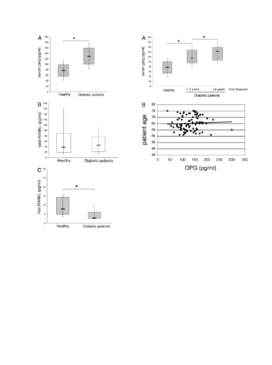
were clustered into two subgroups, ie, newly diagnosed
diabetic patients (within 2 years from diagnosis) and
patients with a longer history of diabetes (
ⱖ6 years from
diagnosis), it is remarkable that recently diagnosed dia-
betic patients also showed significantly (P
⬍ 0.05)
greater OPG serum levels with respect to normal controls
(Figure 2A). Patients with a longer history of diabetes
mellitus showed higher levels of OPG with respect to
either recently diagnosed patients (P
⬍ 0.05) and normal
controls (Figure 2A). Of note, patients with a long history
of diabetes mellitus comprised most of the patients with
documented microvascular and macrovascular compli-
cations (Table 1). The possibility that the increased se-
rum OPG levels detected in patients with a long history of
diabetes mellitus merely reflected an advanced age of
these patients was excluded, because no significant cor-
relation was observed between serum OPG and age of
the patients (r
⫽ 0.05, P ⬎ 0.05) (Figure 2B).
Correlation between OPG Serum Levels and
Hyperglycemia in Diabetic ApoE-Null Mice and
C57Black Littermates
Elevated levels of serum OPG have been reported in pa-
tients affected by either type 1 or type 2 diabetes melli-
tus.
13–18
Therefore, to further analyze whether the serum
levels of OPG were affected by hyperglycemia and/or by
other aspect of the metabolic disorders associated to dia-
betes mellitus, such as hypercholesterolemia, the next ex-
periments were performed in the apoE-null mice,
26
which
are characterized by elevated levels of total serum choles-
terol (mean
⫾ SD, 14.27 ⫾ 2.1 mmol/L) with respect to
littermate mice (mean
⫾ SD, 1.81 ⫾ 0.42 mmol/L). In these
animals, STZ-induced diabetes mellitus was associated to
the development of widespread aortic atherosclerotic le-
sions, starting from 10 to 12 weeks from diabetes induction
onwards (Figure 3A). Hyperglycemia started to become
significantly (P
⬍ 0.05) increased from the 2nd week after
the beginning of STZ treatment (Figure 3B).
Because we were interested in characterizing the OPG
and RANKL levels in the early phases of diabetes melli-
tus, serum OPG and RANKL were measured in apoE-null
mice starting from early time points from STZ treatment. A
Figure 1. Serum OPG and RANKL levels in diabetic patients and healthy
individuals. Levels of OPG (A), total (B), and free (C) RANKL were determined
by ELISA in sera from diabetic patients and from healthy patients. Horizontal bars
are median, upper, and lower edges of box are 75th and 25th percentiles; lines
extending from box are 10th and 90th percentiles. *P
⬍ 0.05.
Figure 2. Relation between serum OPG levels and years from diagnosis in
diabetic patients. Serum levels of OPG were analyzed in patient subgroups,
based on the indicated years from diagnosis (A) and in relation with patient
age (B). Horizontal bars are median, upper, and lower edges of box are 75th
and 25th percentiles; lines extending from box are 10th and 90th percentiles.
*P
⬍ 0.05. Coefficient of correlation is reported in the text.
Increase of Serum OPG in Diabetes Mellitus
2239
AJP December 2006, Vol. 169, No. 6
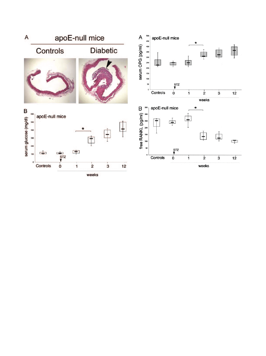
significant (P
⬍ 0.05) increase in OPG levels was mea-
sured in diabetic mice with respect to nondiabetic apoE-
null mice starting the 2nd week from diabetes induction
(Figure 4A). At variance to the ELISA for human RANKL,
which allowed us to measure either free or total (free plus
OPG-bound) RANKL, the available ELISA for mouse
RANKL only recognizes free, uncomplexed RANKL. As
shown in Figure 4B, free RANKL was significantly (P
⬍
0.05) decreased in apoE-null diabetic mice with respect
to nondiabetic controls, in keeping with the concomitant
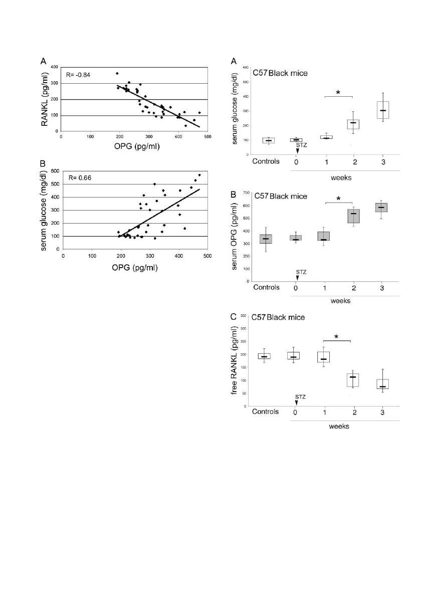
elevation of serum OPG. Of note, OPG serum levels
showed a significant inverse correlation (r
⫽ ⫺0.84, P ⬍
0.05) with the levels of free RANKL (Figure 5A) and a
significant positive correlation (r
⫽ 0.66, P ⬍ 0.05) with
the levels of glycemia (Figure 5B).
To investigate further the findings illustrated above,
pointing to a role of hyperglycemia in the early induction
of OPG elevation during diabetes mellitus, additional ex-
periments were performed on C57Black littermates. As
shown in Figure 6A, STZ treatment induced an increase
of glycemia with kinetics similar to that previously ob-
served in apoE-null mice. Concomitantly with the eleva-
tion of glycemia, OPG serum levels showed a significant
(P
⬍ 0.05) increase also in C57Black whereas the levels
of free RANKL showed a concomitant decrease (Figure
6, B and C).
OPG Is Released
in Vitro by Endothelial Cells in
Response to Inflammatory Cytokines but Not to
High Glucose Levels
Next experiments were performed in vitro to investigate
whether endothelial cells might contribute to the serum
OPG elevation observed in diabetic environment. For this
purpose, HUVECs were exposed to high glucose con-
centrations (30 mmol/L), insulin (1
mol/L), or inflamma-
tory cytokines (1 to 100 pg/ml), which are known to be
elevated in the sera of diabetic patients.
28,29
As shown in
Figure 7A, HUVECs spontaneously secreted measurable
levels of OPG in culture. Neither high glucose levels nor
insulin were able to significantly modulate basal OPG
release. On the other hand, recombinant TNF-
␣ dose
dependently up-regulated the release of OPG in the cul-
ture medium after 24 hours of exposure. It is particularly
noteworthy that TNF-
␣ induced a twofold (P ⬍ 0.05)
increase of OPG release at concentrations as low as 10
pg/ml (Figure 7A). On the other hand, a significant in-
crease of OPG release in response to IL-1
 was ob-
served only after stimulation with 100 pg/ml IL-1
 (Figure
7A), a concentration that might be beyond the physiolog-
ical levels of this cytokine in the plasma of diabetic pa-
tients. The association between high concentrations of
Figure 3. Atherosclerotic aortic lesions and serum glucose levels in apoE-
null mice after induction of diabetes. A: Representative H&E-stained histo-
logical cross-sectional sections from aorta of control and diabetic (3 months)
apoE-null mice. Arrowhead, a wide atherosclerotic plaque. B: Levels of
glucose were determined in serum samples from control and diabetic apoE-
null mice at different weeks after diabetes induction by STZ treatment.
Horizontal bars are median, upper, and lower edges of box are 75th and 25th
percentiles; lines extending from box are 10th and 90th percentiles. *P
⬍
0.05. Original magnifications,
⫻10.
Figure 4. Serum OPG and RANKL levels in diabetic and control apoE-null
mice. Levels of OPG (A) and free RANKL (B) were determined in sera from
control and diabetic apoE-null mice at different weeks after induction of diabe-
tes. Horizontal bars are median, upper, and lower edges of box are 75th and 25th
percentiles; lines extending from box are 10th and 90th percentiles. *P
⬍ 0.05.
2240
Secchiero et al
AJP December 2006, Vol. 169, No. 6
glucose and TNF-
␣ did not result in a further increase of
OPG release, even after 72 hours of exposure (Figure
7B), suggesting that inflammatory cytokines, rather than
hyperglycemia per se, might mediate the up-regulation of
OPG release observed in both humans and mice. Fur-
thermore neither HUVECs nor primary human PBMCs
produced OPG in response to increasing concentrations
of STZ, excluding the possibility that STZ has a direct
effect on OPG production, at least in these cell types
(Figure 7B). In parallel, OPG release was also measured
in the culture supernatants of primary human macro-
phages, taking into account that macrophage infiltration
represents a major event in both microvascular and ma-
crovascular complications associated to diabetes melli-
tus. OPG was not detected in the supernatant of cultured
macrophages in any tested condition, even after stimu-
lation with inflammatory cytokines (ie, TNF-
␣ and IL-1)
(data not shown). Finally, neither endothelial cells nor
PBMCs released detectable amounts of soluble RANKL,
either spontaneously or after addition of high glucose
levels, insulin, TNF-
␣, or IL-1 (data not shown).
OPG Blocks the Intracellular Signaling Induced
by RANKL in Endothelial Cells
It has been previously shown that soluble RANKL, by
interacting with its cognate transmembrane receptor
RANK, triggers a variety of intracellular signal transduc-
tion pathways in endothelial cells, which result in protec-
tive effects, such as promotion of endothelial cell survival
and angiogenesis.
7–9
To ascertain whether the enhanced
OPG production/release observed in sera of diabetic
patients might interfere with the beneficial biological ac-
tivity of RANKL on endothelial cells, we investigated the
activation of ERK/mitogen-activated protein kinase and
eNOS intracellular pathways in endothelial cells after
treatment with RANKL in the presence or absence of
recombinant OPG. As shown in Figure 8, RANKL induced
Figure 5. Relation between serum levels of OPG and free RANKL or glucose.
Relation between serum levels of OPG and free RANKL (A) and between
serum levels of OPG and glucose (B) in diabetic apoE-null mice. Coefficients
of correlation are indicated.
Figure 6. Serum OPG and RANKL levels in diabetic and control C57 litter-
mate mice. Diabetes was induced by STZ injection in C57Black littermate
mice. Levels of glucose (A), OPG (B), and free RANKL (C) were measured
in sera from control and diabetic mice at different weeks after STZ treatment.
Horizontal bars are median, upper, and lower edges of box are 75th and 25th
percentiles; lines extending from box are 10th and 90th percentiles. *P
⬍ 0.05.
Increase of Serum OPG in Diabetes Mellitus
2241
AJP December 2006, Vol. 169, No. 6
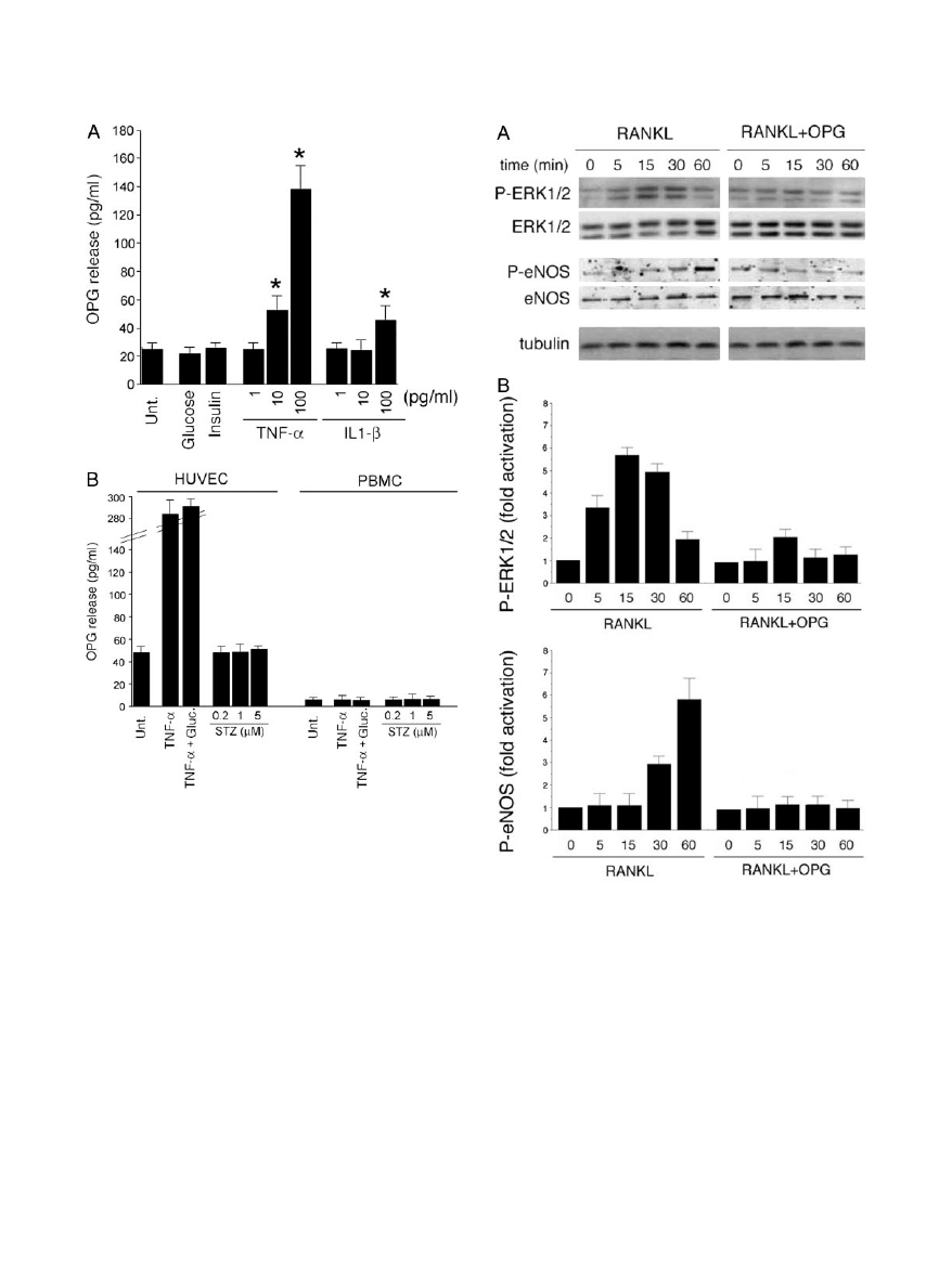
phosphorylation of ERK1/2 and of eNOS. The activation
of both pathways was completely abrogated by preincu-
bation of RANKL with OPG, clearly indicating that OPG
was effective in inhibiting the biological activity of RANKL
in endothelial cells. As expected on the basis of the
Western blot data illustrated above, we next investigated
whether eNOS-expressing cells were able to generate
bioactive NO. For this purpose, we measured the forma-
tion of cGMP, a good proxy for NO, because soluble
guanylate cyclase is activated by nM concentrations of
the gas.
30
Exposure to RANKL resulted in a significant
(P
⬍ 0.05) increase in cGMP over controls (652 ⫾ 40 and
263
⫾ 30 fmol/10
6
cells, respectively; n
⫽ 3), which was
inhibited by the preincubation with OPG (302
⫾ 39 fmol/
10
6
cells, n
⫽ 3). The presence of high glucose concen-
trations in culture medium did not determine any signifi-
cant modulation of these results (data not shown).
Discussion
We have here demonstrated that serum OPG levels are
increased in the early onset of diabetes mellitus and
progressively increase with the duration of the disease.
Figure 7. OPG release in endothelial cells and PBMC cultures. A: HUVECs
were either left untreated or stimulated with glucose, insulin, TNF-
␣, or IL-1.
After 24 hours, the levels of OPG released in culture supernatant were
measured by ELISA. B: HUVECs and PBMCs were either left untreated or
stimulated with TNF-
␣ ⫾ glucose or STZ. After 72 hours, the levels of OPG
released in culture supernatant were measured by ELISA. Results are ex-
pressed as means
⫾ SD of three to four independent experiments, each
performed in triplicate. *P
⬍ 0.05.
Figure 8. Effect of OPG on RANKL-induced intracellular signaling in endo-
thelial cells. Quiescent HUVEC cultures were stimulated with RANKL or
RANKL
⫹ OPG for the indicated time intervals (0 to 60 minutes). A: Cell
lysates were analyzed for ERK1/ERK2 and eNOS activation by Western blot
of total and phosphorylated (P) proteins using specific antibodies. Equal
loading of protein in each lane was confirmed by staining with the antibody
to tubulin. B: Protein bands were quantified by densitometry, and levels of
P-ERK1/2 and P-eNOS were calculated for each time point, after normaliza-
tion to ERK1/2, and eNOS, respectively. Unstimulated basal expression was
set as unity. One of three experiments with similar results is shown.
2242
Secchiero et al
AJP December 2006, Vol. 169, No. 6

Diabetic vasculopathy has an underlying low-grade in-
flammatory component, manifesting itself in the up-regu-
lation of genes responsive to inflammatory process-
es.
27–31
In this respect, it should be emphasized that
OPG is an NF-
B-inducible gene,
32
whose release in
endothelial cell culture is significantly increased by in-
flammatory cytokines as also shown in this study. Be-
cause of the enormous surface area of the endothelium
throughout the body as well as the relatively substantial
levels of constitutive and regulated OPG produced not
only by human endothelial cells but also by vascular
smooth muscle cells,
33,34
vascular cells likely signifi-
cantly contribute to circulating OPG levels measured in
serum. On the other hand, our in vitro data do not support
for a significant role of endothelial cells, PBMCs, or mac-
rophages in releasing significantly amounts of RANKL.
Other authors have reported a local expression of OPG
and RANKL in the tunica media of big arteries during the
process of vascular calcification,
10
but the relative con-
tribution of these cytokines to the vascular calcification is
still incompletely understood.
Although we have analyzed a cohort of type 2 diabetic
patients, it is noteworthy that a couple of recent studies
have demonstrated that serum OPG is also elevated in
patients affected by type 1 diabetes,
13,14
suggesting that
hyperinsulinemia and insulin-resistance are unlikely to
play a key role in OPG induction. Consistently, in vitro
data have demonstrated that insulin rather down-regu-
lates OPG expression in vascular smooth muscle cells.
12
In line with the hypothesis that insulin is not involved in the
induction of OPG expression and secretion, we have also
demonstrated that OPG release is significantly up-regu-
lated in the sera of diabetic apoE-null mice early after the
induction of diabetes mellitus by STZ treatment. Of note,
OPG serum levels in diabetic apoE-null mice positively
correlated with the glycemic levels whereas they were
inversely correlated to the levels of free RANKL. Elevated
levels of OPG were also observed in C57Black littermates
concomitantly with the induction of diabetes mellitus,
suggesting that hypercholesterolemia, characterizing
apoE-null mice, did not play a major role in the up-
regulation of serum OPG associated to diabetes mellitus.
Although we cannot exclude the possibility that autoim-
mune responses associated to STZ treatment are in-
volved in the increase of OPG expression/release, the
fact that OPG serum levels started to increase in both
apoE-null mice and control littermates not during STZ
treatment but only subsequent to the rise of glycemia
renders this possibility unlikely.
Despite the in vivo data obtained in the mouse models
of STZ-induced diabetes, in which we have demon-
strated the existence of a positive correlation between
OPG and glycemic serum levels, high glucose levels per
se were insufficient to modulate OPG release in endothe-
lial cells, PBMCs, and macrophages. On the other hand,
the proinflammatory cytokine TNF-
␣, which is known to
be elevated in the sera of diabetes mellitus,
28 –30
dose
dependently up-regulated OPG secretion by endothelial
cells. Importantly, the concentrations of TNF-
␣ (10 pg/ml)
required to induce a significant (approximately twofold)
increase in OPG, a situation mimicking the OPG rise
observed in the serum of diabetic patients, were in the
range of plasma concentrations reported in diabetic pa-
tients.
28 –30
These in vitro findings, coupled to the data
obtained in the diabetic mouse models, clearly suggest
that the inflammation-driven hyperglycemia, rather than
the high glucose levels per se, is involved in the increase
of OPG observed in both diabetic patients and diabetic
mice.
It is possible that the imbalance of OPG versus RANKL
serum levels in both diabetic patients and diabetic apoE-
null mice might contribute to endothelial cell dysfunction
by blocking RANKL signaling, which is able to activate
protective intracellular pathways in endothelial cells,
such as the eNOS pathway. In this respect, it should be
emphasized that diabetic vascular dysfunction is a major
clinical problem that predisposes patients to a variety of
cardiovascular diseases. In fact, diabetic patients fre-
quently suffer from macroscopic and microscopic vascu-
lopathy and accelerated atherosclerosis. The early im-
pairment of nitric oxide release is a key feature of
endothelial dysfunction, which invariably precedes per-
manent vascular alterations.
35
Our results also suggest a
mechanism to explain why altered serum OPG levels
have been shown to reflect the development or status of
vascular disease in both diabetic and nondiabetic pa-
tients,
13–24
indicating that therapeutic strategies aimed to
decrease the OPG serum levels may be suitable for
improving the vascular function in diabetes mellitus and
possibly in other vascular pathologies characterized by a
chronic inflammatory state.
References
1. Anderson DM, Maraskovsky E, Billingsley WL, Dougall WC, Tometsko
ME, Roux ER, Teepe MC, DuBose RF, Cosman D, Galibert L: A
homologue of the TNF receptor and its ligand enhance T-cell growth
and dendritic-cell function. Nature 1997, 390:175–179
2. Lacey DL, Timms E, Tan HL, Kelley MJ, Dunstan CR, Burgess T, Elliott
R, Colombero A, Elliott G, Scully S, Hsu H, Sullivan J, Hawkins N, Davy
E, Capparelli C, Eli A, Qian YX, Kaufman S, Sarosi I, Shalhoub V,
Senaldi G, Guo J, Delaney J, Boyle WJ: Osteoprotegerin ligand is a
cytokine that regulates osteoclast differentiation and activation. Cell
1998, 93:165–176
3. Hsu H, Lacey DL, Dunstan CR, Solovyev I, Colombero A, Timms E,
Tan HL, Elliott G, Kelley MJ, Sarosi I, Wang L, Xia XZ, Elliott R, Chiu L,
Black T, Scully S, Capparelli C, Morony S, Shimamoto G, Bass MB,
Boyle WJ: Tumor necrosis factor receptor family member RANK me-
diates osteoclast differentiation and activation induced by osteopro-
tegerin ligand. Proc Natl Acad Sci USA 1999, 96:3540 –3545
4. Dougall WC, Glaccum M, Charrier K, Rohrbach K, Brasel K, De
Smedt T, Daro E, Smith J, Tometsko ME, Maliszewski CR, Armstrong
A, Shen V, Bain S, Cosman D, Anderson D, Morrissey PJ, Peschon JJ,
Schuh J: RANK is essential for osteoclast and lymph node develop-
ment. Genes Dev 1999, 13:2412–2418
5. Kong YY, Yoshida H, Sarosi I, Tan HL, Timms E, Capparelli C, Morony
S, Oliveira-dos-Santos AJ, Van G, Itie A, Khoo W, Wakeham A, Dun-
stan CR, Lacey DL, Mak TW, Boyle WJ, Penninger JM: OPGL is a key
regulator of osteoclastogenesis, lymphocyte development and
lymph-node organogenesis. Nature 1999, 397:315–323
6. Wong BR, Josien R, Choi Y: TRANCE is a TNF family member that
regulates dendritic cell and osteoclast function. J Leukoc Biol 1999,
65:715–724
7. Kim YM, Kim YM, Lee YM, Kim HS, Kim JD, Choi Y, Kim KW, Lee SY,
Kwon YG: TNF-related activation-induced cytokine (TRANCE) in-
duces angiogenesis through the activation of Src and phospholipase
Increase of Serum OPG in Diabetes Mellitus
2243
AJP December 2006, Vol. 169, No. 6

C (PLC) in human endothelial cells. J Biol Chem 2002, 277:
6799 – 6805
8. Kim HE, Shin HS, Kwak HJ, Ahn KY, Kim JH, Lee HJ, Lee MS, Lee ZH,
Koh GY: RANKL regulates endothelial cell survival through the phos-
phatidylinositol 3
⬘-kinase/Akt signal transduction pathway. FASEB J
2003, 17:2163–2165
9. Min JK, Kim YM, Kim YM, Kim EC, Gho YS, Kang IJ, Lee SY, Kong YY,
Kwon YG: Vascular endothelial growth factor up-regulates expression
of receptor activator of NF-kappa B (RANK) in endothelial cells.
Concomitant increase of angiogenic responses to RANK ligand.
J Biol Chem 2003, 278:39548 –39557
10. Collin-Osdoby P: Regulation of vascular calcification by osteoclast
regulatory factors RANKL and osteoprotegerin. Circ Res 2004,
95:1046 –1057
11. Schoppet M, Al-Fakhri N, Franke FE, Katz N, Barth PJ, Maisch B,
Preissner KT, Hofbauer LC: Localization of osteoprotegerin, tumor
necrosis factor-related apoptosis-inducing ligand, and receptor acti-
vator of nuclear factor-kB ligand in Monckeberg’s sclerosis and ath-
erosclerosis. J Clin Endocrinol Metab 2004, 89:4104 – 4112
12. Olesen P, Ledet T, Rasmussen LM: Arterial osteoprotegerin: in-
creased amounts in diabetes and modifiable synthesis from vascular
smooth muscle cells by insulin and TNF-alpha. Diabetologia 2005,
48:561–568
13. Galluzzi F, Stagi S, Salti R, Toni S, Piscitelli E, Simonini G, Falcini F,
Chiarelli F: Osteoprotegerin serum levels in children with type 1
diabetes: a potential modulating role in bone status. Eur J Endocrinol
2005, 153:879 – 885
14. Rasmussen LM, Tarnow L, Hansen TK, Parving HH, Flyvbjerg A:
Plasma osteoprotegerin levels are associated with glycaemic status,
systolic blood pressure, kidney function and cardiovascular morbidity
in type 1 diabetic patients. Eur J Endocrinol 2006, 154:75– 81
15. Browner WS, Lui LY, Cummings SR: Associations of serum osteopro-
tegerin levels with diabetes, stroke, bone density, fractures, and
mortality in elderly women. J Clin Endocrinol Metab 2001,
86:631– 637
16. Knudsen ST, Foss CH, Poulsen PL, Andersen NH, Mogensen CE
Rasmussen LM: Increased plasma concentrations of osteoprotegerin
in type 2 diabetic patients with microvascular complications. Eur J
Endocrinol 2003, 149:39 – 42
17. Avignon A, Sultan A, Piot C, Elaerts S, Cristol JP, Dupuy AM: Osteo-
protegerin is associated with silent coronary artery disease in high-
risk but asymptomatic type 2 diabetic patients. Diabetes Care 2005,
28:2176 –2180
18. Anand DV, Lahiri A, Lim E, Hopkins D, Corder R: The relationship
between plasma osteoprotegerin levels and coronary artery calcifi-
cation in uncomplicated type 2 diabetic subjects. J Am Coll Cardiol
2006, 47:1850 –1857
19. Jono S, Ikari Y, Shioi A, Mori K, Miki T, Hara K, Nishizawa Y: Serum
osteoprotegerin levels are associated with the presence and severity
of coronary artery disease. Circulation 2002, 106:1192–1194
20. Schoppet M, Sattler AM, Juergen R, Schaefer JR, Herzum M, Maisch
B, Hofbauer LC: Increased osteoprotegerin serum levels in men with
coronary artery disease. J Clin Endocrinol Metab 2003, 88:
1024 –1028
21. Ueland T, Jemtland R, Godang K, Kjekshus J, Hognestad, Omland T,
Squire IB, Gullestad L, Bollerslev J, Dickstein K, Aukrust P: Prognostic
value of osteoprotegerin in heart failure after acute myocardial infarc-
tion. J Am Coll Cardiol 2004, 44:1970 –1976
22. Barreto DV, Barreto FC, Carvalho AB, Cuppari L, Cendoroglo M,
Draibe SA, Moyses RM, Neves KR, Jorgetti V, Blair A, Guiberteau R,
Fernandes Canziani ME: Coronary calcification in hemodialysis
patients: the contribution of traditional and uremia-related risk factors.
Kidney Int 2005, 67:1576 –1582
23. Moran CS, McCann M, Karan M, Norman P, Ketheesan N, Golledge
J: Association of osteoprotegerin with human abdominal aortic aneu-
rysm progression. Circulation 2005, 111:3119 –3125
24. Schoppet M, Schaefer JR, Hofbauer LC: Low serum levels of soluble
RANK ligand are associated with the presence of coronary artery
disease in men. Circulation 2003, 107:e76
25. Ueland T, Gullestad L, Simonsen S, Endresen K, Scott H, Froland SS,
Geiran O, Fiane AE, Aukrust P: Decreased endomyocardial RANKL
expression in transplant coronary artery disease. Transplantation
2006, 81:1467–1470
26. Candido R, Allen TJ, Lassila M, Cao Z, Thallas V, Cooper ME, Jan-
deleit-Dahm KA: Irbesartan but not amlodipine suppresses diabetes-
associated atherosclerosis. Circulation 2004, 109:1536 –1542
27. Secchiero P, Corallini F, di Iasio MG, Gonelli A, Barbarotto E, Zauli G:
TRAIL counteracts the proadhesive activity of inflammatory cytokines
in endothelial cells by down-modulating CCL8 and CXCL10 chemo-
kine expression and release. Blood 2005, 105:3413–3419
28. Joussen AM, Poulaki V, Mitsiades N, Kirchhof B, Koizumi K, Dohmen
S, Adamis AP: Nonsteroidal anti-inflammatory drugs prevent early
diabetic retinopathy via TNF-alpha suppression. FASEB J 2002,
16:438 – 440
29. Bulotta S, Barsacchi R, Rotiroti D, Borgese N, Clementi E: Activation
of the endothelial nitric-oxide synthase by tumor necrosis factor-
␣.
J Biol Chem 2001, 276:6529 – 6536
30. Fujiwara K, Nagasaka A, Nagata M, Yamamoto K, Imamura S, Oda N,
Sawai Y, Hayakawa N, Suzuki A, Itoh M: A stable prostacyclin ana-
logue reduces high serum TNF-alpha levels in diabetic patients. Exp
Clin Endocrinol Diabetes 2004, 112:390 –394
31. Sjo¨holm A, Nystrom T: Endothelial inflammation in insulin resistance.
Lancet 2005, 365:610 – 612
32. Collin-Osdoby P, Rothe L, Anderson F, Nelson M, Maloney W, Os-
doby P: Receptor activator of NF-kappa B and osteoprotegerin ex-
pression by human microvascular endothelial cells, regulation by
inflammatory cytokines, and role in human osteoclastogenesis. J Biol
Chem 2001, 276:20659 –20672
33. Zhang J, Fu M, Myles D, Zhu X, Du J, Cao X, Chen YE: PDGF induces
osteoprotegerin expression in vascular smooth muscle cells by mul-
tiple signal pathways. FEBS Lett 2002, 521:180 –184
34. Fu M, Zhang J, Lin Y, Zhu X, Willson TM, Chen YE: Activation of
peroxisome proliferator-activated receptor
␥ inhibits osteoprotegerin
gene expression in human aortic smooth muscle cells. Biochem
Biophys Res Commun 2002, 294:597– 601
35. Landmesser U, Hornig B, Drexler H: Endothelial function: a critical
determinant in atherosclerosis? Circulation 2004, 109(Suppl 1):
II27–II33
2244
Secchiero et al
AJP December 2006, Vol. 169, No. 6
Wyszukiwarka
Podobne podstrony:
Dynamic gadolinium enhanced subtraction MR imaging – a simple technique for the early diagnosis of L
Dr Gary L Glum Calling Of An Angel The True Story Of Rene Caisse And An Indian Herbal Medicine Cal
T M P Mahadevan The Early Life of Bhagavan Ramana (13p)
The?onomic Emergence of China, Japan and Vietnam
Nukariya; Religion Of The Samurai Study Of Zen Philosophy And Discipline In China And Japan
Robert Stevenson The Strange Case of Dr Jekyll and Mr Hyde
The United Kingdom of Great Britain and Northern Ireland
the garden and story a contribution to the theory of garden narrative Content File PDF
Goel, Dolan The Functional anatomy of H segregating cognitive and affective components
01 Aramaic (The Ancient Languages of Syria Palestine and Arabia)
The New Age of Russia Occult and Esoteric Dimensions ed by B Menzel & M Hagemeister & BG Rosenthal
Ecumeny and Law 2015 Vol 3 Welfare of the Child Welfare of Family Church and Society
Nazi Spatial Theory; The Dark Geographies of Carl Schmitt and Walter Christaller
Hossam El Dien The acoustical influence of balcony depth and parapet form
Use of exponential, Page’s and diffusional models to simulate the drying kinetics of kiwi fruit
Julius Fast The Body Language of Sex, Power, and Aggression
[Robert Louis Stevenson] The Strange Case of Dr Jekyll and Mr Hyde
Winch The Idea of a Social Science And its Relation to Philosophy
The ultimate goal of all types od advertising is to persuade the consumer to purchase w product
więcej podobnych podstron