
Review Article
Theme: siRNA and microRNA: From Target Validation to Therapy
Guest Editor: Song Li
MicroRNA Regulation of Cancer Stem Cells and Therapeutic Implications
Jeffrey T. DeSano
1
and Liang Xu
1,2,3
Received 1 May 2009; accepted 21 September 2009; published online 20 October 2009
Abstract. MicroRNAs (miRNAs) are a class of endogenous non-protein-coding RNAs that function as
important regulatory molecules by negatively regulating gene and protein expression via the RNA
interference (RNAi) machinery. MiRNAs have been implicated to control a variety of cellular,
physiological, and developmental processes. Aberrant expressions of miRNAs are connected to human
diseases such as cancer. Cancer stem cells are a small subpopulation of cells identi
fied in a variety of
tumors that are capable of self-renewal and differentiation. Dysregulation of stem cell self-renewal is a
likely requirement for the initiation and formation of cancer. Furthermore, cancer stem cells are a very
likely cause of resistance to current cancer treatments, as well as relapse in cancer patients.
Understanding the biology and pathways involved with cancer stem cells offers great promise for
developing better cancer therapies, and might one day even provide a cure for cancer. Emerging
evidence demonstrates that miRNAs are involved in cancer stem cell dysregulation. Recent studies also
suggest that miRNAs play a critical role in carcinogenesis and oncogenesis by regulating cell proliferation
and apoptosis as oncogenes or tumor suppressors, respectively. Therefore, molecularly targeted miRNA
therapy could be a powerful tool to correct the cancer stem cell dysregulation.
KEY WORDS: cancer stem cells; microRNAs; oncogenes; tumor suppressors.
INTRODUCTION
In this review, we discuss the acknowledged functions
and characteristics of microRNAs and cancer stem cells,
focusing on the potential roles of the stem cell related
microRNAs (miRNAs) in cancer stem cells regulation and
the implications in developing novel and more effective
molecular cancer therapies.
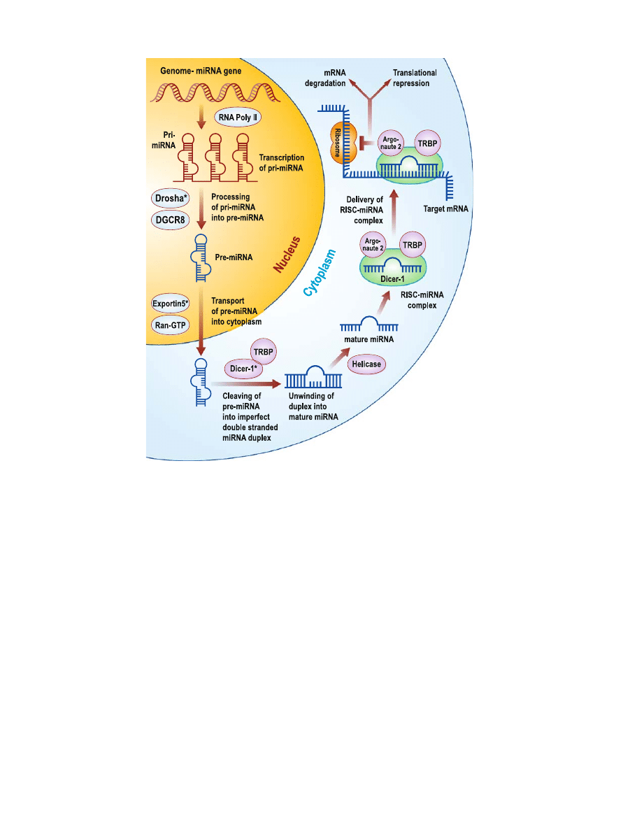
MicroRNA Biogenesis
MiRNAs and small interfering RNAs (siRNAs) are two
key components of RNA interference within cells. siRNAs
are derived by processing of long double-stranded RNAs and
are often of exogenous origin and degrade mRNAs bearing
fully complementary sequences (
). In contrast, miRNAs are
endogenously encoded small noncoding RNAs, derived by
processing of short RNA hairpins, which can inhibit the
translation of mRNAs bearing partially complementary target
sequences (
). miRNAs are endogenous and naturally
generated in animal cells. For this reason, the use of miRNAs
is more applicable in developing therapeutics that can
regulate mRNA in animal cells.
MiRNA biogenesis has been studied by many investi-
gators. A schematic overview of miRNA biogenesis is given
in Fig.
. MiRNAs are transcribed by RNA polymerase II
enzyme producing a long primary-miRNA (pri-miRNA) (
).
These pri-miRNAs contain a cap structure at the 5
′ end and
are poly-adenylated at the 3
′ end, suggesting that pri-
miRNAs are structurally and functionally similar to mRNAs
(
). Also, pri-miRNAs contain speci
fic hairpin-shaped stem-
loop structures of ~70 nucleotides that are recognized and
cleaved by a ~650-kDa nuclear microprocessor complex
consisting of the RNase III endonuclease Drosha and the
essential DiGeorge syndrome critical region gene 8 (DGCR8)
binding protein (
). The resulting ~70 nucleotide hairpin
intermediate (pre-miRNA) is transported out of the nucleus
and into the cytoplasm by Exportin-5 and its cofactor Ran-
GTP (
). In the cytoplasm, the pre-miRNAs are further
cleaved by a second RNase III endonulease Dicer-1 and its
essential transactivating response RNA binding protein
(TRBP) producing a short imperfect double-stranded
miRNA duplex. The imperfect miRNA duplex is then
unwound into a mature miRNA by helicase. Next, TRBP
recruits the catalytic Argonaute 2 to the Dicer complex with
the mature miRNA forming the RNA-induced silencing
complex (RISC) (
,
). RISC then regulates gene expression
by mRNA degradation or translational repression (
–
).
Therefore, miRNAs negatively regulate gene and protein
1
Department of Radiation Oncology, Division of Cancer Biology,
University of Michigan, 4424E Med Sci I, 1301 Catherine St., Ann
Arbor, MI 48109-5637, USA.
2
Comprehensive Cancer Center, University of Michigan, Ann Arbor,
MI 48109, USA.
3
To whom correspondence should be addressed. (e-mail: liangxu@
umich.edu)
The AAPS Journal, Vol. 11, No. 4, December 2009 (
#
2009)
DOI: 10.1208/s12248-009-9147-7
682
1550-7416/09/0400-0682/0 # 2009 American Association of Pharmaceutical Scientists
expression via the RNA interference (RNAi) pathway.
miRNA is different from siRNA in that miRNA represses
mRNA with complementary sequence in the 3
′-untranslated
region (3
′-UTR) (
), although miRNA may also target
coding regions of mRNA, at least in animals (
).
MicroRNA and Regulation of Stem Cells
MiRNAs have been proposed to be important factors in
stem cell function. One reason for this is that expression
levels of certain miRNAs in stem cells are different from
other normal tissues (
). This implies that miRNAs may
have a unique role in stem cell regulation. In order to con
firm
that miRNAs do indeed regulate stem cell function, many
investigators have used Dicer-1 (dcr-1) mutants. Dicer-1 plays
a specialized role in the biogenesis of miRNAs and therefore
can offer great insight into the role of miRNAs in stem cells.
Loss of dcr-1 function in mouse models resulted in animal
death early in development and depletion of stem cells in
mouse embryos, suggesting that the disrupted miRNA path-
way plays a large role in maintaining the stem cell population
(
). Mutated dcr-1 gene in embryonic stem cells in mice
leads to a reduced expression of miRNAs and displayed
severe defects in embryonic stem cell differentiation in vivo
and in vitro. Re-expression of Dicer-1 in the knockout cells
rescued the phenotypes (
). Another study observed a
reduction in cyst production in Drosophila germline stem cell
mutants for dcr-1 and a delay in the transition from G1 to S
phase, which is dependent on the cyclin-dependent kinase
inhibitor Dacapo (
). This
finding that miRNAs are required
for stem cell division suggests that miRNAs are needed to
make stem cells insensitive to environmental signals in order
to overcome the normal G1/S checkpoint (
). The use of
dcr-1 mutants has resulted in a great multitude of data and
findings that implicate the important role that miRNAs play
in stem cell function.
Cancer Stem Cell Hypothesis
Stem cells are de
fined by their ability to undergo self-
renewal, as well as multi-lineage differentiation (
). Adult
stem cells are found in numerous tissues of the body and play
a role in tissue development, replacement, and repair (
). In
this review, cancer cells are de
fined as cells that are part of a
Fig. 1. An overview of miRNA biogenesis. MiRNAs are a class of endogenous non-
protein-coding RNAs that negatively regulate gene and protein expression via the RNAi
pathway. Figure modi
fied from Chen, C.Z., et al. N Engl J Med, 2005; 353(17):1768–71
683
MicroRNA Regulation of Cancer Stem Cells and Therapeutic Implications

malignancy. Recently, there has been a dramatic increase in
research geared towards a small subpopulation of cells
identi
fied in cancers that have stem cell properties. The
cancer stem cell hypothesis proposes that cancers are derived
from a small fraction of cancer cells that constitute a reservoir
of self-sustaining cells with the exclusive ability to self-renew
and initiate/maintain the tumor (
). Thus, according to the
cancer stem cell hypothesis, these cancer stem cells are
tumor-initiating cells that proliferate through their unique
self-renewal ability. Cancer stem cells were
first identified in
leukemia (
). Recently, many investigators have identi-
fied cancer stem cells in solid tumors including breast, brain,
pancreas, colon, and head and neck cancers (
–
). These
new discoveries provide further support for the cancer stem
cell hypothesis. In some types of human cancers, such as
melanoma, such tumorigenic cells may not be rare (
).
The cancer stem cell hypothesis established a basis for
future studies, as well as presented a better understanding of the
biology and intricacies of cancer and tumor formation. To be
maximally effective, cancer therapy must also be directed
against both the resting cancer stem cells and the proliferating
cancer cells (
). This may be possible if speci
fic stem cell signals
are inhibited using molecular therapy, while at the same time
attacking proliferating cells by conventional therapies (
,
).
Self-Renewal
Using different systems, many investigators have dem-
onstrated that only a small minority of cells in human cancers
are capable of self-renewal. Self-renewal is distinguished from
other proliferating processes in that at least one of the
progeny is identical to the initial stem cell (
). Speci
fically,
asymmetric stem cell self-renewal produces two different
progenies. The
first progeny is identical to the original stem
cell
—thus, maintaining stem cell number—and the other
progeny produced is a committed progenitor cell, which
undergoes cellular differentiation (
). Both the self-renewal
and differentiation of normal stem cells are regulated by the
stem cell microenvironment, which has been termed the stem
cell niche (
,
). In order to study the self-renewal potential,
a mammosphere assay has been developed that plates cells
in a serum-free medium with growth factor supplementation
on a non-adherent substrata followed by quanti
fication of
sphere formation (
). Using this method, one study found
that secondary mammospheres from the human breast cancer
Lin
−
CD29
H
CD24
H
cell subgroup as determined from seven
independent tumors were larger in size and number
compared with all other subpopulations, suggesting the
ability for tumor-initiating cells to undergo self-renewal (
Therefore, a certain subpopulation of cancer cells is able to
self-renew and initiate tumor formation, thus coining the term
“cancer stem cells.” The central feature of cancer stem cells is
this relatively unlimited asymmetric self-renewal (
). Self-
renewal of cancer stem cells could be a likely cause of
resistance of current cancer treatment, as well as relapse in
cancer patients. One recent study provided the
first clinical
evidence for the implication of a
“glioma stem cell” or “self-
renewal
” phenotype in treatment resistance of glioblastoma
(
). It is believed that genetic alterations cause dysregulation
in cancer stem cells, resulting in unlimited self-renewal
capabilities. Abnormal stem cell self-renewal is a likely
requirement for the initiation, formation and resistance of
cancer.
Signaling Pathways of the
“Stem Cell Genes”
There is growing evidence that illustrates that many
pathways classically connected with cancer may also regulate
normal stem cell development (
). The pivotal signaling
pathways of the
“stem cell genes” Notch, Hedgehog, Wnt/β-
catenin, HMGA2, Bcl-2, and Bmi-1 are involved in the
regulation of self-renewal, differentiation, and survival of
cancer stem cells (
). These key signaling pathways,
which may be dysregulated in cancer stem cells, offer great
promise for future cancer therapies and treatments.
Notch
The Notch signaling pathway is a short-range communica-
tion transducer that is involved in regulating many cellular
processes during development and renewal of adult tissues.
Notch signaling has been highlighted as a pathway that aids in
development of the breast and is frequently dysregulated in
invasive breast cancer (
). It was also demonstrated that Notch
signaling can act on mammary stem cells to promote self-
renewal and on early progenitor cells to promote their
proliferation (
). These effects were also shown to be
completely inhibited by either a Notch 4 antibody or a gamma
secretase inhibitor that blocks Notch processing (
). These
findings suggest that atypical Notch signaling could lead to
dysregulation of the self-renewal properties of cancer stem cells,
thus resulting in carcinogenesis and oncogenesis (
Hedgehog
The importance of Hedgehog signaling in carcinogenesis
revolves around Hedgehog
’s effect on cancer stem cell self-
renewal. Hedgehog (speci
fically Sonic Hedgehog) signaling
has been implicated in the regulation of self-renewal charac-
teristics by the
finding that populations enriched for human
hematopoietic stem cells exhibit increased self-renewal in
response to Sonic Hedgehog stimulation in vitro, albeit in
combination with other growth factors (
,
). In humans,
several distinctive cancers, including basal-cell carcinoma,
result from mutations that aberrantly activate Hedgehog
signal transduction (
). It has been shown that Drosophila
ovarian stem cells cannot proliferate as stem cells in the absence
of Hedgehog signaling, whereas excessive Hedgehog signaling
produces supernumerary stem cells, implying that Hedgehog is a
stem-cell factor (
). This suggests that human cancers due to
excessive Hedgehog signaling might result from dysregulated
self-renewal properties of cancer stem cells.
Wnt/
β-catenin
Another pathway that regulates both self-renewal and
oncogenesis in different tissues is the Wnt/
β-catenin signaling
pathway (
). Activation of the Wnt receptor causes an
accumulation of
β-catenin and other Wnt gene family proteins
in the cytoplasm, which eventually translocates into the
nucleus. The nuclear translocation of
β-catenin drives the
expression of genes associated with self-renewal. Over-
684
DeSano and Xu

expression of activated
β-catenin expands the pool of stem
cells (
). Dysregulation in the Wnt/
β-catenin signaling path-
way contributes to the onset of cancer. Gain or loss-of-
function mutations of several members of this pathway have
been found in many types of human tumors (
,
). Another
study showed that, in chronic myelogenous leukemia,
β-
catenin accumulates in the nuclei of granulocyte
–macrophage
progenitors, seemingly enhancing the self-renewal activity
and leukemic potential of these cells (
). Thus, dysregulation
of this pathway within cancer stem cells may be associated
with the acquisition of self-renewal properties.
HMGA2
HMGA2 has also been implicated in survival and self-
renewal of cancer stem cells. HMGA2 is thought to play a
role in modulating macromolecule complexes that are
involved in many biological processes, including binding
directly to the DNA and aiding in the regulation of many
genes (
). The expression of HMGA proteins during
embryogenesis suggests that they have important functions
in development (
). Moreover, the HMGA2 gene is
suggested to control growth, proliferation, and differentiation
(
). HMGA2 has also been implicated in cancer. HMGA2
overexpression has been found in lung and pancreatic
carcinomas (
,
). HMGA2 protein overexpression is
usually met with the presence of metastasis and reduced
survival of the cancer patient (
). Thus, HMGA2
’s role in
embryogenesis and aggressive cancers suggests that human
cancers due to excessive HMGA2 signaling might result from
dysregulated cell survival and self-renewal properties of
cancer stem cells.
Bmi-1
The signi
ficance of Bmi-1 signaling in carcinogenesis
revolves around Bmi-1
’s effect on cancer stem cell self-
renewal. Bmi-1 was shown to be expressed in neural stem
cells and proliferating progenitor cells, but not in differ-
entiated cells (
). Loss of Bmi-1 resulted in a drastic
decrease in neural stem cell proliferation and self-renewal
(
). This suggests that Bmi-1 is necessary for stem cell self-
renewal. Bmi-1 has also been implicated in cancer. Bmi-1 was
identi
fied to promote the generation of lymphomas (
).
This demonstrates that Bmi-1 plays a role in cancer develop-
ment. Bmi-1 seems to be important in both stem cells and
cancer. Bmi-1 was found to be activated in human breast
“cancer stem cells” characterized as CD44
+
CD24
−/low
Lin
−
(
). Furthermore, Bmi-1 was found to mediate the
mammosphere-initiating cell number and mammosphere
size, supporting a role in the regulation of self-renewal of
normal and tumorigenic human mammary stem cells (
).
Therefore, dysregulation of this Bmi-1 pathway within cancer
stem cells may be associated with the acquisition of self-
renewal properties.
Bcl-2
Bcl-2 has been researched by many investigators because
of its role within cancer cells as a proto-oncogene. Bcl-2 is
over-expressed in many cancers, leading to a prevention of
apoptosis. It has been shown that this obstacle to apoptosis
due to over-expression of Bcl-2 results in an increased
number of stem cells in vivo (
). This suggests that apoptosis
plays a role in regulating the microenvironments of stem cells
(
). Therefore, the Bcl-2 signaling pathway is very important
to the survival of stem cells, especially cancer stem cells,
because of the overexpression of Bcl-2 in cancers (
Link Between miRNA and Cancer Stem Cells
Aberrant expressions of miRNAs are connected to human
diseases, such as cancer. Tumors analyzed by miRNA pro
filing
have shown signi
ficantly different miRNA profiles (for mature
and/or precursor miRNAs) compared with normal cells from
the same tissue (
). It has also been shown by convincing
evidence that miRNAs are important factors in stem cell
biology. Thirty-six unique miRNAs (from 32 stem-loops) have
been identi
fied by cDNA cloning to be specifically expressed in
human embryonic stem cells relative to their differentiated
embryoid bodies (
). The obvious parallel that can be drawn
is that undifferentiated stem cells display miRNA expression
pro
files reminiscent of cancer cells (
). There is also over-
whelming evidence that a distinct subpopulation of cancer cells
have the ability of self-renewal, and thus act as cancer stem
cells within tumors (
). Knowing that aberrant gene function
and expression are key characteristics in cancer, it is thought
that acquired epigenetic abnormalities participate in genetic
alterations, causing dysregulation in cancer stem cells (
). This
dysregulation allows them to escape the restrictions of the stem
cell niche, resulting in unlimited self-renewal ability and
potential. It is believed that microenvironmental factors or
signals account for the epigenetic abnormalities in cancer stem
cells. These signals interfere with gene expressions, resulting in
the silencing of some genes. Therefore, there must be some
underlying sub-cellular process that accounts for this dysregu-
lation in cancer stem cells.
One pathway of investigative relevance is the RNAi
pathway. The RNAi pathway is important because it silences
gene expression at transcription or translation. MiRNAs have
been implicated in the RNAi pathway by negatively regulat-
ing gene and protein expression at the post-transcriptional
level. Altered expression of speci
fic miRNA genes contrib-
utes to the initiation and progression of cancer (
).
Disruption of miRNA expression levels in tumor cells may
result from distorted epigenetic regulation of miRNA expres-
sion, abnormalities in miRNA processing genes or proteins,
and the location of miRNAs at cancer-associated genomic
regions (
). Clearly, miRNAs play a critical role in carcino-
genesis and oncogenesis. Emerging evidence suggests that
certain abnormal miRNA expression levels cause cancer stem
cell dysregulation, resulting in unlimited self-renewal and
cancer progression. Therefore, miRNA expression is a vital
key to cancer stem cell dysregulation.
In addition, a number of miRNAs have been identi
fied
within cancers to function as either oncogenes or tumor
suppressors (
). These miRNAs offer great promise for
cancer therapy because they might have the potential to
regulate aberrant miRNA expression. Therefore, miRNA
therapy could be a powerful tool to address cancer stem cell
685
MicroRNA Regulation of Cancer Stem Cells and Therapeutic Implications

dysregulation and its resulting self-renewal and cancer pro-
gression in patients.
Examples of Potential Links Between miRNA and Cancer
Stem Cells
Oncogenes
Many events can trigger cancer formation within the
body. Over the past few years, research in the area of cancer
has shown that there are aberrant levels of certain miRNAs
in cancer stem cells, resulting in dysregulation of these cancer
stem cells. This cancer stem cell dysregulation may explain
how carcinogenesis and oncogenesis progress. MiRNAs
either act as oncogenes or tumor suppressors in cancer cells.
Oncogenic miRNAs are often called oncomiRs and are up-
regulated in the cancer cells.
It has been shown that miRNA miR-21 is over-expressed
in breast tumor tissues and functions as an oncogene by
modulating tumorigenesis through the regulation of Bcl-2 and
Programmed Cell Death 4 (PDCD4) (
). Thus, miR-21
over-expression leads to up-regulation of Bcl-2, which results
in increased tumor growth and decreased apoptosis (
The miR-17-92 cluster, which is comprised of seven
miRNAs, is markedly over-expressed in lung cancers and
could play a role as an oncogene (
). Enforced expression of
the mir-17-92 cluster acted with c-Myc expression to accel-
erate tumor development in a mouse B-cell lymphoma model
(
). Introduction of miR-17-92 into hematopoietic stem cells
was shown to signi
ficantly accelerate the formation of
lymphoid malignancies (
). Other evidence has proposed
the potential targets of miR-17-92 include E2F1 (which
promotes cell proliferation) and the tumor-suppressor genes
PTEN (which promotes apoptosis) and RB2 (
). Interest-
ingly, a functional relationship between miR-17-92 and the
Sonic Hedgehog signaling pathway was studied. In engi-
neered medulloblastomas, miR-17-92-induced tumors were
shown to have activated the Sonic Hedgehog signaling
pathway (
). This is thought to result in increased self-
renewal. Taken together, these studies implicate the miR-17-
92 cluster as a potential human oncogene that plays a role in
cancer stem cells.
miR-135 has an oncogenic role within cancer as well.
miR-135a and miR-135b were found to be greatly up-
regulated in colorectal adenomas and carcinomas (
APC, a gene found to lead to truncated proteins that have
lost their
β-catenin binding sites, was down-regulated in
cancers with increased expression of miR-135a&b (
). If
APC is not expressed to the proper level, an accumulation of
β-catenin would occur, leading to the activation of self-
renewal genes. Thus, these oncogenes miR-135a&b play a
vital role in controlling Wnt signaling pathway. Consequently,
miR-135a and miR-135b may play vital roles in cancer stem
cells themselves. Over-expression of these oncomiRs leads to
further cancer progression.
Tumor Suppressors
In oncogenesis, some miRNAs
’ expression is decreased in
cancerous cells (
). These miRNAs are called tumor suppressor
miRNAs. In this review, tumor suppressor miRNAs are termed
TSmiRs. TSmiRs are supposed to prevent tumor development;
however, their expression in cancer is down-regulated, resulting
in increased progression of the disease.
One example of tumor suppressor miRNAs is let-7. Let-7
expression levels are reduced in various lung cancer cell lines
and pulmonary tumors, relative to normal lung samples
(
). Let-7 expression is lower in lung tumors than in
normal lung tissue, while RAS protein is signi
ficantly higher
in lung tumors, suggesting that let-7 negatively regulates RAS
protein (
). RAS appears to be important for self-renewal
since silencing RAS reduces mammosphere formation, clonal
expansion, and tumorigenicity (
). Chromosomal transloca-
tions previously associated with human tumors disrupt
repression of HMGA2 by let-7 miRNA, suggesting that let-7
also negatively regulates HMGA2 (
). The let-7 family is not
expressed in breast tumor-initiating cells (
). By expressing let-
7 in breast tumor-initiating cells, it was found that let-7 regulates
the key features of breast cancer stem cells
—self renewal in
vitro, multipotent differentiation, and the ability to form tumors
(
). Thus, let-7 is a tumor suppressor that negatively regulates
RAS protein and HMGA2 and plays an important role in the
self-renewal potential of cancer stem cells.
miR-15a and miR-16-1 are both tumor suppressors. In the
majority of leukemic cells, both miR-15a and miR-16-1 were
expressed at low levels and Bcl-2 was over-expressed (
). It was
also shown that down-regulation of Bcl-2 by miR-15a and miR-16-
1 triggers apoptosis and that the levels of expression of these two
miRNAs are important (
). Also in prostate cancer cells, down
regulation of these two miRNAs resulted in an up regulation of
WNT3A, a Wnt gene family protein (
). WNT3A help promote
cancer cell proliferation and invasiveness of their respective
tumors (
). These TSmiRs are critical to suppressing tumor
growth and progression. Thus, miR-15a and miR-16-1 play
extremely vital roles in both the Bcl-2 and Wnt signaling pathways,
which are essential for self-renewal potential in cancer stem cells.
MiRNA-128 is also a tumor suppressor involved in
cancer stem cells. In high-grade gliomas, miR-128 levels were
signi
ficantly reduced, suggesting tumor suppressor properties
). Glioma cell proliferation and growth were inhibited by
miR-128 introduction (
). Later, the mechanism behind
miR-128
’s tumor suppressor characteristics was found.
Expression of miR-128 caused a down regulation of Bmi-1
signaling pathway levels (
). Thus, miR-128 speci
fically
blocked glioma self-renewal via Bmi-1 down-regulation.
miR-128
’s regulation of the Bmi-1 signaling pathway demon-
strates the importance of it in the self-renewal capacity in
cancer stem cells.
MiRNA-199b-5p is a tumor suppressor of great intrigue.
Expression of miR-199b-5p was shown to be lost in metastatic
cancer patients (
). In medulloblastoma cells, miR-199b-5p
was found to down-regulate the expression of HES1, a
transcription factor of the Notch signaling pathway (
).
Thus, miR-199b-5p should lead to a decrease of the self-
renewal properties of cancer stem cells. Introduction of an
over-expression of miR-199b-5p did indeed block Notch
signaling, as well as decrease the medulloblastoma stem-cell-
like (CD133+) subpopulation of cells (
). Therefore, the
tumor suppressor miR-199b-5p is extremely important to the
cancer stem cell self-regulation potential via the Notch
signaling pathway.
686
DeSano and Xu
Furthermore, miR-125b, miR-326, and miR-324-5p are
all tumor suppressors. Using miRNA pro
file screening of
human medulloblastoma cells, these miRNAs were found to
be down-regulated where the Hedgehog signaling pathway
was elevated (
). miR-125b, miR-326, and miR-324-5p were
all shown to suppress Smo, an activator component of the
Hedgehog pathway, and only miR-324-5p was also found to
suppress Gli-1, another activator component of the Hedgehog
signaling pathway (
). All three of these miRNAs were
found to inhibit cancer cell growth (
). This is consistent
with the fact that the Hedgehog signaling pathway is involved
in the self-renewal potential of cancer stem cells.
MiRNA-34 is a tumor suppressor of great interest. This
TSmiR is down-regulated in several types of cancer (
). We
found that, in p53-de
ficient human gastric and pancreatic
cancer cells, restoration of functional miR-34 inhibits cell
growth and induces G1 block and apoptosis, indicating that
miR34 may restore p53 function (
). miR-34 restoration
inhibited tumorsphere growth in vitro and tumor initiation in
vivo, which is reported to be correlated to the self-renewal of
cancer stem cells (
). The mechanism of miR-34-mediated
suppression of self-renewal appears to be related to the direct
modulation of downstream targets
—Bcl-2, Notch, and
HMGA2
—indicating that miR-34 may be involved in gastric
cancer cells
’ self-renewal/differentiation decision-making
(
). Thus, miR-34 is a signi
ficant tumor suppressor of
cancer stem cells by regulating both apoptosis and self-renewal
properties. Decreased expression of these TSmiRs leads to
further cancer progression.
Examples Support the Role of
“Stem Cells miRNAs”
in Oncogenesis and the Implications in Molecular
Cancer Therapy
It is clear that these
“stem cell miRNAs” play a vital
purpose in the regulation of the discussed
“stem cell genes”
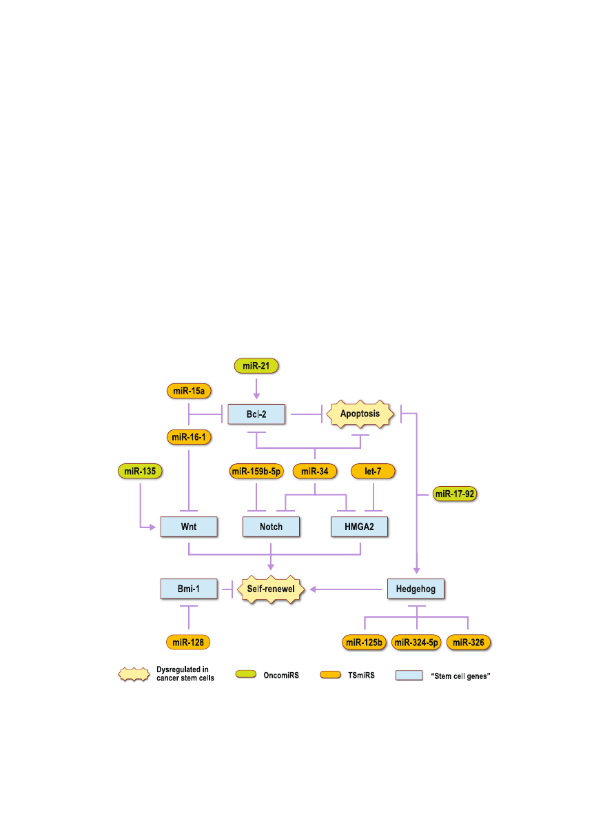
and their subsequent signaling pathways in cancer. Figure
provides a schematic view of these
“stem cell miRNAs” and
their interactions with
“stem cell genes” in cancer stem cells.
These
“stem cell miRNAs” support the potential link
between miRNAs and cancer stem cells. Figure
outlines this
potential link between miRNAs and cancer stem cells. All of
these examples suggest that miRNAs have a pivotal function
in carcinogenesis and oncogenesis by regulating self-renewal
and apoptosis via cancer stem cell signaling pathways as
oncogenes or tumor suppressors, respectively. These onco-
genic and tumor suppressor miRNAs lead to a better under-
standing of cancer stem cell biology, and thus, a greater
knowledge of how cancer starts and progresses into malignant
tumor formation. Dysregulation of cancer stem cells allows
Fig. 2. Potential
“stem cell miRNAs” that modulate “stem cell genes” related to cancer stem cells. Certain
miRNAs have been shown to be aberrantly expressed in cancer. OncomiRs, which initiate cancer
development, are over-expressed. TSmiRs, which prevent tumor development, are decreased. These
miRNAs regulate genes that are implicated in stem cells. The aberrant expression of these potential
“stem
cell miRNAs
” in cancer suggests that dysregulation of “stem cell genes” leads to increased levels of self-
renewal and decreased levels of apoptosis within cancer stem cells. This results in further cancer
progression
687
MicroRNA Regulation of Cancer Stem Cells and Therapeutic Implications
them to escape the restrictions of the stem cell niche, resulting
in unlimited self-renewal ability and potential, which results
in further cancer development and resistance to current
treatments. It has been demonstrated that aberrant expres-
sion of certain miRNAs are not only connected to cancers in
general, but these aberrant miRNA expressions are involved
in cancer stem cell dysregulation. Functional studies of
speci
fic miRNAs within the cancer stem cells of various
cancers are crucial for the elucidation of the mechanisms
behind oncogenesis in various cancers (
). Some miRNAs
are up-regulated in cancer stem cells and act as oncogenes.
These oncogenes should be targeted with treatments that
knockdown their expression. Other miRNAs suppress cell
proliferation by nature, acting as tumor suppressors, but are
down-regulated in cancer stem cells, resulting in cancer
progression. These tumor suppressors should be targeted
with therapies that restore their tumor suppressor capabilities
within the cancer stem cells. Therefore, addressing these
abnormal miRNA expression levels with molecular miRNA
therapy could be a powerful tool to tackle cancer stem cell
dysregulation and, hence, oncogenesis.
MicroRNA Therapeutics
MiRNAs are very promising as therapeutic targets for
anti-cancer treatments because their aberrant expressions are
linked to cancer stem cell dysregulation and, thus, onco-
genesis. MiRNA-based molecular cancer therapy should
eliminate the self-renewal capabilities of the cancer stem cells
and greatly reduce the resistance of current cancer treatment,
as well as relapse in cancer patients.
Development of miRNA/RNAi-based therapeutics
requires several critical experimental steps, which include:
(1) miRNA pro
filing of cancer versus healthy tissue, and
especially cancer stem cells versus the differentiated cells, (2)
functional analysis of dysregulated miRNAs, and (3) in vivo
studies with use of different RNAi-based therapeutic methods
address aberrant miRNA expressions (
For oncogenic miRNAs, which promote cancer when
over-expressed, an antagomiR should be used to block the
effects of the oncomiR (
). The antagomiR knocks down the
oncogenic properties of the miRNA, resulting in cancer
suppression and decreased progression. For example, to
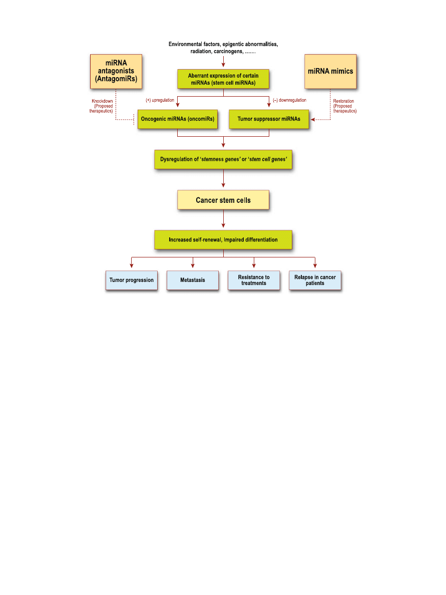
Fig. 3. Link between miRNA and cancer stem cells. Aberrant expressions of miRNAs, either as oncogenic
or tumor suppressor miRNAs, can lead to dysregulation of stem cell genes, causing increased self-renewal
potential and impaired differentiation in cancer stem cells. This dysregulation subsequently results in
carcinogenesis and oncogenesis. It is proposed that miRNA antagonists can knockdown the effects of
oncogenic miRNAs and miRNA mimics can restore the capabilities of tumor suppressor miRNAs.
Therefore, miRNA could be a vital tool in addressing cancer stem cell dysregulation. MiRNA-based
molecular therapy could hold great therapeutic potential against cancer progression, resistance, and
relapse
688
DeSano and Xu

knock down the expression of the oncogene miR-21, an anti-
miR-21 oligonucleotide was transfected into breast cancer
MCF-7 cells (
). It was demonstrated that the anit-miR-21
suppressed both cell growth in vitro and tumor growth in a
xenograft mouse model by increasing apoptosis and decreas-
ing cell proliferation (
). Therefore, antagomiRs are prom-
ising as therapeutic targets for oncogenic miRNA-based
cancer stem cell dysregulation.
For tumor suppressor miRNAs, which promote cancer
when under-expressed, miRNA mimics or lentiviruses should
be used to restore the tumor suppressors
’ natural potential,
resulting in decreased cancer development. For example, to
re-introduce miR-34 and its tumor suppressor capabilities, we
transfected miR-34 mimics into cancer cells, and the mimic
was shown to block the cell cycle in the G1 phase,
signi
ficantly increase activation of caspase-3, and knock down
its down
field targets of bcl-2, Notch, and HMGA2 (
). The
miRNA mimic, thus, restored miR-34 with its tumor suppres-
sor potential; however, the transfection of the miR-34 mimics
can only last a couple of days and the long-term biological
effects were not observed very effectively. To overcome this
dilemma, the cancer cells were infected with a lentivirus that
expressed miR-34a. This generated stable cells expressing
miR-34a. The lentiviral miR-34a was found to be able to
inhibit cancer cell growth and tumorsphere formation (
).
The lentiviral system restored the tumor suppressor effect of
miR-34 in pancreatic cancer stem cells as well (
). There-
fore, miRNA mimics and lentiviral miRNAs show great
potential in restoring tumor suppressor miRNAs to correct
the dysregulation of
“stem cell genes” in cancer stem cells.
The Challenge of miRNA
—Therapeutics: Delivery, Delivery,
Delivery
However, from a clinical/translational research point of
view, for the miRNA-based therapeutics to be effective, the
ef
ficient and functional delivery of miRNA mimics and/or
antagonists to tumor remains a great challenge.
Current approaches to deliver gene- and RNAi-based
therapeutics employ either viral or non-viral vector systems
(
). Viral vector-directed methods show high gene transfer
ef
ficiency but are deficient in several areas. The limitations of
a viral approach are related to their lack of tumor targeting
and to residual viral elements that can be immunogenic,
cytopathic, or recombinogenic (
). Non-viral gene transfer
vectors could circumvent some of the problems associated
with viral vectors. Progress has been made toward developing
non-viral, pharmaceutical formulations of gene therapeutics
for in vivo human therapy, particularly cationic liposome-
mediated gene transfer systems (
). Cationic liposomes
are composed of positively charged lipid bilayers and can be
complexed to negatively charged, naked DNA by simple
mixing of lipids and DNA such that the resulting complex
(lipoplex) has a net positive charge (
). The lipoplex is easily
bound and taken up by cells with relatively high transfection
ef
ficiency. Features of cationic liposomes that make them
versatile and attractive for DNA delivery include: simplicity
of preparation; the ability to complex large amounts of DNA;
versatility in use with any type and size of DNA or RNA; the
ability to transfect many different types of cells, including non-
dividing cells; and lack of immunogenicity or biohazardous
activity (
). There are multiple clinical trials now under-
way using cationic liposomes for gene delivery, and liposomes
for delivery of chemotherapeutics such as doxorubicin are
already on the market for breast cancer chemotherapy.
One disadvantage of cationic liposomes is that they lack
tumor speci
ficity and have relatively low transfection efficien-
cies as compared to viral vectors. However, this can be
dramatically increased when the lipoplexes bear a ligand
recognized by a cell surface receptor (
,
). Receptor-
mediated endocytosis represents a highly ef
ficient internal-
ization pathway in eukaryotic cells. The presence of a ligand
on a lipoplex facilitates the entry of DNA into cells through
initial binding of ligand by its receptor on the cell surface
followed by internalization of the bound lipoplex (
). Once
internalized, suf
ficient DNA escapes the endocytic pathway
to be expressed in the cell nucleus. A variety of ligands have
been examined for their lipoplex-targeting ability (
Recently, we developed tumor-speci
fic, ligand-targeting,
self-assembled, nanoparticle
–DNA lipoplex systems designed
for systemic gene therapy of cancer (US Patent No. 6,749,863,
European Patent No. EP 1,154,756) (
). These nano-
vector systems employ transferrin (Tf) or scFv against trans-
ferrin receptor (TfR), which is over-expressed in the majority
of human cancers, as tumor-targeting ligand (
,
). When
using Tf as a targeting ligand, we obtained the self-assembled
nanovectors at the sizes of 50
–90 nm, with highly compact
structure and favorite surface charge (
). These nanovectors
have novel nanostructure that resembles a virus particle with
a dense core enveloped by a membrane coated with Tf
molecules spiking on the surface (
). This nanovector system
shows promising ef
ficiency and specificity in targeted delivery
of various genes and anti-sense oligonucleotides to cancer in
vivo but not normal tissues (
). Systemic p53 gene
therapy using these nanovector systems demonstrated long-
term therapeutic ef
ficacy in animal models of human cancers
(
–
,
). Tf- and TfR-scFv-targeted nanovectors were
recently approved by the FDA for clinical testing, and the
first Phase-I clinical trial for non-viral systemic p53 gene
therapy is ongoing (
). The success of
these nanovectors for systemic p53 gene therapy, and more
recently Her-2 siRNA therapy (
–
), provide a promising,
tumor-targeted delivery system for novel RNAi-based thera-
pies, such as miRNA-therapeutics discussed above.
CONCLUSIONS
Abnormal miRNA expressions are connected to cancer
stem cell dysregulation. This dysregulation leads to the
initiation, development, and progression of cancer. Conse-
quently, molecular miRNA therapy is very important to
addressing oncogenesis linked with cancer stem cell dysregu-
lation. For this reason, future research should be aimed at
validating the link between miRNAs and cancer stem cells,
investigating miRNAs
’ role in cancer stem cells’ self-renewal
pathways, as well as studying therapeutic potential of
miRNAs against cancer progression, resistance, and relapse.
ACKNOWLEDGEMENTS
We wish to thank Mr. Steven Kronenberg for graphical
support and expertise in producing the
figures. This review was
689
MicroRNA Regulation of Cancer Stem Cells and Therapeutic Implications

supported in part by NIH grants CA121830, CA128220, and
CA134655 (to L. X.). J. D. is a University of Michigan Under-
graduate Research Opportunity Program (UROP) student.
REFERENCES
1. Zeng Y, Yi R, Cullen BR. MicroRNAs and small interfering
RNAs can inhibit mRNA expression by similar mechanisms.
Proc Natl Acad Sci U S A. 2003;100:9779
–84.
2. Lee Y, Kim M, Han J, Yeom KH, Lee S, Baek SH, et al.
MicroRNA genes are transcribed by RNA polymerase II. Embo
J. 2004;23:4051
–60.
3. Cai X, Hagedorn CH, Cullen BR. Human microRNAs are
processed from capped, polyadenylated transcripts that can also
function as mRNAs. RNA. 2004;10:1957
–66.
4. Han J, Lee Y, Yeom KH, Kim YK, Jin H, Kim VN. The Drosha-
DGCR8 complex in primary microRNA processing. Genes Dev.
2004;18:3016
–27.
5. Yi R, Qin Y, Macara IG, Cullen BR. Exportin-5 mediates the
nuclear export of pre-microRNAs and short hairpin RNAs.
Genes Dev. 2003;17:3011
–6.
6. Haase AD, Jaskiewicz L, Zhang H, Laine S, Sack R, Gatignol A,
et al. TRBP, a regulator of cellular PKR and HIV-1 virus
expression, interacts with Dicer and functions in RNA silencing.
EMBO Rep. 2005;6:961
–7.
7. Chendrimada TP, Gregory RI, Kumaraswamy E, Norman J,
Cooch N, Nishikura K, et al. TRBP recruits the Dicer complex to
Ago2 for microRNA processing and gene silencing. Nature.
2005;436:740
–4.
8. Zhang B, Pan X, Cobb GP, Anderson TA. microRNAs as
oncogenes and tumor suppressors. Dev Biol. 2007;302:1
–12.
9. Liu J, Valencia-Sanchez MA, Hannon GJ, Parker R. MicroRNA-
dependent localization of targeted mRNAs to mammalian P-
bodies. Nat Cell Biol. 2005;7:719
–23.
10. Chekanova JA, Belostotsky DA. MicroRNAs and messenger
RNA turnover. Methods Mol Biol. 2006;342:73
–85.
11. Croce CM, Calin GA. miRNAs, cancer, and stem cell division.
Cell. 2005;122:6
–7.
12. Rigoutsos I. New tricks for animal microRNAS: targeting of
amino acid coding regions at conserved and nonconserved sites.
Cancer Res. 2009;69:3245
–8.
13. Suh MR, Lee Y, Kim JY, Kim SK, Moon SH, Lee JY, et al.
Human embryonic stem cells express a unique set of micro-
RNAs. Dev Biol. 2004;270:488
–98.
14. Bernstein E, Kim SY, Carmell MA, Murchison EP, Alcorn H, Li
MZ, et al. Dicer is essential for mouse development. Nat Genet.
2003;35:215
–7.
15. Kanellopoulou C, Muljo SA, Kung AL, Ganesan S, Drapkin R,
Jenuwein T, et al. Dicer-de
ficient mouse embryonic stem cells are
defective in differentiation and centromeric silencing. Genes
Dev. 2005;19:489
–501.
16. Hat
field SD, Shcherbata HR, Fischer KA, Nakahara K, Carthew
RW, Ruohola-Baker H. Stem cell division is regulated by the
microRNA pathway. Nature. 2005;435:974
–8.
17. Dontu G, Al-Hajj M, Abdallah WM, Clarke MF, Wicha MS.
Stem cells in normal breast development and breast cancer. Cell
Prolif. 2003;36(Suppl 1):59
–72.
18. Farnie G, Clarke RB. Mammary stem cells and breast cancer
—
role of Notch signalling. Stem Cell Rev. 2007;3:169
–75.
19. Papagiannakopoulos T, Kosik KS. MicroRNAs: regulators of
oncogenesis and stemness. BMC Med. 2008;6:15.
20. Dick JE. Normal and leukemic human stem cells assayed in
SCID mice. Semin Immunol. 1996;8:197
–206.
21. Jin L, Hope KJ, Zhai Q, Smadja-Joffe F, Dick JE. Targeting of
CD44 eradicates human acute myeloid leukemic stem cells. Nat
Med. 2006;12:1167
–74.
22. Al-Hajj M, Clarke MF. Self-renewal and solid tumor stem cells.
Oncogene. 2004;23:7274
–82.
23. Collins AT, Berry PA, Hyde C, Stower MJ, Maitland NJ.
Prospective identi
fication of tumorigenic prostate cancer stem
cells. Cancer Res. 2005;65:10946
–51.
24. Lawson DA, Witte ON. Stem cells in prostate cancer initiation
and progression. J Clin Invest. 2007;117:2044
–50.
25. Tang DG, Patrawala L, Calhoun T, Bhatia B, Choy G,
Schneider-Broussard R, et al. Prostate cancer stem/progenitor
cells: identi
fication, characterization, and implications. Mol
Carcinog. 2007;46:1
–14.
26. Patrawala L, Calhoun-Davis T, Schneider-Broussard R, Tang
DG. Hierarchical organization of prostate cancer cells in
xenograft tumors: the CD44+alpha2beta1+ cell population is
enriched in tumor-initiating cells. Cancer Res. 2007;67:6796
–805.
27. Li H, Chen X, Calhoun-Davis T, Claypool K, Tang DG. PC3
human prostate carcinoma cell holoclones contain self-renewing
tumor-initiating cells. Cancer Res. 2008;68:1820
–5.
28. Dontu G, Jackson KW, McNicholas E, Kawamura MJ, Abdallah
WM, Wicha MS. Role of Notch signaling in cell-fate determi-
nation of human mammary stem/progenitor cells. Breast Cancer
Res. 2004;6:R605
–15.
29. Ginestier C, Hur MH, Charafe-Jauffret E, Monville F, Dutcher J,
Brown M, et al. ALDH1 is a marker of normal and malignant
human mammary stem cells and a predictor of poor clinical
outcome. Cell Stem Cell. 2007;1:555
–67.
30. Prince ME, Sivanandan R, Kaczorowski A, Wolf GT, Kaplan
MJ, Dalerba P, et al. Identi
fication of a subpopulation of cells
with cancer stem cell properties in head and neck squamous cell
carcinoma. Proc Natl Acad Sci U S A. 2007;104:973
–8.
31. Li C, Heidt DG, Dalerba P, Burant CF, Zhang L, Adsay V, et al.
Identi
fication of pancreatic cancer stem cells. Cancer Res.
2007;67:1030
–7.
32. Szotek PP, Pieretti-Vanmarcke R, Masiakos PT, Dinulescu DM,
Connolly D, Foster R, et al. Ovarian cancer side population
de
fines cells with stem cell-like characteristics and Mullerian
Inhibiting Substance responsiveness. Proc Natl Acad Sci U S A.
2006;103:11154
–9.
33. Ricci-Vitiani L, Lombardi DG, Pilozzi E, Biffoni M, Todaro M,
Peschle C, et al. Identi
fication and expansion of human colon-
cancer-initiating cells. Nature. 2007;445:111
–5.
34. Vescovi AL, Galli R, Reynolds BA. Brain tumour stem cells. Nat
Rev Cancer. 2006;6:425
–36.
35. Bussolati B, Grange C, Sapino A, Camussi G. Endothelial Cell
Differentiation of Human Breast Tumor Stem/Progenitor Cells. J
Cell Mol Med. 2009;13:309
–19.
36. Quintana E, Shackleton M, Sabel MS, Fullen DR, Johnson TM,
Morrison SJ. Ef
ficient tumour formation by single human
melanoma cells. Nature. 2008;456:593
–8.
37. Rich JN. Cancer stem cells in radiation resistance. Cancer Res.
2007;67:8980
–4.
38. Al-Hajj M. Cancer stem cells and oncology therapeutics. Curr
Opin Oncol. 2007;19:61
–4.
39. Wicha MS. Cancer stem cells and metastasis: lethal seeds. Clin
Cancer Res. 2006;12:5606
–7.
40. Morrison SJ, Spradling AC. Stem cells and niches: mechanisms
that promote stem cell maintenance throughout life. Cell.
2008;132:598
–611.
41. Dontu G, Abdallah WM, Foley JM, Jackson KW, Clarke MF,
Kawamura MJ, et al. In vitro propagation and transcriptional
pro
filing of human mammary stem/progenitor cells. Genes Dev.
2003;17:1253
–70.
42. Zhang M, Behbod F, Atkinson RL, Landis MD, Kittrell F,
Edwards D, et al. Identi
fication of tumor-initiating cells in a p53-
null mouse model of breast cancer. Cancer Res. 2008;68:4674
–82.
43. Murat A, Migliavacca E, Gorlia T, Lambiv WL, Shay T, Hamou
MF, et al. Stem cell-related
“self-renewal” signature and high
epidermal growth factor receptor expression associated with
resistance to concomitant chemoradiotherapy in glioblastoma. J
Clin Oncol. 2008;26:3015
–24.
44. Reya T, Morrison SJ, Clarke MF, Weissman IL. Stem cells,
cancer, and cancer stem cells. Nature. 2001;414:105
–11.
45. Zencak D, Lingbeek M, Kostic C, Tekaya M, Tanger E,
Hornfeld D, et al. Bmi1 loss produces an increase in astroglial
cells and a decrease in neural stem cell population and
proliferation. J Neurosci. 2005;25:5774
–83.
46. Fasano CA, Dimos JT, Ivanova NB, Lowry N, Lemischka IR,
Temple S. shRNA knockdown of Bmi-1 reveals a critical role for
p21-Rb pathway in NSC self-renewal during development. Cell
Stem Cell. 2007;1:87
–99.
690
DeSano and Xu

47. Hambardzumyan D, Becher OJ, Holland EC. Cancer stem cells
and survival pathways. Cell Cycle. 2008;7:1371
–8.
48. Bhardwaj G, Murdoch B, Wu D, Baker DP, Williams KP,
Chadwick K, et al. Sonic hedgehog induces the proliferation of
primitive human hematopoietic cells via BMP regulation. Nat
Immunol. 2001;2:172
–80.
49. Zhang Y, Kalderon D. Hedgehog acts as a somatic stem cell
factor in the Drosophila ovary. Nature. 2001;410:599
–604.
50. Reya T, Duncan AW, Ailles L, Domen J, Scherer DC, Willert K,
et al. A role for Wnt signalling in self-renewal of haematopoietic
stem cells. Nature. 2003;423:409
–14.
51. Zhao RC, Zhu YS, Shi Y. New hope for cancer treatment:
Exploring the distinction between normal adult stem cells and
cancer stem cells. Pharmacol Ther. 2008;119:74
–82.
52. Luu HH, Zhang R, Haydon RC, Rayburn E, Kang Q, Si W, et al.
Wnt/beta-catenin signaling pathway as a novel cancer drug
target. Curr Cancer Drug Targets. 2004;4:653
–71.
53. Jamieson CH, Ailles LE, Dylla SJ, Muijtjens M, Jones C,
Zehnder JL, et al. Granulocyte-macrophage progenitors as
candidate leukemic stem cells in blast-crisis CML. N Engl J
Med. 2004;351:657
–67.
54. Fusco A, Fedele M. Roles of HMGA proteins in cancer. Nat Rev
Cancer. 2007;7:899
–910.
55. Abe N, Watanabe T, Suzuki Y, Matsumoto N, Masaki T, Mori T,
et al. An increased high-mobility group A2 expression level is
associated with malignant phenotype in pancreatic exocrine
tissue. Br J Cancer. 2003;89:2104
–9.
56. Meyer B, Loeschke S, Schultze A, Weigel T, Sandkamp M,
Goldmann T, et al. HMGA2 overexpression in non-small cell
lung cancer. Mol Carcinog. 2007;46:503
–11.
57. Sparmann A, van Lohuizen M. Polycomb silencers control cell
fate, development and cancer. Nat Rev Cancer. 2006;6:846
–56.
58. Haupt Y, Alexander WS, Barri G, Klinken SP, Adams JM. Novel
zinc
finger gene implicated as myc collaborator by retrovirally
accelerated lymphomagenesis in E mu-myc transgenic mice. Cell.
1991;65:753
–63.
59. Liu S, Dontu G, Mantle ID, Patel S, Ahn NS, Jackson KW, et al.
Hedgehog signaling and Bmi-1 regulate self-renewal of normal
and malignant human mammary stem cells. Cancer Res.
2006;66:6063
–71.
60. Domen J, Gandy KL, Weissman IL. Systemic overexpression of
BCL-2 in the hematopoietic system protects transgenic mice
from the consequences of lethal irradiation. Blood. 1998;91:
2272
–82.
61. Domen J, Cheshier SH, Weissman IL. The role of apoptosis in
the regulation of hematopoietic stem cells: Overexpression of
Bcl-2 increases both their number and repopulation potential. J
Exp Med. 2000;191:253
–64.
62. Ji Q, Hao X, Zhang M, Tang W, Yang M, Li L, et al. MicroRNA
miR-34 inhibits human pancreatic cancer tumor-initiating cells.
PLoS ONE. 2009;4:e6816.
63. Calin GA, Croce CM. MicroRNA signatures in human cancers.
Nat Rev Cancer. 2006;6:857
–66.
64. Hirschmann-Jax C, Foster AE, Wulf GG, Nuchtern JG, Jax TW,
Gobel U, et al. A distinct
“side population” of cells with high
drug ef
flux capacity in human tumor cells. Proc Natl Acad Sci
U S A. 2004;101:14228
–33.
65. Wiemer EA. The role of microRNAs in cancer: no small matter.
Eur J Cancer. 2007;43:1529
–44.
66. Frankel LB, Christoffersen NR, Jacobsen A, Lindow M, Krogh
A, Lund AH. Programmed Cell Death 4 (PDCD4) is an
important functional target of the MicroRNA miR-21 in breast
cancer cells. J Biol Chem. 2008;283:1026
–33.
67. Asangani IA, Rasheed SA, Nikolova DA, Leupold JH, Colburn
NH, Post S, et al. MicroRNA-21 (miR-21) post-transcriptionally
downregulates tumor suppressor Pdcd4 and stimulates invasion,
intravasation and metastasis in colorectal cancer. Oncogene.
2008;27:2128
–36.
68. Yan LX, Huang XF, Shao Q, Huang MY, Deng L, Wu QL, et al.
MicroRNA miR-21 overexpression in human breast cancer is
associated with advanced clinical stage, lymph node metastasis
and patient poor prognosis. RNA. 2008;14:2348
–60.
69. Si ML, Zhu S, Wu H, Lu Z, Wu F, Mo YY. miR-21-mediated
tumor growth. Oncogene. 2007;26:2799
–803.
70. Hayashita Y, Osada H, Tatematsu Y, Yamada H, Yanagisawa K,
Tomida S, et al. A polycistronic microRNA cluster, miR-17
–92, is
overexpressed in human lung cancers and enhances cell prolif-
eration. Cancer Res. 2005;65:9628
–32.
71. He L, Thomson JM, Hemann MT, Hernando-Monge E, Mu D,
Goodson S, et al. A microRNA polycistron as a potential human
oncogene. Nature. 2005;435:828
–33.
72. Lu Y, Thomson JM, Wong HY, Hammond SM, Hogan BL.
Transgenic over-expression of the microRNA miR-17
–92 cluster
promotes proliferation and inhibits differentiation of lung
epithelial progenitor cells. Dev Biol. 2007;310:442
–53.
73. Uziel T, Karginov FV, Xie S, Parker JS, Wang YD, Gajjar A, et
al. The miR-17 92 cluster collaborates with the Sonic Hedgehog
pathway in medulloblastoma. Proc Natl Acad Sci U S A.
2009;106:2812
–7.
74 Nagel R, le Sage C, Diosdado B, van der Waal M, Oude Vrielink
JA, Bolijn A, et al. Regulation of the adenomatous polyposis coli
gene by the miR-135 family in colorectal cancer. Cancer Res.
2008;68:5795
–802.
75. Johnson CD, Esquela-Kerscher A, Stefani G, Byrom M, Kelnar
K, Ovcharenko D, et al. The let-7 microRNA represses cell
proliferation pathways in human cells. Cancer Res. 2007;67:
7713
–22.
76. Esquela-Kerscher A, Trang P, Wiggins JF, Patrawala L, Cheng
A, Ford L, et al. The let-7 microRNA reduces tumor growth in
mouse models of lung cancer. Cell Cycle. 2008;7:759
–64.
77. Johnson SM, Grosshans H, Shingara J, Byrom M, Jarvis R,
Cheng A, et al. RAS is regulated by the let-7 microRNA family.
Cell. 2005;120:635
–47.
78. Yu F, Yao H, Zhu P, Zhang X, Pan Q, Gong C, et al. let-7
regulates self renewal and tumorigenicity of breast cancer cells.
Cell. 2007;131:1109
–23.
79. Mayr C, Hemann MT, Bartel DP. Disrupting the pairing between
let-7 and Hmga2 enhances oncogenic transformation. Science.
2007;315:1576
–9.
80. Cimmino A, Calin GA, Fabbri M, Iorio MV, Ferracin M,
Shimizu M, et al. miR-15 and miR-16 induce apoptosis by
targeting BCL2. Proc Natl Acad Sci U S A. 2005;102:13944
–9.
81. Bonci D, Coppola V, Musumeci M, Addario A, Giuffrida R,
Memeo L, et al. The miR-15a-miR-16
–1 cluster controls prostate
cancer by targeting multiple oncogenic activities. Nat Med.
2008;14:1271
–7.
82. Godlewski J, Nowicki MO, Bronisz A, Williams S, Otsuki A,
Nuovo G, et al. Targeting of the Bmi-1 oncogene/stem cell
renewal factor by microRNA-128 inhibits glioma proliferation
and self-renewal. Cancer Res. 2008;68:9125
–30.
83. Garzia L, Andolfo I, Cusanelli E, Marino N, Petrosino G, De
Martino D, et al. MicroRNA-199b
–5p impairs cancer stem cells
through negative regulation of HES1 in medulloblastoma. PLoS
ONE. 2009;4:e4998.
84. Ferretti E, De Smaele E, Miele E, Laneve P, Po A, Pelloni M, et
al. Concerted microRNA control of Hedgehog signalling in
cerebellar neuronal progenitor and tumour cells. Embo J.
2008;27:2616
–27.
85. He X, He L, Hannon GJ. The guardian
’s little helper: micro-
RNAs in the p53 tumor suppressor network. Cancer Res.
2007;67:11099
–101.
86. Ji Q, Hao X, Meng Y, Zhang M, Desano J, Fan D, et al.
Restoration of tumor suppressor miR-34 inhibits human p53-
mutant gastric cancer tumorspheres. BMC Cancer. 2008;8:266.
87. Hat
field S, Ruohola-Baker H. microRNA and stem cell function.
Cell Tissue Res. 2008;331:57
–66.
88. Krutzfeldt J, Rajewsky N, Braich R, Rajeev KG, Tuschl T,
Manoharan M, et al. Silencing of microRNAs in vivo with
‘antagomirs’. Nature. 2005;438:685–9.
89. Pirollo KF, Xu L, Chang EH. Non-viral gene delivery for p53.
Curr Opin Mol Ther. 2000;2:168
–75.
90. Xu L, Pirollo KF, Chang EH. Tumor-targeted p53-gene therapy
enhances the ef
ficacy of conventional chemo/radiotherapy. J
Control Release. 2001;74:115
–28.
91. Xu L, Frederik P, Pirollo KF, Tang WH, Rait A, Xiang LM, et al.
Self-assembly of a virus-mimicking nanostructure system for
ef
ficient tumor-targeted gene delivery. Hum Gene Ther.
2002;13:469
–81.
691
MicroRNA Regulation of Cancer Stem Cells and Therapeutic Implications

92. Xu L, Huang CC, Huang W, Tang WH, Rait A, Yin YZ, et al.
Systemic tumor-targeted gene delivery by anti-transferrin recep-
tor scFv-immunoliposomes. Molecular Cancer Therapeutics.
2002;1:337
–46.
93. Xu L, Pirollo KF, Tang WH, Rait A, Chang EH. Transferrin-
liposome-mediated systemic p53 gene therapy in combination
with radiation results in regression of human head and neck
cancer xenografts. Hum Gene Ther. 1999;10:2941
–52.
94. Xu L, Pirollo KF, Chang EH. Transferrin-liposome-mediated p53
sensitization of squamous cell carcinoma of the head and neck to
radiation in vitro. Hum Gene Ther. 1997;8:467
–75.
95. Xu L, Tang WH, Huang CC, Alexander W, Xiang LM, Pirollo
KF, et al. Systemic p53 gene therapy of cancer with immunoli-
poplexes targeted by anti-transferrin receptor scFv. Mol Med.
2001;7:723
–34.
96. Pirollo KF, Rait A, Zhou Q, Hwang SH, Dagata JA, Zon G, et al.
Materializing the potential of small interfering RNA via a tumor-
targeting nanodelivery system. Cancer Res. 2007;67:2938
–43.
97. Pirollo KF, Zon G, Rait A, Zhou Q, Yu W, Hogrefe R, et al.
Tumor-targeting nanoimmunoliposome complex for short inter-
fering RNA delivery. Hum Gene Ther. 2006;17:117
–24.
98. Hogrefe RI, Lebedev AV, Zon G, Pirollo KF, Rait A, Zhou Q, et
al. Chemically modi
fied short interfering hybrids (siHYBRIDS):
nanoimmunoliposome delivery in vitro and in vivo for RNAi of
HER-2. Nucleosides Nucleotides Nucleic Acids. 2006;25:889
–
907.
692
DeSano and Xu
Document Outline
- MicroRNA Regulation of Cancer Stem Cells and Therapeutic Implications
- Abstract
- INTRODUCTION
- MicroRNA Biogenesis
- MicroRNA and Regulation of Stem Cells
- Cancer Stem Cell Hypothesis
- Self-Renewal
- Signaling Pathways of the “Stem Cell Genes”
- Link Between miRNA and Cancer Stem Cells
- Examples of Potential Links Between miRNA and Cancer Stem Cells
- Examples Support the Role of “Stem Cells miRNAs” in Oncogenesis and the Implications in Molecular Cancer Therapy
- MicroRNA Therapeutics
- The Challenge of miRNA—Therapeutics: Delivery, Delivery, Delivery
- CONCLUSIONS
- References
- INTRODUCTION
- Abstract
Wyszukiwarka
Podobne podstrony:
Theory of Varied Consumer Choice?haviour and Its Implicati
Non Intrinsic Differential Mode Noise of Switching Power Supplies and Its Implications to Filter Des
Ferrell Gerson Therapy for Those Dying of Cancer
Cancer Proposed Common Cause and Cure for All Forms of Cancer David W Gregg, PhD
Tea polyphenols prevention of cancer and optimizing health
Ebsco Martin Cognitive emotion regulation in the prediction of depression, anxiety, stress, and an
Ebsco Martin Cognitive emotion regulation in the prediction of depression, anxiety, stress, and an
Eleswarapu, Thompson And Venkataraman The Impact Of Regulation Fair Disclosure Trading Costs And Inf
Working hypothesis on the nature of voices and therapies
Dos Santos Ferreira D , Staubach W Global and local regularity of Fourier integral operators on weig
Electrical Properties of Cancer Cells
How Can We Stop Our Children from Hurting Themselves Stages of Change, Motivational Interviewing, a
BSAVA Manual of Rabbit Surgery Dentistry and Imaging
Functional Origins of Religious Concepts Ontological and Strategic Selection in Evolved Minds
Guide to the properties and uses of detergents in biology and biochemistry
EurRad Ultrasound of thyroid, parathyroid glands and neck lymph nodes
więcej podobnych podstron