58023
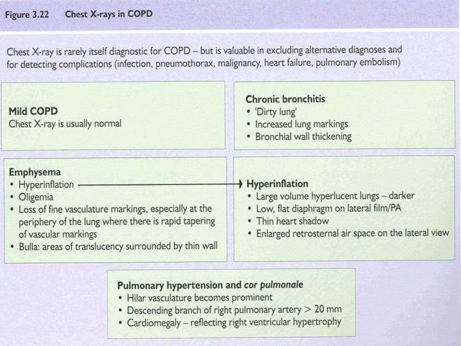
Figurę 3.22 Chest X-rays in COPD
Chest X-ray is rarely itself diagnostic for COPD - but is valuable in excluding alternative diagnoses and for detecting complications (infection, pneumothorax. malignancy, heart failure. pulmonary embolism)
|
Mild COPD Chest X-ray is usually normal |
Chronić bronchitis • 'Dirty lung' • Increased lung markings • Bronchial wali thickening |
|
Emphysema | |
|
• Oligemia • Loss of fine vasculature markings. especially at the periphery of the lung where there is rapid tapering of vascular markings • Bulla: areas of translucency surrounded by thin wali |
f nypenmiation • Large volume hyperlucent lungs - darker • Low, fiat diaphragm on lateral film/PA • Thin heart shadow • Enlarged retrosternal air space on the lateral view |
Pulmonary hypertension and cor pulmonale
• Hilar va$culature becomes prominent
• Descending branch of right pulmonary artery > 20 mm
• Cardiomegaly - reflecting right ventricular hypertrophy
Wyszukiwarka
Podobne podstrony:
6 22 Figurę 6-22 Cross-fiber stroking of rotatores in thoracic region using support fingertips (Drap
P1080002 (2) Tytuł oryginału: Chest X-Ray Madę Easy Second edition Autorzy: Jonathan Come, Mary Carr
227 (21) 22 : Diagnostic approach to otitis extema Figurę 22 :17: Invasive mast celi tumour in the e
22 (669) 38 The Viking Age in Denmark Figurę 8 B. ‘Aftcr Jelling’ Stones in southernmost Jylland. Si
Figurę 3.21 Biood tests in COPD ct
fig22 Figurę 22 Mantle
figure37b • —I P ! ^ CO CO IN T CD CO ucunaN ucunaN rM ^ to co
00456 fb2f4b1a62a95fe3cc8d4c408500a0c A Graphical Aid for Analyzing Autocorrelated Dynamical System
image004 HjC - (CHj), HjC - CHj - Clij - CH2 - CH2 - CHj _CK2 _ CB2 -OHj -CHj Figurę 2: Undecune. A
Document 2 (11) Figurę 2.13: Pronounced gynecomastia in a man with ReifensteirTs syndrome. For cosme
więcej podobnych podstron