5 29
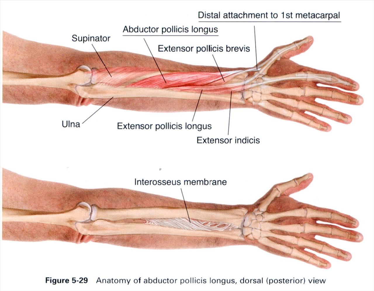

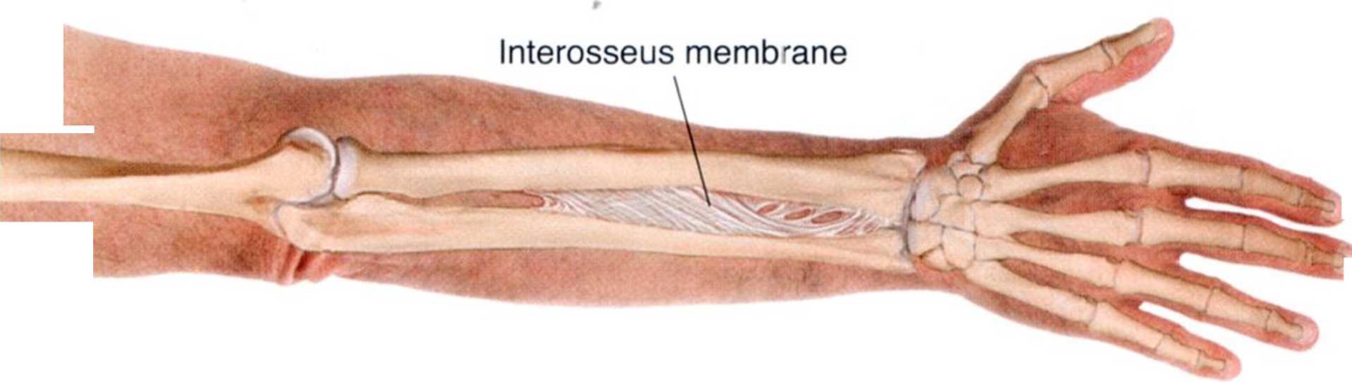
Figurę 5-29 Anatomy of abductor pollicis longus, dorsal (posterior) view
Wyszukiwarka
Podobne podstrony:
5 28 Extensor indicis Interossęus membranę Figurę 5-28 Anatomy of extensor pollicis longus, dorsal (
5 28 Extensor incicis Figurę 5-28 Anatomy of extensor pollicis longus, dorsal (posterior) view
5 29 Distal attachment to 1 st metacarpal Supinator Abductor pollicis longus Extensor pollicis
5 22 Brachioradialis Distal attachment to 2nd metacarpal Lateral epicondyle Extens
5 22 Brachioradialis Distal attachment to 2nd metacarpal Supracondylar ridge Extensor retinaculum Ex
5 26 Supinator Abductor pollicis longus Extensor pollicis longus Extensor indicis Figurę 5-26 Anatom
5 49 Extensor tendon expansion First interosseus Metacarpals Radius Figurę 5-49 Anatomy of the dorsa
5 35 Distal attachment to base of 2nd & 3rd metacarpal Flexor carpi ulnaris Palmaris longus Pron
3 37 Occipitalis Clavicle Figurę 3-36 Anatomy of SCM Manubrium of sternum Attachment to superior nuc
5 36 Brachioradialis Flexor carpi radialis Distal attachment to pisiform bonę, Flexor carpi ulnaris
5 17 Attachment to the ulna Figurę 5-17 Anatomy of pronator guadratus, volar (anterior) view
5 23 Brachioradialis Extensor carpi radialis longus Extensor digiti minimi Distal attachment to 5th
5 27 Supinator Abductor pollicis longus Proximal attachment to dorsal radius and interosseus membran
5 26 Figurę 5-26 Anatomy of extensor indicis, dorsal (posterior) view
5 50 Proximal phalanx First palmar interosseus Metacarpals Ulna Radius Figurę 5-50 Anatomy of the pa
Fully-constructed unit before attachment to a Gunn source. Please notę the placement of the 1N9
7 15 Ouadratus lumborum Emerging spinał nerve roots lliac crest Figurę 7-15 Anatomy of guadratu
5 17 Figurę 5-17 Anatomy of pronator guadratus, volar (anterior) view
5 23 Brachioradialis Extensor carpi radialis longus Figurę 5-23 Anatomy of extenso
więcej podobnych podstron