3 37

Occipitalis
Clavicle
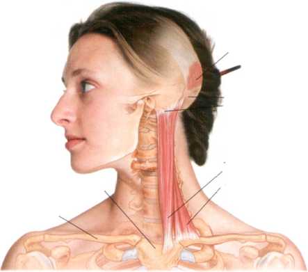
Figurę 3-36 Anatomy of SCM
Manubrium of sternum
Attachment to superior nuchal linę and mastoid process
Sternoc eidomastoid sternal head clavicular head
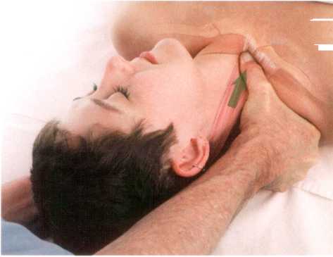
Figurę 3-37 Stripping of sternal head of SCM
Wyszukiwarka
Podobne podstrony:
3 37 Occipitalis sternal head clavicular head Figurę 3-36 Anatomy of SCM Manubrium of slernum
3 52 Occipital bonę (occiput) Semispinalis capitis Middle scalene Posterior scalene Figurę 3-52 Anat
7 15 Ouadratus lumborum Emerging spinał nerve roots lliac crest Figurę 7-15 Anatomy of guadratu
5 17 Figurę 5-17 Anatomy of pronator guadratus, volar (anterior) view
5 23 Brachioradialis Extensor carpi radialis longus Figurę 5-23 Anatomy of extenso
5 24 Brachioradialis Extensor carpi radialis longus Anconeus Figurę 5-24 Anatomy of extensor digiti
5 26 Figurę 5-26 Anatomy of extensor indicis, dorsal (posterior) view
5 28 Extensor indicis Interossęus membranę Figurę 5-28 Anatomy of extensor pollicis longus, dorsal (
10 1 Fascia lata Medial collateral ligament Patellar ligament Extensor retinacula Figurę 10-1 Anatom
5 17 Attachment to the ulna Figurę 5-17 Anatomy of pronator guadratus, volar (anterior) view
5 26 Supinator Abductor pollicis longus Extensor pollicis longus Extensor indicis Figurę 5-26 Anatom
5 28 Extensor incicis Figurę 5-28 Anatomy of extensor pollicis longus, dorsal (posterior) view
5 29 Distal attachment to 1 st metacarpalExtensor indicis Figurę 5-29 Anatomy of abductor pollicis l
5 34 Figurę 5-34 Anatomy of palmaris longus, volar (anterior) view
5 38 Figurę 5-38 Anatomy of flexor digitorum superficialis, volar (anterior) view
5 49 Extensor tendon expansion First interosseus Metacarpals Radius Figurę 5-49 Anatomy of the dorsa
5 50 Proximal phalanx First palmar interosseus Metacarpals Ulna Radius Figurę 5-50 Anatomy of the pa
7 10 11 th rib 12th rib Figurę 7-10 Anatomy of external obligue Rectus sheath (removed over
10 7 Figurę 10-7 Anatomy of the plantar fascia
więcej podobnych podstron