
CHAPTER
DIAGNOSIS AND
TREATMENT OF
STROKES
Ischemic Lesions and
Intraparenchymal
Hemorrhages
of the Brain
Dennis E. McDonnell
Marshall B. Alien, Jr.
Cerebrovascular disease is the third leading cause of death
among adults in the United States, ranking behind cancer
and heart diseased Although on the decline, it is the most
important cause of chronic disability.
2
'
3
Ischemic cerebro-
vascular disease accounts for about 75 percent of the cases
while lesions resulting from hemorrhage into the brain or
subarachnoid space account for about 16 percent.
4
-
5
The role
of surgery in the prevention of stroke and the treatment of
various clinical afflictions is becoming clearly defined. In
this chapter, we will discuss ischemic lesions and spontane-
ous intraparenchymal hemorrhages of the brain. Cerebrovas-
cular diseases have been classified and detailed both clini-
cally and pathologically.
6
that are occluded or when there is a global reduction of bulk
flow resulting from systemic hypoperfusion. This critical
level is approximately 18 mVlOO g per minute compared to
a normal average resting flow rate of 60 ml/100 g per
minute.
7
Emboli originating from intraluminal lesions of the
extracranial carotid artery account for two-thirds of infarcts
in the middle cerebral artery distribution (Fig. 14-1).
8
Sur-
gery may be utilized to excise or repair constriction, remove
lesions that might be a source of emboli. bypass occlusions,
or augment collateral flow.
9
-
12
Surgical attempts to reopen
complete occlusions often fail clinically.
13
Bypass revascu-
larizadons, once popular, are presently much more re-
stricted-
14
-
15
Decompressive resections of intracranial hema-
toma or edematous or infarcted brain may be life-saving, by
relieving mass effect and preventing tentorial hemiation.
MECHANISMS OF
CEREBRAL ISCHEMIA
LOCALIZATION
Cerebral infarction occurs when blood flow drops below
critical levels in regions of brain irrigated by specific vessels
Cerebral infarction occurs in the distribution of tissue sup-
plied by one of the four major arteries that supply the brain,
251
252
CHAPTER 1
4
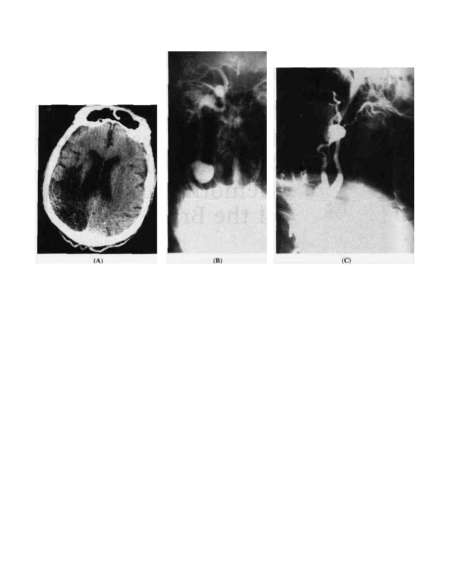
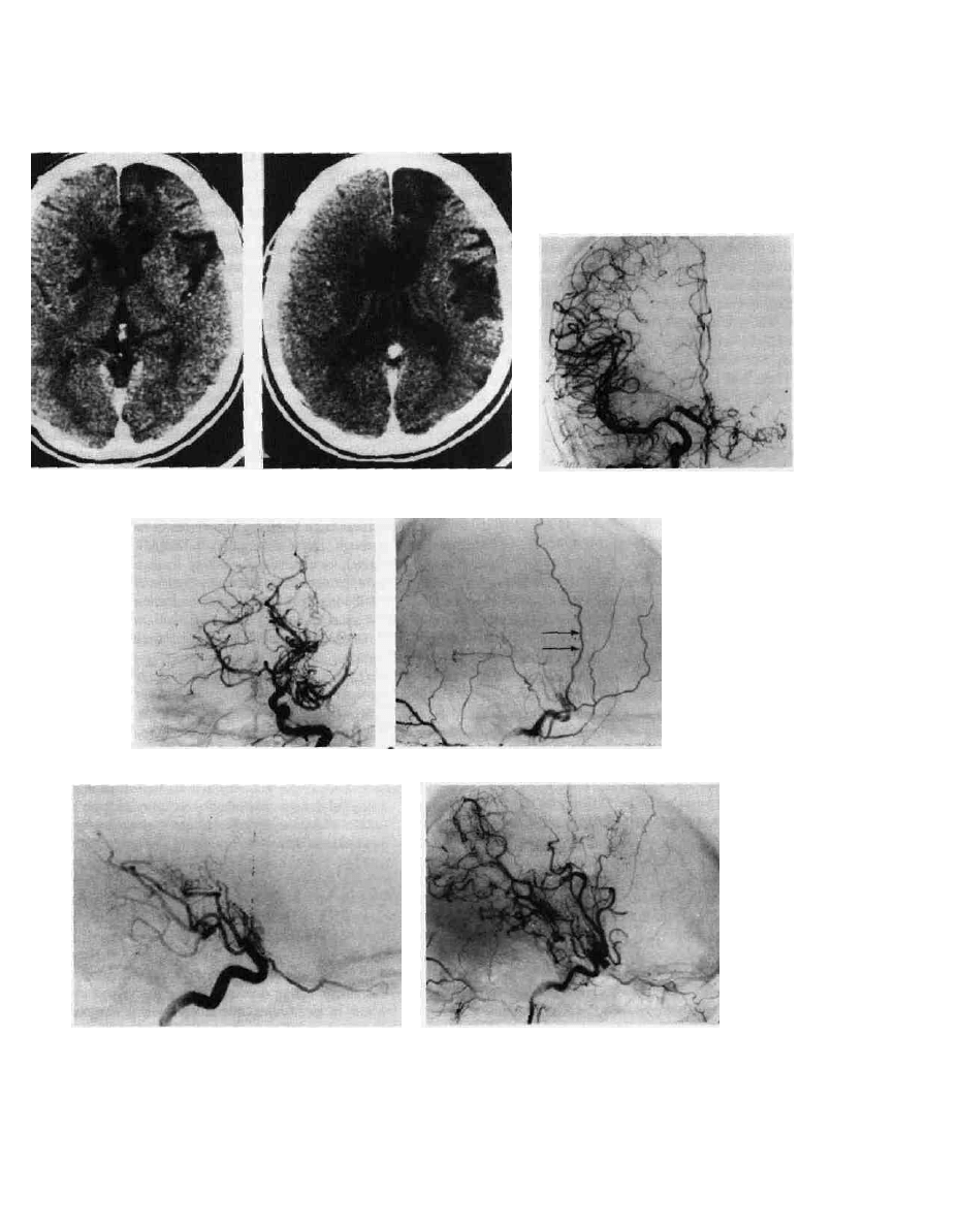
Figure 14-1 Infarction of the right temporal and occipital lobes
due to emboli from an aneurysm of the right extracranial internal
carotid artery. (A) CT scan shows decreased attenuation due to
encephalomalacia of infarct; (B) right carotid angiogram fronta-'
projection; and (C) lateral projection shows the aneurysm and muck
thrombus, source of emboli, within it. .1:
a major arterial branch such as the middle cerebral artery or
posterior inferior cerebellar artery, or a cortical or penetrat-
ing branch. Infarclion may occur in zones of limited perfu-
sion within the watershed distribution between areas sup-
plied by two cerebral arteries—for example, parenchyma at
the peripheral zone of supply by the middle cerebral artery
(Fig. 14-2). Infarction in the primary distribution of a major
vessel is most likely the result of occlusion of that vessel.
Most occlusions of major vessels supplying the brain result
from emboli, but other causes include propagation of an
intraluminal clot, dissection of the wall of a vessel, throm-
bosis within a vessel containing a plaque, or progressive
narrowing of the vessel.
The occurrence, size, and distribution of the infarction
may be assured or even expanded by the development and
propagation of an antegrade intraluminal thrombus which
progresses to compromise collateral conduits. On the other
hand, an efficient collateral circulation may limit the size
and distribution of an infarct. Thus occlusion of a carotid or
a vertebral artery may be of no clinical consequence in the
presence of a competent circle of Willis or other collaterals.
In many instances, occlusion of a proximal segment of the
middle cerebral artery proves to be inconsequential in the
presence of adequate collateral circulation from the anterior
and posterior cerebral arteries.
Most strokes that occur in the distribution of the middle
cerebral artery are not due to a local lesion in the middle
cerebral artery but arise from propagation of a thrombus or
an embolus released from a proximal extracranial artery
endocardium.
13
In some cases of occlusion of the circle
Willis, the entire cerebrum may be irrigated by collates
often supplied from the vertebral arteries. When the colL.
eral circulation is less efficient, clinical consequences of
vascular obstructions may be severe. When acute occlusioc
of the carotid artery produces a stroke, only 2 to 12 percent
of patients will recover, 40 to 69 percent will be disabled
with severe neurological deficits, and 16 to 55 percent will
die.'
3
Penetrating arteries are generally "end" arteries with no
collateral circulation, and occlusion is likely to result in
"lacunar" infarcts, i.e., small infarcts deep within the white
matter^
6
'
17
Infarction in a watershed area is usually the result of
reduced bulk flow from a devastating cardiovascular eveni
such as cardiac arrest, hypovolemic shock, occlusion of an
extracranial internal carotid artery, or widespread distur-
bances in the microvascular circulation.
3
PATHOGENESIS
Emboli may originate from the endocardium within the
heart, from plaques on the endothelial walls of proximal
arteries, from fresh thrombi originating in acutely occluded
major proximal arteries, or from systemic sources passing
through defects in the heart (paradoxical emboli) such as
DIAGNOSIS AND TREATMENT OF STROKES 253
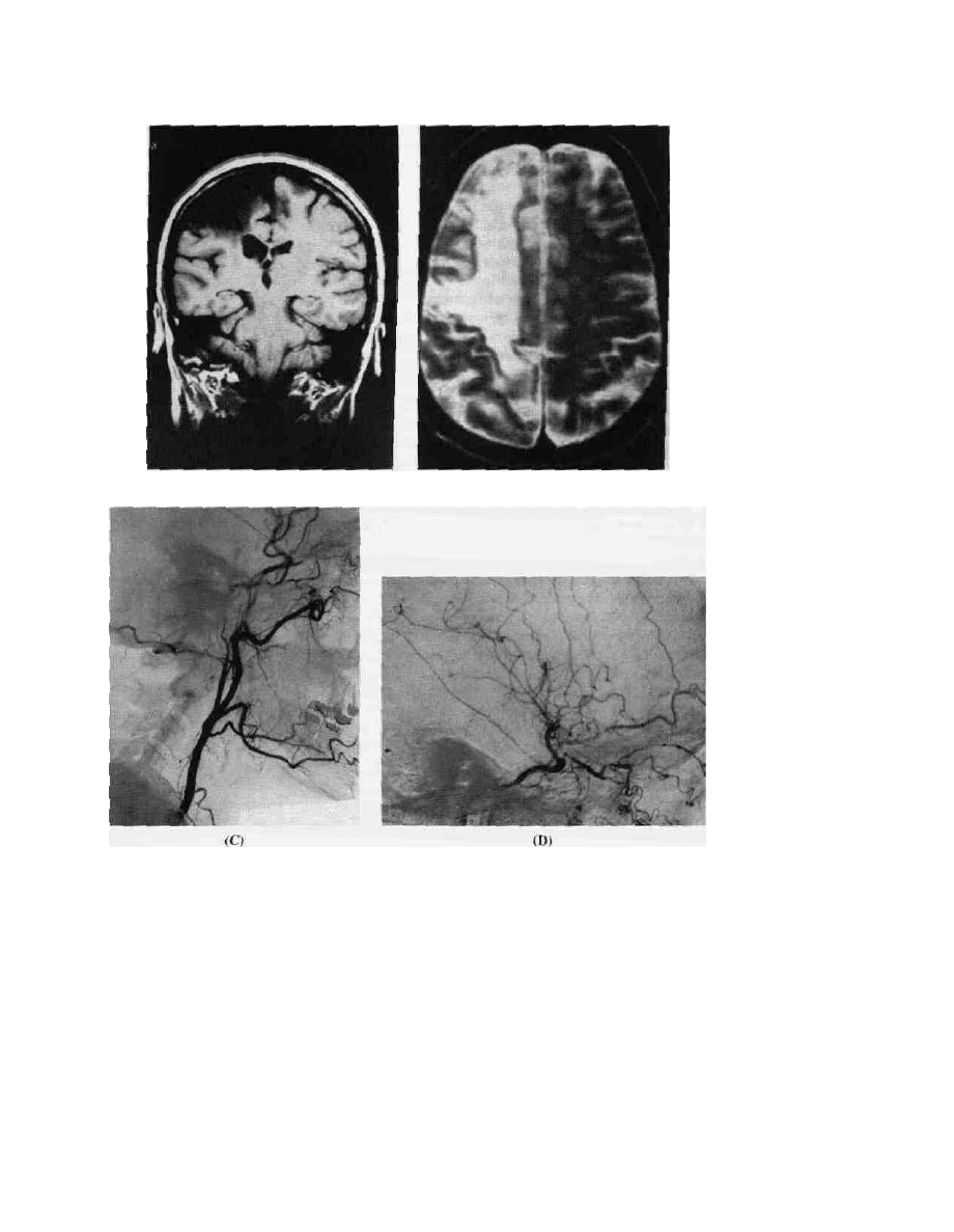
Figure 14-2 Infarction within the right cerebral hemisphere in a
36-year-old female who suffered an acute left hemiparesis due to
sudden occlusion of the right internal carotid artery while using
cocaine 10 years previously. (A) MRI Tl-weighted image coronal
view and (B) MRI T2-weighted image axial view show location
and extent of the infarct. The right carotid angiogram lateral view
shows (C) no filling of the extracranial internal carotid artery and
prominent meningeal collaterals from ascending pharyngeal and
internal maxillary branches of the external carotid artery
reconstituting the petrous and cavernous segments of the internal
carotid artery. (D) additional collateral flow from the ophthalmic
artery augmenting iniracranial internal carotid flow with
reconstitution of the middle cerebral artery.
occur in the course of pulmonary embolization. Emboli may
be bland or septic. Bland emboli may be platelet aggregates,
clots, or acellular debris having developed in proximal seg-
ments of the vascular tree, as in the case of thrombi occur-
ring within the heart following myocardial infarction or in
blood vessels that have been partially occluded, or they may
be the result of atheromatous calcification or ulcerated
plaques in proximal vessels. Periarterial inflammatory reac-
tion to an atheroma is often intense enough to produce dense
adherence between the carotid artery and the adjacent tis-
sues.
12
Other sources of emboli are vegetations or calcine
deposits on the mitral valves. Emboli may also be iatrogenic,
due to thrombi forming on intravascular catheters used for
infusions and shunts or from thrombi released during the
course of intracardiac or intravascular injections, monitoring,
or surgery.
Emboli pass distally, usually lodging at bifurcations.
There they may occlude the vessel temporarily and then
resolve. They may break up into smaller fragments and pass
more distally. After a vessel is occluded, an infarction may

254 CHAPTER 14
result and yet the embolus resolve, leaving a patent vessel
supplying an area of infarction. Arteritis with a resulting
aneurysm or brain abscess is often the end result of a septic
embolus.
Occlusion of penetrating vessels is usually the result of
thickening of the walls by hyalin degeneration, most com-
monly the result of long-standing hypertension.
3
'
16
-
17
The brain is somewhat resistant to ischemia and can
recover if blood flow is reestablished before irreversible
neuronal damage occurs.
18
A focal volume of infarcted
cerebral tissue is surrounded by an ischemic region that is
nonfunctional but still viable and potentially recoverable,
the penumbra.
19
Complex interactions occur at the cellular
level under conditions of acute ischemia. Over time, they
lead to irreversible damage of cells. Neuronal membrane
depolarization occurs with loss of detectable electrical ac-
tivity within 20 sec. Several minutes after this, major ionic
shifts occur with influx of sodium into the cells and efflux
of potassium into the extracellular space. Calcium is re-
leased from mitochondria and endoplastic reticulum, fur-
ther impairing cellular energy transformation. Extracellular
edema and astrocytic swelling results, which further im-
pedes oxygen transfer. Additionally there is depletion of
energy stores with loss of phosphocreatine and adenosine
triphosphate (ATP), leading to build up of anaerobic meta-
bolites and lactic acid. These changes evolve over several
hours and can potentially be reversed if oxygenated blood
flow is reestablished. However, ischemia tends to potentiate
more ischemia because of edema, reduced cellular oxygen
delivery, and increased metabolic demand of the ischemic
tissue.
20
.
21
The main cause of neuronal dysfunction and cell death in
ischemia is failure of ATP synthesis with loss of cellular ion
homeostasis and acid-base balance. Ion fluxes with buildup
of intracellular Ca
24
-, Na
4
', and H+ are associated with a
leaking cell membrane so that ATP is wasted and dissipated
further by futile cycling of ions. Acidosis is caused by
increased glycolysis over oxidative phosphorylation where
pyruvate is converted to lactate rather than CO; and H^O by
the normal oxidative reactions in the mitrochondria. This is
followed by lipolysis, proteolysis, and inhibition of protein
synthesis. Cell death rapidly follows.
20
-
22
The metabolites of arachidonic acid (AA) also have a
direct effect on cell membrane integrity, platelet aggrega-
tion, and microvascular patency in cerebral ischemia. These
include prostaglandins (PGs), thromboxane (TXA), and leu-
kotrienes (LTs), which are compounds derived from 20-car-
bon polyunsaturated fatty acids collectively known as eico-
sanoids.
23
Prostaglandins and thromboxane have opposing
actions on platelets and vessel walls. Leukotrienes are che-
motactic agents that increase cell permeability. PGs and LTs
interact to cause edema. Several reactions catalyze the con-
version of AA to PGs, TXA, and LTs. Free oxygen radicals
are also generated by these reactions, which have direct
deleterious effects on cell membrane integrity. PG conver-
sions can be inhibited by aspirin and other nonsteroidal
anti-inflammatory drugs. Thromboxane is a major AA meta-
bolite in platelets and named for its platelet aggregating
effects; it is also a potent vasoconstrictor. PGs tend to inhibit
platelet aggregation and are vasodilators. Leukotrienes are
released in immune reactions and anaphylaxis. They read
slowly, stimulate smooth muscle contraction, and increase
vascular permeability. LTs stimulate TXA synthesis, and
PGs tend to inhibit synthesis of LTs.
23
In cerebral ischemia, cell breakdown leads to release or
fatty acids, including AA. These are converted to eicosan-
oids. A predominance of TXA synthesis contributes to
platelet microthrombi and impaired cerebral microcircula-
tory perfusion as well as aggravate ischemia.
23
Intervention
to impede or reverse these reactions offers modes of ther-
apy.
CLINICAL FEATURES
Clinical features of ischemic cerebrovascular disease depend
upon the site of the vascular obstruction, its duration, and
the severity of the damage produced. The term transient
ischemic attack (TIA) has been applied to episodes of neuro-
logical deficits of vascular origin, lasting for less than 24 h,
When the duration of such episodes exceeds 24 h but lasts
for less than 3 weeks, the name reversible ischemic neuro-
logical deficit (RIND) is applied. The name stroke in evolu-
tion, or progressing stroke, is applied to a neurological
deficit thought to be of ischemic origin which progresses for
6 h or more. The differential etiology of such clinical expe-
riences must include infarction, hemorrhage, and neoplasm.
The name completed stroke is applied when the neurological
deficit has been stable for 72 h or longer.
The incidence of cerebral infarction is greatly increased in
patients with TIAs although TIAs precede infarction in less
than 10 percent a/cases.
2
* They are likely to be indicative
of carotid lesions when the neurological symptoms are lim-
ited to an arm, a leg, or aphasia rather than when a combina-
tion of these is involved (Fig. 14-3).
25
The risk of stroke is
highest during the month after the first TIA.
22
Headache is a
common premonitory symptom of cerebral ischemia and is
prominent in 25 percent of patients with TIAs.
26
When
symptoms and deficits build over several hours, this implies
an acute unstable cerebral ischemic event that is termed
crescendo TIA; this is an uncommon event and may be
reversed with urgent medical or surgical treatment. Unfortu-
nately, there is a tendency for patients suffering stroke to
delay seeking medical care. The majority of patients with
infarcts (64 percent) and subarachnoid hemorrhage (54 per-
cent) do not present within 24 h of stroke onset.
27
This delay
may preclude the benefits of some treatments for acute
stroke.
Ischemic lesions in the distribution of one of the internal
carotid arteries result in hemiparesis—most marked in me
upper extremity—and hemianopsia. Aphasia with its asso-
ciated features of acalculia and right-left disorientation is
associated with lesions in the dominant hemisphere while
dyspraxia, loss of initiative, and parietal lobe signs are
DIAGNOSIS AND TREATMENT OF STROKES 255
Isolated monocular transient visual loss, amaurosis fitgax
(AF), is the ocular equivalent of a TIA. The majority (79
percent) of patients with AF have plaques in the ipsilateral
carotid artery, although only 16 percent of these are severe
enough to impede flow.
28
Even though the prognosis in this
group is much better, they should be evaluated by carotid
ultrasonography.
Occlusions of the anterior cerebral artery result in motor
and sensory disturbances of the lower extremities on the
contralateral side. There may be behavioral changes as well.
Bilateral anterior cerebral artery occlusion results in akinetic
mutism with infarction of the septal nuclei, medial head of
caudate nuclei, anterior cingulate gyri, and corpus callo-
sum.
29
Epilepsy complicates cerebral infarction in 3 percent of
cases in the early period after infarction.
30
Seizures may be a
late consequence of infarction, commencing as late as 5
years after the infarct. An incidence of 9.5 percent has been
reported.
31
Hemorrhage may complicate acute infarction when the
involved tissue is exposed to normal perfusion pressures.
32
^
This is rare in the natural state but is a potential complica-'
tion that has limited the use of fibrinolytic therapy and
revascularization procedures.
Massive cerebral infarction frequently results in death,
usually as a result of hemispheric edema and tentorial her-
niation if death occurs within the first week.
33
Massive
edema may result in compartmental shifts or further decrease
in cerebral perfusion. Death after a week is likely to result
from a complication such as pneumonia, renal infection, or
septicemia.
RISK FACTORS
Habits, lifestyles, and diseases that potentiate cerebrovascu-
lar disease are sought in the clinical history. Behavioral
factors include smoking cigarettes, drinking alcohol, and
abusing drugs (particularly cocaine).
34
-
35
Constitutional fac-
tors include age, sex, race, familial factors, abnormal serum
lipids, diabetes mellitis, hypertension, sickle cell disease,
elevated fibrinogen, polycythemia, migraine, hypothyroid-
ism, and cardiac disease (congenital, atherosc|ero0c,_oir_dys_
rhythmic).
4
-
24
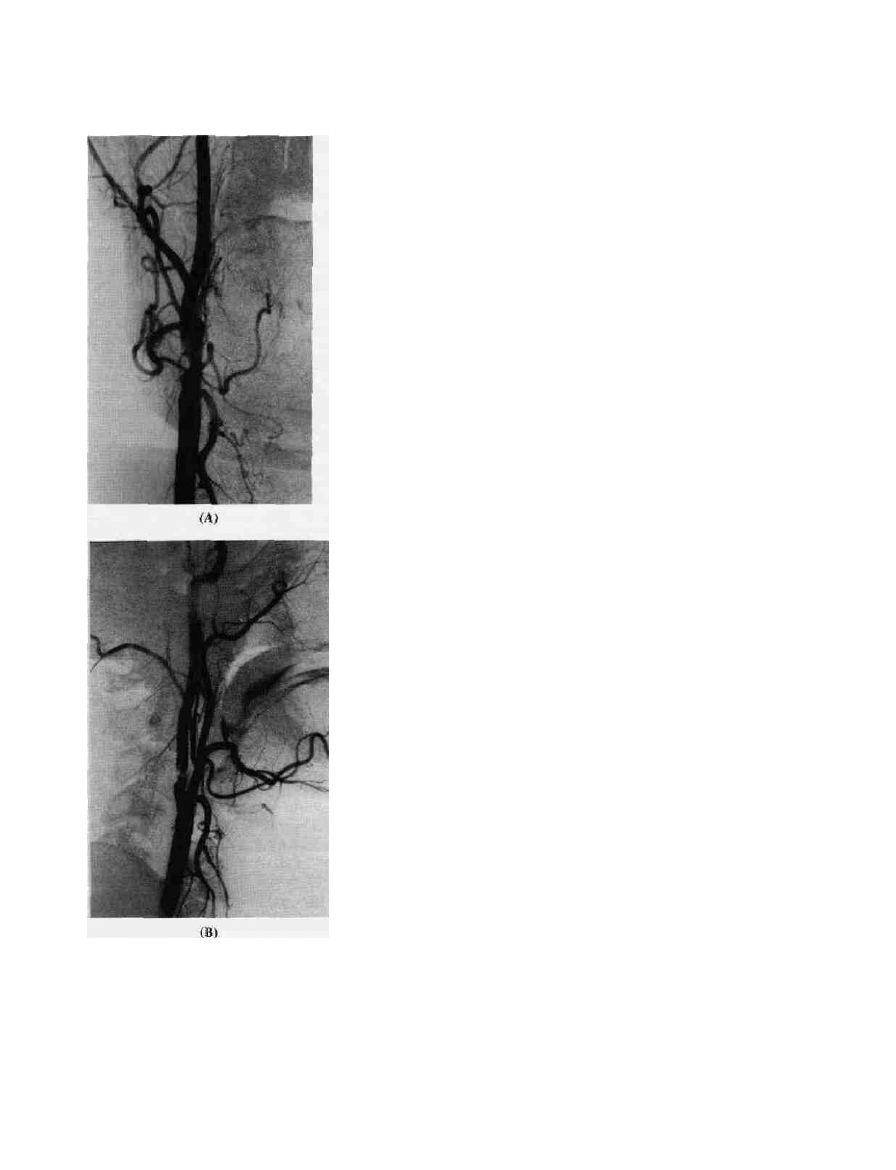
Figure 14-3 Atherosclerotic plaque with intraluminal thrombus
in the proximal internal carotid artery is seen on (A) frontal and
(B) lateral views of a right carotid angiogram, as a source of emboli
causing multiple focal TIAs.
prominent when lesions affect the nondominant hemisphere.
Consciousness is impaired in 25 percent of patients with
acute infarction in the distribution of the carotid arteries.
3
Involvement of the middle cerebral artery at the level of the
trifurcation causes a faciobrachial motor and sensory distur-
bance with dysphasia or dyspraxia, depending on the side.
EVALUATION
Indications for evaluating patients with respect to stroke
prevention and treatment include episodes of temporary def-
icit (TIAs or RINDs) and bruits in the neck of those over 40
years of age. Similarly, patients who have recently experi-
enced clinically evident ischemic episodes should be evalu-
ated as they often have risk factors for vasculopathy and
hypercoagulability, which potentiate strokes.
Workup includes computed tomography (CT) with and
without contrast enhancement, magnetic resonance imaging

256 CHAPTER 1-i
(MRI) with and without gadolinium, electroencephalography
(EEG), direct Doppler or B-scan ultrasound examinations of
the neck, angiography, cardiac investigation, and hematolo-
gical studies.
The evaluation of patients who have recently experienced
strokes has been greatly simplified by the advent of CT.
36
CT
identifies the site and size of areas of infarction so that it
readily differentiates lacunar infarcts from those produced by
cortical vessels. It differentiates those that are in the primary
distribution of a major feeding vessel from those that are
located in watershed areas. CT with contrast enhancement
shows areas of compromise of the blood brain barrier (BBB).
CT also demonstrates the site and degree of edema and com-
partmental shift, which may be a major factor in determining
the need for surgery (Fig. 14-4). In the posterior fossa, cerebel-
lar infarction with swelling or hemorrhage and presence of
acute hydrocephalus are readily detected by CT; this is very
important in determining the need for surgical intervention.
Occasionally, CT may demonstrate an unexpected mass lesion
such as a neoplasm or intracranial hematoma.
MRI is very sensitive to the presence of edema and may
demonstrate evidence of infarction even before CT.
37
There
is less often need for contrast media with MRI than there is
with CT. MRI may indicate whether blood is flowing in
large vessels supplying the brain by the "flow void" image.
The primary disadvantages of MRI relate to the length of
time required for scanning, its cost, and its interference with
cardiac pacemakers.
EEG is normal in only 25 percent of patients having
recently experienced a stroke.
3
A slow-wave focus is the
most common abnormality and may last for several weeks
after a stroke, even in a patient who has not experienced
seizures. A seizure focus is more likely to be present when
seizures occur late or are remote to the ictus. Seizures may
be helpful in differentiating lacunar from '"striatocapsular
lesions.
38
Although ophthalmodynamometry and orbital Doppler in-
vestigations can demonstrate the laterality of severe stenosis
or occlusion of the carotid artery, duplex Doppler and color
Doppler ultrasonic examinations of the cervical carotid ar-
tery have proved to be accurate, noninvasive indicators of
the local site of pathology in these vessels.
3
-
39
Accuracy
depends on the skill of the technician and diagnostician. The
Doppler indicates the speed of blood flow and evidence of
turbulence, from which the presence and amount of stenosis
within the carotid artery can be determined. Noninvasive
procedures are preferred for screening and initial evaluation.
Duplex Doppler ultrasonographic examination of the cervi-
cal carotid artery is accurate and specific for detecting le-
sions that obstruct flow.
28
.
40
Transcranial Doppler (TCD) has
been shown to be as accurate in evaluating obstructive
lesions in the intracranial arteries. The most important clini-
cal application is detection of moderate to severe stenosis in
asymptomatic but high-risk patients, such as those with
sickle cell disease.
41
Angiography remains the most definitive indicator of arte-
rial lesions of the neck that might be the cause of ischemic
cerebral disease. However, angiography in the patient who'
has recently experienced an acute cerebrovascular ischemic
event is associated with increased morbidity and is not
advocated as a routine study. It is clearly indicated in the
presence of: (1) a stenotic lesion in the cervical carotid
artery imaged by ultrasound, (2) cerebral infarction follow-
ing trauma, (3) symptoms of cerebral ischemia suggesting
intimal dissection, (4) symptoms occurring in a youth, and
(5) symptoms of stroke that progress atypically. Angiogr&-
phy is not necessary for lacunar strokes or when a cardia;
source for emboli has been demonstrated, and it may also be
contraindicated by age, debility, or associated diseases
Venous digital subtraction angiography may be used as the
first angiographic study since it avoids manipulating the
vessel housing an obstructive lesion.
42
However, image res-
olution is poor using this modality, making it difficult ic
assess vascular pathology accurately. It also requires large-
amounts of contrast, with resultant adverse effects in patien:'
who have impaired renal function. Arterial contrast injec-
tions usually provide clearer images of arterial lesions re-
quired when surgery is anticipated.
42
'
43
Generally it is desir-
able to limit the amount of contrast material used for
angiography so that intraarterial injections are preferable tc
venous digital subtraction angiography.
Routine cardiac evaluation for a patient who has recently
had a stroke includes a 12-channel electrocardiogram and
chest x-ray. Abnormalities will be seen in about 50 to 60
percent of patients who have had a recent cerebral infarc-
tion.
3
An indicator of a cardiac origin for a recent cerebro-
vascular embolus is the presence of atrial fibrillation or
evidence of a recent myocardial infarction.
44
Evidence of an
old, large anterior and apical myocardial infarction may
indicate an adynamic wall from which mural thrombi might
develop. Most patients also undergo a treadmill stress test or
a thallium isotope test with dipyridamole cardiac imaging to
define concomitant coronary arterial atherosclerosis.
Other investigations that may provide indication of a
cardiac origin of stroke include echocardiogram and pro-
longed ECG cardiac monitoring. Echocardiograms can be
used to evaluate the heart valves, motility in cardiomegaly.
and evidence of myxoma.
45
Extended ECG monitoring using
a Holter-type cardiac monitor can detect intermittent ar-
rhythmias such as episodes of prolonged asystole or tran-
sient tachycardia that affect cardiac output, resulting in im-
paired cerebrovascular perfusion.
Common hematological disorders associated with cerebral
infarction are: abnormalities in clot lysis, presence of lupus
anticoagulants, abnormal platelet function, hyperviscosity,
and hemoglobin abnormalities. Specific cellular aberrations
linked to stroke include sickle cell disease, erythrocytosis,
thrombocytosis, and high white cell counts (leukemia). The
presence of most of these is determined by routine blood
counts and a coagulation profile. An occult hematological
disorder should be considered when a stroke occurs in a very
young individual, when infarction occurs in more than one
arterial territory, and when a cause for arterial occlusive
disease or embolic source cannot be determined.
3
DIAGNOSIS AND TREATMENT OF STROKES
(B)
(D)
(A)
(C)
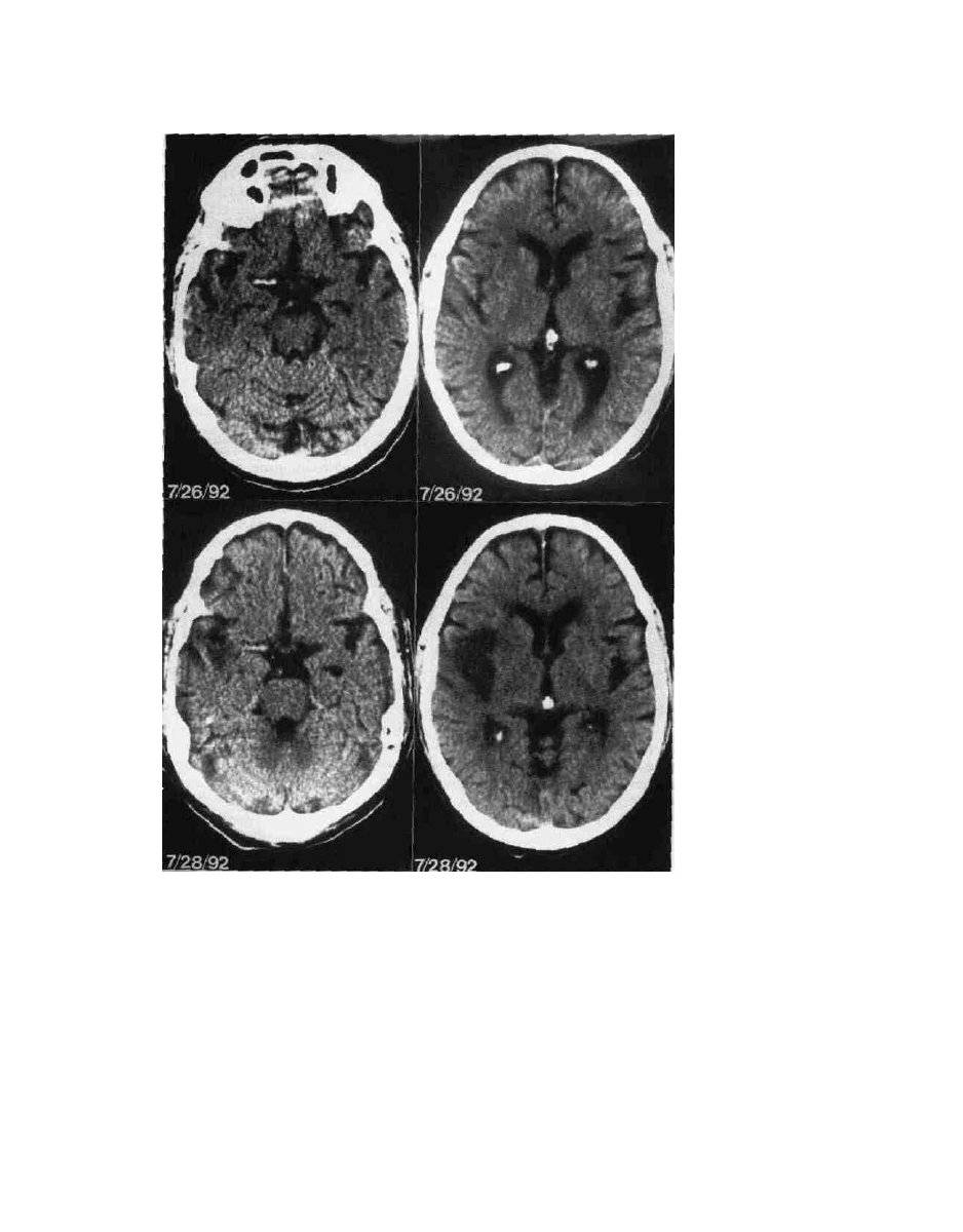
Figure 14-4 Developing infarction with positive "cord sign" in a 58-year-old male presenting
with acute onset of left hemiplegia as seen on noncontrasted CT; (A) and (B) on 7/26/92 show I
thrombosis of the M-l segment of the middle cerebral artery along with diffuse atrophy. Repeat
study 48 h later on 7/28/92 at comparable levels (C) and (D) respectively shows decreased
attenuation in the right temporal and frontal lobes as well as the strianun. The smaller frontal horn
of the right lateral ventricle in the later CT suggests associative edema-

258 CHAPTER 14
Lupus anticoagulants are frequently associated with a
hypercoagulable state.
46
They are a group of antibodies
found in only 5 to 10 percent of patients with lupus erythe-
matosus and commonly occur in patients with autoimmune
disorders and malignant neoplasms. These antibodies are
seen in patients receiving drugs such as phenothiazine, peni-
cillin derivatives, hydralazine, phenytoin, procainamide, and
isoniazid. These antibodies are immunoglobulins with anti-
body specificity to phospholipids found in several spontane-
ous and drug-induced autoimmune states. They may be
associated with thrombosis of deep veins, recurrent intravas-
cular thrombosis, and myocardial and/or cerebral infarction.
These antibodies may cause prolongation of the partial
thromboplastin time, false-positive serological tests, and
mild thrombocytopenia. Such reactions result from specific
phospholipids that are activity-directed.
Other common systemic disorders that are complicated by
an increased incidence of stroke include diabetes mellitus,
essential hypertension, peripheral vascular arteriosclerosis,
and the group of hyperlipidoses. Uncommon conditions
such as fibromuscular dysplasia, inflammatory arteritis, Ta-
kayasu's syndrome, and moyamoya disease will occasion-
ally present.
SOME SPECIFIC SYNDROMES
ASSOCIATED WITH
CEREBROVASCULAR INSUFFICIENCY
MOYAMOYA DISEASE
Moyamoya disease is a progressive cerebrovascular occlu-
sive disease involving the siphon and proximakintracranial
portion of the carotid arteries (Fig. 14-5). There is a bimodal
age incidence in children and adolescents and again in adults
peaking in the fourth decade. It was first described in Asians
by Takeuchi in 1957.
50
Occlusion of vessels supplying the
circle of Willis results in the development of a variety of
transdural, leptomeningeal anastomotic vessels, as well as
dilated perforating vessels that supply the basal ganglia. The
angiographic appearance of these vessels inspired the name
of the disease, moyamoya—Japanese for mist, fog, or puffs
of smoke. There also may be a history of inflammation in
the head or neck.
47
Patients experience repeated attacks of motor weakness,
speech disturbance, alteration in mental status, and organic
mental syndromes. These may be in the form of TIAs but
more commonly are completed strokes. Intermittent ocular
symptoms of amaurosis, impaired acuity, scotomata, diplo-
pia, and hemianopsia can accompany the symptoms of cere-
bral ischemia; however, intraocular findings are rare.
48
Older
patients may present with intracranial hemorrhage. Aneu-
rysms occur frequently and can be a source of hemor-
rhage.
49
-
50
The more common source of bleeding is from
collateral vessels that develop near the ventricular walls.
47
Surgical treatment is directed to augmenting collateral
blood flow by intracranial-extracranial bypass. Dural synan-
giosis is technically simple and is reported to be an effective
procedure but not without complications.
51
-
52
Nevertheless,
moyamoya is a progressive disease, and repeated TIAs and
seizures may eventually result in permanent severe deficits
unless there is surgical intervention.
53
FIBROMUSCULAR DYSPLASIA
Fibromuscular dysplasia is a nonatherosclerotic and nonin-
flammatory stenotic arterial disease commonly affecting the
renal and internal carotid arteries. It occurs most frequently
in young women, and the trait appears to be inherited.
Patients are often asymptomatic but may have hypertension,
stroke, or other evidences of vascular insufficiency, depend-
ing on the severity of stenosis and the arteries affected. It
involves different parts of the arterial wall, the intima,
media, or adventitia. Medial fibromuscular dysplasia is the
most-frequent form, representing 70 to 90 percent of cases.
The angiographic appearance of fibromuscular dysplasia,
characteristically, is corrugated or beaded. The internal caro-
tid artery is usually involved at the level of C2, where the
artery can be mechanically irritated by repeated stretching
with extension or rotation of the neck. Intimal fibroplasia
has a similar angiographic appearance to the medial type,
but it may present as a long tubular narrowing of the artery,
particularly in young patients.
54
Fibrous dysplasia may remain stable, but the form seen in
renal arteries has been shown to progress in 35 percent of
patients. It may also be aggravated by smoking. Ergotamines
have produced similar arterial changes. Although oral con-
traceptives may induce some increased stenosis, pregnancy
does not worsen it. Renal artery aneurysms may occur alone
or in combination with stenosis and hypertension. Angioten-
sin-converting enzyme inhibitors are reportedly effective in
managing associated hypertension.
54
In the cerebrovascular
form, patients complain of headache, vertigo, tinnitus, or
fatigue. More serious TIAs, stroke, and subarachnoid hem-
orrhage occur in about 30 percent of patients, and spontane-
ous dissections can occur.
55
-
56
The long-term prognosis for
these patients is good, as they tend not to progress over the
years.
54
This benign course should be remembered when
managing these patients.
VERTEBROVASCULAR INSUFFICIENCY
Stenosis in the vertebrobasilar system most commonly arises
at the vertebral artery origin. Thrombosis can occur at the
site of stenosis and extend distally or act as a source of
emboli. Since signs of ischemia present distal to the site of
occlusion, the site of the vascular lesion cannot be localized.
Also, an intact contralateral vertebral artery does not neces-
sarily protect against a brainstem or cerebellar stroke.
57
Vertebral arteries may be compromised within the serial
3SIS AND TREATMENT OP STROKES 259
(A) (B) (C)
(D) (E)
(F) (G)
Figure 14-5 A 19-year-old female who had transient episodes of
confusion, aphasia, and sialorrhea along with progressive intellectual
decline; her left supraclinoid internal carotid artery was found to be
occluded. (A) CT shows a right striatal lacunar infarct along with left
frontal and sylvian infarcts. (B) CT shows extension of the left frontal
lobe infarcts; (C) anterior-posterior (AP) view right carotid angiogram
shows tight stenosis of proximal A-l segment that may explain the
lacunar infarct in the striatum; (D) AP view of the left vertebral artery
reconstitutes the left middle cerebral artery via the posterior
and other collaterals; (E) lateral view left
carotid angiogram shows retrograde filling of the occluded internal
carotid artery and prominent meningeal vessels. The large superficial
temporal artery (arrows) was subsequently used for a dural
synangiosis procedure as an additional source to augment collateral
flow. (F) Left internal carotid is occluded; "whispery" collaterals are
typical for moyamoya disease. (G) Lateral view left carotid angiogram
shows reconstituted middle cerebral artery with leptomeningeal,
dural, and ophthalmic artery collaterals. The patient recovered speech
and intellectual function after additional augmentation of collateral
flow with a scalp to dura arterial synangiosis procedure.

260 CHAPTER 14
foramina transversae through which they course rostrally.
This may be the result of traumatic dislocation of the cervi-
cal spine or encroachment on the vessels by flaring vertebral
osteophytes. Generally, vertebrobasilar insufficiency results
more often from intracranial vasculopathy as opposed to the
extracranial vascular disease of the carotid system. Stenosis
of the vertebral or basilar arteries results in hemodynamic
insufficiency rather than emboli, and there is less collateral
flow beyond the vertebral arteries than in the carotid system.
The etiology can be confirmed by angiography. Symp-
toms of vertebrobasilar insufficiency are often characterized
by transient "dizziness" and/or a loss of consciousness.
There may be transient visual loss or dizziness when extend-
ing the neck or turning the head. Clinical examination alone
fails to localize the vascular pathology. Symptoms of verte-
brobasilar insufficiency have multiple causes; stenotic and
occlusive lesions have been found at every level of the
vertebrobasilar system. To study it adequately, at present,
requires four-vessel angiography.
SUBCLAVIAN STEAL
The transience and variability of vertebrobasilar insuffi-
ciency is seen in the manifestation of flow changes with
subclavian steal syndrome. Here the subclavian or innomin-
ate artery is occluded proximal to the origin of the vertebral
artery, and the distal subclavian artery is supplied by retro-
grade flow from the vertebral artery that fills from the
contralateral vertebral artery. Vertigo occurs in over one-half
of the patients, while about one-third have binocular visual
disturbances, paresis of a limb, and/or paresthesias. Ataxia,
diplopia, syncope, and monocular visual changes are fre-
quent complaints.
58
BASILAR ARTERY OCCLUSION
Basilar artery occlusion was described in 1946.
59
Many
patients have chronic hypertension. The prognosis is usually
grave, and anticoagulant treatment is probably ineffective.
60
Circulatory characteristics of the penetrating branches of the
basilar artery are the critical factors determining the severity
of the symptoms and outcome of this disease. These are
100 u-m or less and arise at right angles from the basilar
artery; they are also vulnerable to a reduction in flow, as
well as to atherosclerosis at their origin from the basilar
artery. Although they are considered end arteries, there are
collateral channels between adjacent territories.
61
Patients usually have a prodrome of vertigo, headache,
nausea, hemiparesis, diplopia, dysarthria, and other symp-
toms in various combinations that clear before the definitive
clinical onset, which is often sudden. Patients often deterio-
rate to a "locked-in" syndrome or to coma, indicating an
advanced form of the disease. Noninvasive evoked potentials
and transcranial Doppler can assist in early recognition of
this disease.
TRAUMATIC EXTRACRANIAL
VASCULAR OCCLUSION
Trauma to the head and neck can result in significant injun
to the carotid and/or vertebral arteries along their extracran-
ial course. Such injuries may result from blunt or penetrating
injuries. The morbidity of neurological deficits from trauma
lesions is high, about 52 percent, and mortality is 40 per-
cent.
62
The carotid artery is most commonly involved proximo
to the base of the skull at C2. With proximity to the
transverse process of C2, the internal carotid artery is
stretched over it by any injury involving hyperextension
and rotation of the head and neck. This may result in
intimal disruption with platelet aggregation and occlusion
or embolization or the development of an intimal flap with
dissection and occlusion, rupture and hemorrhage, or de-
velopment of a pseudoaneurysm with embolism.
63
'
64
The
carotid artery can also be injured by a direct blow or blunt
intraoral trauma, or it can be affected in conjunction with a
basilar skull fracture. The vertebral arteries in their course
through the paravertebral foramina are also subject to
stretch or contusion, resulting in dissection, pseudoaneur-
ysm, or occlusion. This may lead to focal ischemic lesions
of the cervical spinal cord, brainstem, cerebellum, and/or
occipital lobes.
Diagnosing traumatic vascular occlusions before symp-
toms of cerebral ischemia occur is difficult because the
diagnosis is made by angiography, which is rarely performed
in head and neck trauma, having been replaced by CT and
MRI. One should remain suspicious of such injuries in
appropriate cases. Auscultation is a poor screening tool. The
presence of Homer's syndrome and focal deficits are promi-
nent clues to diagnosing carotid injury. The initial CT scan
is frequently normal. Any trauma patient having focal neu-
rological deficits that cannot be explained from the imaging
studies should undergo early cerebral angiography to diag-
nose carotid artery dissection.
65
-
66
TREATMENT OF
CEREBRAL ISCHEMIA
Treatment of transient neurological deficits is directed
toward providing dependably adequate blood supply to the
brain. In most cases, this involves eradicating or bypassing
lesions that cause cerebral ischemia. The heart is the most
frequent source of cerebral emboli, so an embolic stroke
should be considered an indication of heart disease. Cardiac
or vascular sources of bland emboli may be treated by
antiplatelet factors or anticoagulants. Intensive antibiotic
therapy is required for septic cardiovascular disease. Surgi-
cal correction of intracardiac masses or valvular lesions may
be required. Endarterectomy may be indicated for stenotic
lesions or eroded plaques in major arteries supplying the
brain.
9
-
12
Treatment of cerebral ischemia must be catego-

DIAGNOSIS AND TREATMENT OP STROKES
•=———,261
rized into prophylaxis against potential infarction and treat-
ment of the completed infarction.
Symptoms that point to the need for prophylaxis usually
relate to episodes of temporary neurological deficits such as
TIAs or RINDs. The occurrence of these symptoms is indi-
cation enough for determining the cause. Demonstration of a
source of emboli should lead to treatment of that source. A
cardiac cause of emboli may require anticoagulation, correc-
tion of arrhythmias, or repair of valves. Extracranial carotid
stenosis becomes hemodynamically significant when the
constriction is 80 percent or greater, and it may require
surgical reconstruction to remove impedance of flow.
Ulcerated plaques in the cervical carotid artery are a
common source of cerebral embolism and may be treated
medically by anticoagulant and antiplatelet agents or by
surgical endarterectomy. Septic embolism from subacute
bacterial endocarditis, pulmonary or other systemic infec-
tion, or other sources of septicemia usually requires aggres-
sive parenteral antibiotic therapy for specific organisms.
MEDICAL TREATMENT
A significant advancement in understanding cerebrovascular
disease is the recognition and management of risk factors
such as hypertension, diabetes, and hyperlipidemia; as well
as control of smoking, drug abuse, and obesity. Once the
cerebrovascular event occurs, methods of management are
determined by the presence and amount of cerebral tissue
damage incurred, with the object of limiting or preventing
infarction. Directions of treatment are toward the heart,
blood, arterial wall, and brain tissue. Treatment of lesions
that may be producing symptoms of cerebral ischemia is
usually accomplished by the person or team most experi-
enced with the treatment of the particular lesion.
Cardiac sources include arrhythmias, myocardial infarc-
tion with global flow deficiency due to low cardiac output,
or mural thrombi which produce emboli, valvular obstruc-
tion due to stenosis, or endocardiac vegetations, which may
produce emboli. Each must be managed individually. Anti-
coagulation may protect against emboli from a prosthesis of
the aortic valve, mitral stenosis, and certain instances of
atrial fibrillation.
70
Anticoagulants should be avoided in the
face of septic emboli because of the increased risk of hemor-
rhage. Patients who are subjects of septic emboli are best
managed by sensitivity-specific antibiotics.
y Blood-clotting characteristics may be the cause of cerebral
ischemia, and treatment by anticoagulation is applied
cautiously because of the risk of complications from bleed-
ing and intracerebral hemorrhage. Therapeutic anticoagula-
tion requires (1) an accurate diagnosis, (2) physician under-
standing of anticoagulants, (3) accurate laboratory clotting
tests, and (4) no contraindication to treatment.
66
-
67
Intrave-
nous heparin by continuous infusion is the generally ac-
cepted treatment for acute TIAs, major arterial thrombosis
without infarction, and emboli of cardiac origin.
71
The acti-
vated partial prothrombin time is maintained at two times
the control values. However, both retrospective and prospec-
tive studies have failed to demonstrate a difference between
patients treated with and without heparin.
68
-
69
Anticoagulants may be prescribed in specific circum-
stances to prevent further embolization or propagation of
thrombi.
70
Recombinant genetic technology has allowed tis-
sue plasminogen activator (TPA) to be available for clinical
use. TPA induces the conversion of plasminogen to active
plasmin, the main fibrinolytic enzyme. The TPA binds to the
surface of fibrin and enhances the affinity and action of
plasminogen, which accelerates the thrombolytic process.
Clinical experience in the treatment of cerebrovascular
thrombi has been limited, but TPA remains an attractive
thrombolytic agent.
71
-
72
It has a half-life of about 4 min. It is
nonimmunogenic and has no hemodynamic effects.
Oral anticoagulants, warfarin compounds, and, specifi-
cally sodium warfarin (Coumadin), are used for chronic
anticoagulation and are effective in eliminating TIAs.
70
The
feared complication of this therapy is intracerebral hemor-
rhage, which occurs in 4 percent of cases.
24
Antiplatelet agents have special application in prophylaxis
against stroke. Aspirin is the current standard medical treat-
ment for patients who are prone to strokes. Aspmn irreversi-
bly inhibits platelet function by blocking cydooxygenase
and the conversion of AA to prostaglandin endopcroiudes
and, ultimately, TXA. Taking 325 mg every other da? re-
duces the risk for myocardial infarction, nonfatal stroke, and
cardiovascular death by 18 percent, although hemonh.tgk
strokes may be increased.
73
Dipyridamole, a phospbodicsBar-
ase inhibitor, when combined with aspirin was found 10
reduce stroke risk by 30 percent.
74
Ticlopidine is a new
powerful platelet suppressor that inhibits the adenoaae di-
phosphate pathway of the platelet membrane. The dose is
250 mg twice a day, and the effect lasts the lifetime of the
platelet. It has been shown to reduce the risk of cerebral
ischemia by 21 to 46 percent, but it can cause diarifaca. dan
rash, and neutropenia.
75
Platelet suppression is effecow in
reducing the incidence of stroke.
Systemic supportive measures are directed wand in-
creasing collateral flow by increasing blood pressure aod
cardiac output. This includes parenteral fluids, laciaied
Ringer's solution, and plasmanate colloid to increase circu-
lating blood volume. The risk of aggravating cerebral edema
and hemorrhage requires close monitoring and balance of
fluids and electrolytes. Hyperglycemia is to be avoided
because it increases lactic acid concentrations in ihe regions
of cerebral ischemia, aggravating edema and tissue infarc-
tion.
22
-
74
Hemodilution and reduction of hematocrit have
been advocated to reduce viscosity and improve rheologic
qualities of blood for better capillary flow.
76
-* These mea-
sures do not increase oxygen metabolism or change clinical
status.
74
Laboratory studies suggest that normovolemic he-
modilution avoids the edema associated with hypervolemic
hemodilution and reduces the size of infarction.
77
This treat-
ment is precluded by signs, symptoms, or CT evidence of
increased intracranial pressure.
80
There is a general consensus that corticosteroids are of no

262 CHAPTER 14
benefit in attenuating either the cytotoxic or vasogenic
edema of cerebral ischemia.
74
MANAGEMENT OF STROKE PATIENTS
Generally, management of stroke patients is directed toward
minimizing neurological deficits, treating complications, and
managing concomitant medical problems. Supplemental 0^
and maintaining the PO^ at or above 100 torr assures ade-
quate oxygenation. Optimizing circulating blood volume
with parenteral fluids (lactated Ringer's Solution) and onco-
tic agents (6% hetastarch) along with exogenous pressor
amines (dopamine) will assist in maintaining adequate car-
diac output. This, in turn, is directed toward maintaining
cerebral perfusion pressure and cerebral blood flow even in
the otherwise normotensive stroke patient. Anticoagulation
is rarely indicated for patients who have recently sustained
cerebral infarction. Patients having cerebral ischemia with-
out infarction due to intraarterial constriction or emboli may
benefit from continuous intravenous heparin while awaiting
endarterectomy, and long-term oral anticoagulation may be
indicated for chronic cardiac conditions such as atrial fibril-
lation, as well as for patients with prosthetic heart valves,
mitral valvular disease, or atrial fibrillation with mitral valve
prolapse. Long-term anticoagulation is rarely recommended
for intracranial vascular occlusive disease.
Activated tissue thromboplastin and urokinase are agents
under investigation for lysis of acute thrombotic vascular
occlusions. They show promise in reestablishing flow in
acutely occluded cerebral arteries, and they may reduce or
prevent infarction. Hemorrhage into the infarction is the
major concern and limiting factor; however, preliminary
results with this mode of therapy are encouraging.
Surgical therapy is usually reserved for endarterectomy,
arterial ligation or reimplantation, and treatment of compli-
cations such as severe intracerebral edema or hemorrhage
when there is a danger of hemiation and brainstem com-
pression. Expeditious evacuation of a hematoma may be
life-saving. Basilar artery occlusion is now potentially treat-
able with local intraarterial fibrinolytic thrombolysis.
78
Pa-
tients who have been in coma for more than 6 h are no
longer candidates for this treatment.
The vertebral artery is compromised by local compression
or distortion in the neck. This can occur with cervical spine
dislocation, spondylotic spurs, or chiropractic manipulation.
Treatment is usually directed toward reestablishing cervical
alignment or surgically opening the foramina transversae.
When vertebrobasilar insufficiency is caused by atheroma-
tous stenosis at the origin of the vertebral arteries, it pro-
duces symptoms similar to those of intermittent postural
hypotension. This can be treated surgically by bypassing the
stenotic lesion with a carotid-vertebral anastomosis.
Treatment of traumatic vascular occlusions is individu-
alized. Most patients can be managed nonoperatively.
Usually, heparinization for 3 weeks followed by oral anti-
coagulation for 6 months prevents progression. Carotid liga-
tion will prevent embolization from intimal flaps, but liga-
tion will not be tolerated unless cross-filling is adequate.
79
Rarely carotid bypass will prevent disabling stroke.
80
Fol-
low-up angiography frequently demonstrates progressive
pseudoaneurysm formation and delayed changes on the con-
tralateral side.
64
Penetrating injuries of the neck may be associated with
severe blood loss. Patients with knife, gunshot, or other
penetrating injuries to the cervical vessels are hemodynami-
cally unstable and demand aggressive blood volume replace-
ment with emergency operative exploration. If the injuries
are in the midcervical region, they should be surgically
explored. If they are above the mandible or below the level
of the cricoid tracheal cartilage and the patients are hemo-
dynamically stable, angiography is performed before any
surgical procedure in order to plan management.
81
CAROTID ENDARTERECTOMY
Endarterectomies remove arteriosclerotic plaques commonly
developing at the bifurcation of the common carotid artery
(see Fig. 14-3). The American Symptomatic Endarterectomy
Trial (NASCET) and the European Carotid Surgery Trial
(ECST) have concluded that carotid endarterectomy reduces
risk of stroke in patients with tight stenosis of the extracranial
carotid artery who are symptomatic with symptoms of cere-
bral ischemia due to the focal stenotic lesion.
81
-
82
There is still
a question regarding patients with tight lesions who are
asymptomatic, and study of this situation is ongoing.
83
Long-
term follow-up of endarterectomy has shown 10 percent res-
tenosis compared to 26 percent progression of stenotic lesions
on the opposite side. The cumulative stroke or ischemic epi-
sode probability was 4 percent at 1 month and 8 percent at 5
years, or less than 1 percent after the first month.
84
Technique of Endarterectomy. The carotid artery is
isolated initially by dissection of the distal 3 to 5 cm of the
common carotid artery for proximal control. The bifurcation
of the common carotid and the proximal 2 to 3 cm of the
external and internal carotid arteries are then dissected. The
patient is anticoagulated with heparin, 100 U/kg body
weight. The common carotid artery is then clamped, fol-
lowed sequentially by clamping the external and internal
carotid arteries. Some surgeons routinely insert a shunt im-
mediately upon opening the artery. Others insert the shunt
only in the event that there is evidence of inadequate blood
supply to the brain when the vessel is occluded. This may be
indicated by altered consciousness when the patient is oper-
ated on under local anesthesia, diminished "back-flow"
when the distal carotid artery is opened, or, most commonly,
focal or hemispheric decline in cortical electrical activity
with continuous intraoperative ERG monitoring.
The atherosclerotic plaque is excised by sharp and blunt
dissection. Most important to the success of carotid endar-

\GNOSIS AND TREATMENT OF STROKES 263
terectomy is the fixation of the distal intimal flap to the
vessel wall with tacking sutures, preventing elevation of an
intimal flap that can be a source of emboli or luminal
oclusion. Heparin may be reversed at the termination of the
procedure with protamine sulfate; however, it is preferable
to allow the anticoagulant effect of heparin to resolve spon-
cutaneously, thus impeding immediate postoperative thrombus
formation. A review of many series indicates that the fewest
complications in carotid endarterectomy occur when opera-
tions are performed by the most experienced surgeons.
Dissections of intimal flaps along the intraluminal course
of the carotid may require permanent ligation to avoid
distal embolism. Excision of the involved segment and
interposition of a graft to maintain intraluminal flow is an
altemative measure when the patient cannot tolerate carotid
occlusion.
Patients presenting with symptomatic carotid stenosis
often have accompanying occlusive disease of the coronary
arteries. Therefore they undergo cardiac evaluation to in-
clude a stress test. Some may present with severe coronary
occlusive disease requiring a coronary artery bypass graft
CABG). Such patients may have carotid Doppler studies to
evaluate risk of stroke occurring during the CABG. Simulta-
neous carotid endarterectomy with CABG has been per-
formed to avoid a perceived risk of stroke. A retrospective
study concludes that carotid stenosis does not increase the
risk of stroke during CABG.
85
CEREBRAL ARTERY BYPASS
cerebral revascularization procedures were introduced in
1967 to reestablish perfusion of regions of the brain with
diminished flow due to proximal arterial obstruction when
the obstruction was not directly accessible. Superficial tem-
poral to middle cerebral artery anastomosis (STA-MCA)
became a popular bypass procedure. However, in 1985, an
international cooperative study concluded that strokes were
not prevented by STA-MCA bypass surgery.
14
This led to a
virtual shutdown of bypass surgery, but considerable con-
troversy regarding the study's design, method, perfor-
mance, and conclusions has followed. The study failed to
separate any subgroup of patients with symptoms of cere-
bral ischemia due to demonstrable perfusion deficits but
having viable residual tissue in the region of ischemia.
Positron emission tomography (PET) offers the capability
of determining the presence of ischemic nonfunctional but
viable cerebral tissue. The response of such tissues to
revascularization is under study. Another question is
whether the standard STA-MCA will deliver enough bulk
flow to correct a clinically significant perfusion deficit. A
conventional STA-MCA bypass delivers initial flows of 25
to 50 mVmin compared to potentially 100 ml/min or more
by a larger vein graft bypass.
86
Thus, the study may not
have separated the population that might have been bene-
fited by a STA-MCA bypass.
There is risk of acute and delayed cerebral ischemia with
carotid ligation: 49 percent after internal carotid and 28
percent after common carotid ligation.
87
Bypass may prevent
ischemic stroke in the cerebral distribution of a major artery,
giving origin to an unclippable giant aneurysm that requires
ligation of the parent artery and distal runoff (trapping the
aneurysm).
88
'
89
It remains the best treatment for moyamoya
disease, particularly in adults, along with other measures
previously discussed here.
53
-
90
A bypass may have a role in limiting the extent of acute
stroke. Use of the procedure is limited by the possibility of
aggravating edema or the chance of precipitating hemor-
rhage by increasing flow to infarcted brain tissue that is no
longer protected by an intact blood-brain barrier. This results
in a "luxury perfusion syndrome."
91
The low flow rates of
STA-MCA may be an advantage in this case and offer
enough flow to preserve the penumbra.
92
Vertebrobasilar
insufficiency with the limited available collateral flow may
be best ameliorated by a bypass when medical treatment
fails.
93
-
95
However a bypass using the proximal superior
cerebellar artery as the recipient should be considered with
caution because of the technical difficulties involved and the
risk of complications.
96
Finally, there may be a small select group of patients with
multiple carotid and vertebral occlusions sustained by iso-
lated collateral flow and suffering recurrent TIAs while on
medical treatment who would benefit from a bypass.
97
Re-
gional cerebral blood flow has been demonstrated to be
improved along with the clinical course in this patient popu-
lation.
98
Limitations of STA-MCA bypass are due to inadequate
donor arteries and insufficient flow through the small diame-
ter of the anastomosis. Placement of the anastomosis within
the sylvian fissure along the proximal middle cerebral artery
close to the trifurcation allows for a larger anastomosis and
greater flow rates.
99
Alternatively, saphenous vein interposi-
tion grafts have been advocated as a solution to these limita-
tions. This eliminates uncertainties by having a donor vessel
of a size large enough to bring ample bulk flows to the
cerebral tissues immediately after construction of the by-
pass.
100
-
101
The drawbacks are that interposition vein grafts
are technically demanding and subject to occlusion because
of a size discrepancy between vein and recipient artery, as
well as kinking or compression along the course of the graft
and endothelial reaction within the vein graft.
102
Technical
advances to limit these impediments include care in dissect-
ing the vein and limiting distension pressure within the graft
while it is harvested.
103
-
104
These measures are effective in
augmenting flow to the brain.
The conclusion of the cerebral artery bypass study is
that it has only limited application, although it may have
dramatic effects in selected circumstances. Meanwhile, a
specific surgical procedure that resulted in 95 percent pa-
tency rate after anastomosis has generally been retired for
lack of purpose except for selected, relatively rare, indica-

264 CHAPTER P'
CAROTID OCCLUSION
STUDIES
Carotid ligation is a classic operation, but indications for it
have changed. Classically, it was used most often to treat
otherwise nonoperable aneurysms of the carotid artery.
About 30 percent of the population will not tolerate carotid
ligation without a stroke because of incompetent collateral
flow through the circle of Willis.
105
Clamps such as the
Crutchfield and Selverstone were designed to be gradually
closed over several days. These theoretically allow collateral
flow to develop so that closure of the carotid can be toler-
ated. Carotid occlusion is performed less often for treatment
of aneurysms now because microsurgical techniques and
multiple designs and forms of clips allow for direct aneur-
ysm obliteration and parent artery reconstruction. With the
advancement of "skull base" surgical techniques and radical
resections of tumors located along the intracranial course of
the internal carotid artery, the carotid artery may require
occlusion for cure or hemostasis. If there is a potential need
to occlude the carotid artery, it is first necessary to predeter-
mine the patient's tolerance to carotid occlusion in order to
plan for bypass or other procedures.
TEMPORARY BALLOON OCCLUSION
After cerebral angiography, a nondetachable balloon is posi-
tioned in the internal carotid artery under local anesthesia.
The patient is anticoagulated with heparin, 100 U/kg of body
weight. Adequacy of anticoagulation is verified by obtaining
serial activated clotting times, which should be twice the
control time. The balloon is expanded and occlusion of flow
verified angiographically. The patient is examined neurolog-
ically throughout the procedure. Additionally, trie scalp EEG
is monitored and any slowing or change in symmetry of
activity is critically observed. A transcranial Doppler is used
to observe changes in direction and velocity of flows
through the major cerebral arteries. Regional cerebral blood
flow studies are performed as additional verification of ade-
quacy of collateral flow. Inhalation xenon gives quantitative
volumetric flow rates. Recently, single-photon emission
computed tomography (SPECT) using the isotope, "'"Tc-
hexamethylpropyleneamine oxime (""Tc-HMPAO) has
given quick and accurate semiquantitative cerebral perfusion
rates during balloon occlusion of the carotid artery.
107
-
108
There has been a 3.7 percent overall complication rate due to
this procedure. Asymptomatic carotid dissection was discov-
ered in 2 percent. The rate of permanent neurologic deficit
was 0.33 percent. Approximately 9 percent of patients fail
the clinical portion of the occlusion test, and they inevitably
would have experienced a stroke if the carotid artery had
been permanently occluded.
106
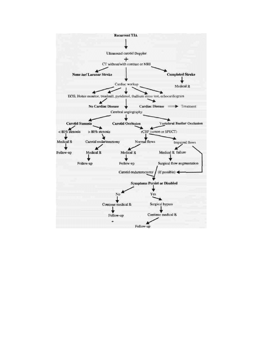
An algorithm is presented that is a synopsis for a clinical
approach to evaluation and management of cerebrovascular
ischemia (Fig. 14-6).
VENOUS SINUS THROMBOSIS
GENERAL CONSIDERATIONS
Cerebral venous drainage is characterized by ample collater-
als so that disorders of cerebral function are uncommon. If
superior sagittal sinus occlusion occurs gradually as by neo-
plastic invasion, many alternate collateral drainage routes
including the scalp veins are recruited, thus avoiding cere-
bral edema and symptoms of elevated intracranial pres-
sure.
109
Acute thrombosis of the cerebral venous outflow is ar-
uncommon but difficult problem to manage, particularly
when complicated by venous infarction, which may be disa-
bling or even life-threatening. Infections and septic phlebitis
such as cavernous sinus thrombosis (cavernous sinus syn-
drome) and lateral sinus thrombosis (otic hydrocephalus)
110
are causes of venous obstruction that can be dramatic. These
conditions are uncommon with modem antibiotic treatment
of intracranial infection. Other etiologies include hypercoa-
gulable states associated with oral contraceptives, preg-
nancy, and protein S deficiency.
111
Surgical occlusion by
jugular vein resection, depression of a skull fracture so that
it protrudes into and occludes a major dural sinus, or surgi-
cal transection of a dominant cortical or deep draining vein
are other mechanisms resulting in venous congestion and
major neurological morbidity. Venous infarction has a
greater tendency for hemorrhage than ischemic infarction of
arterial occlusion.'
12
Surgical management of traumatic in-
jury or neoplastic evolvement is a major challenge requiring
considerable skill to avoid an impairment or even fatal
outcome. Cortical veins may be incorporated in the dura,
forming dural lakes or sinuses before entering the superior
sagittal sinus. Cortical veins can be anastomosed or con-
nected by tubes, but veins 2 to 3 mm in diameter should be
carefully dissected or avoided.
113
SYMPTOMS
Headache is a prominent initial symptom of occlusion of a
venous sinus. This progresses and seizures supervene. Focal
deficits follow, depending on the location. Signs of cavern-
ous sinus occlusion are fever, proptosis, and variable ophthal-
moplegia. Papilledema and altered sensorium may be noted
as the condition advances.
DIAGNOSIS
Multiple findings of edema and hemorrhage on the unen-
hanced and contrast-enhanced CT give strong indications to
dural sinus thrombosis. The "empty delta" sign on enhanced
CT, indicating venous engorgement around a thrombosed
superior sagittal sinus, is the most specific finding on CT
DIAGNOSIS AND TREATMENT OF STROKES 265
Figure 14-6 This algorithm outlines a clinical approach for the evaluation and
management of cerebrovascular ischemic and occlusive disease.
and carries an ominous prognosis.
114
Cerebral angiography,
apart from surgery or autopsy, has been the definitive diag-
nostic procedure for this condition. Digital subtraction arter-
ial angiography is a refinement that nicely demonstrates the
venous system.
115
However, MRI angiography using phase-
sensitive gradient-echo imaging is an accurate noninvasive
method that can measure flow velocities within the dural
sinuses, and it may replace contrast injection angiography as
the preferred method for evaluating the cerebral venous
system.
116
TREATMENT
General supportive care with parenteral fluids, intensive
care monitoring, and systemic anticoagulation with hep-
arm and warfarin are the usual measures of management
for patients with acute venous sinus thrombosis. The dilem-
ma is whether anticoagulation will aggravate the hemor-
rhagic infarction that characteristically accompanies acute
sinus occlusion. Aggressive direct intervention by surgical
clot removal has met with only limited success. More suc-
cess in reestablishing venous sinus patency has been
reported using direct sinus perfusion of thrombolytic en-
zymes, i.e., urokinase or streptokinase.
117
"
120
Activated tis-
sue plasminogen may have some advantage in local treat-
ment of sinus thrombosis, but a definitive conclusion awaits
further trials. Many patients recover with supportive
measures. It is the patient who continues to decline in
spite of these measures and faces a grim prognosis that
may be salvaged by local enzymatic manipulation of the
sinus.

266 CHAPTER 14
SPONTANEOUSINTRACEREBRAL
HEMORRHAGE (HEMORRHAGIC
STROKE)
INCIDENCE
The incidence of primary intraparenchymal hemorrhage into
the brain is probably decreasing, as is the incidence of
strokes from other causes except for cerebral ischemia and
cocaine abuse. However, prevalent use of CT has improved
the accuracy and significantly increased the frequency of
diagnosis of intraparenchymal hemorrhage. The current esti-
mate of hemorrhage as a cause of stroke is about 17 per-
cent.
121
ETIOLOGY
Causes of intraparenchymal hemorrhage include coagula-
tion defects, intracranial neoplasms, vascular abnormalities,
venous thrombosis, arteritis, and drug abuse, but the most
frequent cause of intraparenchymal hemorrhage is chronic
hypertension. Aging is also a contributing factor.
121
.
122
It is hypothesized that degeneration of the proximal pene-
trating branches of the middle cerebral and basilar arteries
occurs because these small arterioles feed directly off me-
dium-sized arteries and are not protected by the usual step-
down in vessel size that protects more distal end arteries of
cortical vessels from the high intraluminal pressure.
121
Hemorrhage within the white matter tends to separate
the fiber tracts without destroying them, although their
function may be impaired. Therefore, as the hematoma
reabsorbs, the function of the fiber tracts recovers. With
large hemorrhages (50 ml or more) and hemorrhagic in-
farcts, significant permanent deficits persist. Hematomas
resolve through macrophage resorption over several
months, leaving a hemosiderin-stained fibroglial slitlike
cavity as a remnant.
121
-
122
The initial clinical course of hypertensive intracerebral
hemorrhage is variable and unpredictable. Usually the hema-
toma is of short duration without recurrence.
124
Unfortu-
nately the initial episode may be massive and destructive.
with high mortality. In 3 percent of cases, it rapidly expands
to twice the initial size hours to days later, with rapid
clinical deterioration and a poor outcome.
125
DISTRIBUTION
The vessels most commonly responsible for intraparenchy-
mal hemorrhage are the deep perforating branches of the
proximal middle and anterior cerebral and posterior commu-
nicating and basilar arteries, which supply the basal nuclei
(Fig. 14-7), thalamus, pons, and cerebellum. The most com-
monly involved sites, comprising 60 percent of spontaneous
intraparenchymal hemorrhages, are the basal nuclei, with the
location of other hematomas, divided between those in the
posterior fossa and those in the subcortical white matter.
Intraparenchymal hemorrhages in the posterior fossa occur
in the pons and cerebellum. Hemorrhages occurring at the
junction of the cortex and white matter are due to malforma-
tions, venous thromboses, coagulopathies, and other causes.
Midbrain hemorrhages are frequently extensions of lesions
in other primary locations or are Duret hemorrhages due to
tentorial hemiation caused by other mass lesions.
121
Table 14-1 compares the various types of cerebral vascu-
lar ischemic lesions and corresponding attributes of these
lesions, as well as differing diagnostic features (adapted
from Caplan
67
).
The sites of spontaneous intracerebral hemorrhages are
shown in Table 14-2.
122
Degenerative lesions characterized by deposition of fibrin-
ous material are found in the walls of the vessels supplying the
basal nuclei associated with long-standing hypertension or
aging. These changes in the vessel walls occur along with focal
thickening and intermingled thinning of the arterial walls.
Thinned regions develop microaneurysms of Charcot and Bou-
chard, which were described 150 years ago.
123
CLINICAL SIGNS AND SYMPTOMS
Clinical symptoms of intraparenchymal hemorrhage vary
according to location, size, and spread of the hemorrhage.
Large hemorrhages into the putamen, thalamus, or pons are
likely to be associated with coma early. Smaller hemor-
rhages in the region of the putamen are often associated with
hemiplegia, often with deviation of the eyes to the side of
the lesion. If the hematoma ruptures into the ventricles and
blood escapes into the subarachnoid space, symptoms may
present as subarachnoid hemorrhage. Headache, nausea, and
vomiting are later in onset when compared to patients with
primary subarachnoid hemorrhage.
126
Hemorrhage into the
thalamus may produce predominantly sensory impairment.
Visual loss in the form of hemianopsia and impaired eye
movements is often evident. Coma or altered sensorium
accompanies large, deep cerebral hemorrhages.
127
Symptoms associated with hemorrhages in the subcortical
areas depend on the location. Motor or sensory impairments
are predominant findings in the posterior frontal and parietal
lobes, respectively. Visual field defects accompany lesions
in the temporal, parietal, or occipital lobes. Seizures may
accompany hemorrhages in the frontal, temporal, or parietal
lobes.
Severe occipital headache and ataxia, often associated
with vomiting with or without nausea, are features of hem-
orrhage into the cerebellum. Altered sensorium with rapid
deterioration due to respiratory depression and decerebrate
posturing may occur early. Similar findings along with
myosis and impaired eye movements characterize pontine
hemorrhages.
Ocular, orbital, or temporal headache followed by pro-
gressive focal deficits such as contralateral motor paresis,
DIAGNOSIS AND TREATMENT OF STROKES 267
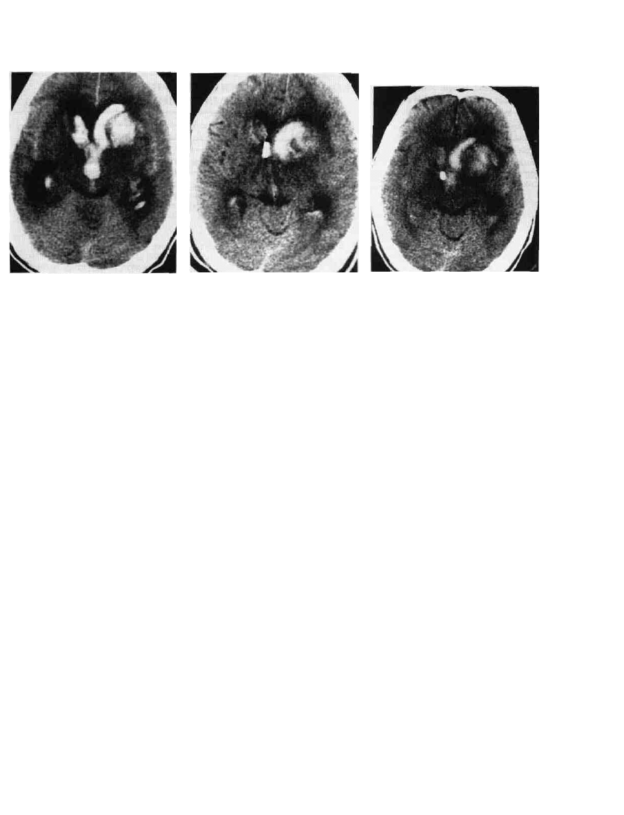
(A) (B) <C)
Figure 14-7 A 53-year-old male with poorly controlled resolving hematoma (B) 9 days after hemorrhage and at (C) 12 days
hypertension sustained an acute putamenal hemorrhage as seen on after hemorrhage. Note the catheter within the third ventricle for
plane CT (A) showing fresh hemorrhage extending into the anterior external drainage in (B) and (C). The patient recovered but
horns of the lateral ventricles and third ventricle. CT shows the remained aphasic.
hemisensory deficits, impaired speech, or hemianopic field
cuts suggests a lobar hemorrhage.
128
DIAGNOSTIC EVALUATION
Diagnostic evaluation of intracerebral hemorrhage centers
around CT scanning, which demonstrates the site, size, pres-
ence of mass effect with shift, and ventricular extension. CT
will also indicate evidence of infarction, which may be a
cause of hemorrhage. Serial CT scans allow observing pro-
gression or regression of the hemorrhage. A CT with can-
trast may reveal the presence of vascular malformations or
other abnormalities as sources of hemorrhage. Systemic hy-
pertension is often the causative factor. Coagulation studies
including platelet count, prothrombin time, and partial
thromboplastin time may indicate defects in the clotting
mechanism. Patients with hemophilia present special man-
agement problems that require consultation with hemotolo-
gists and replacement of specific clotting factors.
129
MRI is less helpful in the acute phase because deoxyhemo-
globin, which is paramagnetic and the blood-formed compo-
nent imaged, has not yet formed.
TREATMENT OF SPONTANEOUS
INTRACRANIAL HEMORRHAGE
General. The treatment of intraparenchymal hemor-
rhage deep within the cerebrum remains controversial. Evac-
uation of large devastating hemorrhages when the patient is
deeply stuporous is rarely helpful. Evacuation of small hem-
orrhages may not be necessary. Some patients exhibit pro-
gressive deterioration, usually a few days after the ictus
caused by further hemorrhage and increasing cerebral
edema. Surgical evacuation of the hematoma may improve
the patient's outcome. Japanese literature advocates aggres-
sive evacuation of such lesions and reports good re-
sults.
125
-
130
This therapy is controversial and is under clinical
trial.
131
'
132
SUPRATENTORIAL HEMORRHAGE
The method for evacuation of supratentorial hematomas is
variable. Stereotaxic evacuation by burr hole aspiration is
minimally destructive and frequently results in a good out-
come. Fresh solid clots are not readily aspirated through
cannulae; therefore, devices using rotors to fragment the clot
have been developed.
122
'
130
More recently, a stereotaxic
method supplemented by streptolysin to dissolve clots has
been recommended.
133
-
135
The commonly accepted tech-
nique is craniotomy with resection of the hematoma under
direct vision.
127
The craniotomy is made over the most
superficial extent of the hematoma, avoiding the primary
motor and sensory regions. While the mass of the hematoma
should be removed, most authors recommend leaving small
bits of clot on vessels in order to avoid new hemorrhage.
121
A variation of this technique which has been recommended
for the treatment of putaminal hemorrhage is to open the
sylvian fissure and enter the area of the putamen through the
operculum, thus minimizing the amount of brain to be tra-
versed in order to evacuate the hematoma.
127
Microsurgical
evacuation with precise delineation and obliteration of the
bleeding sites has also been advocated. This reportedly re-
veals the ruptured stump of the lenticulostriate branch,
which can then be coagulated without damage to the parent
artery, reducing the risk of recurrent hemorrhage.'
25
A large lobar subcortical hematoma—frontal, temporal,
or occipital—can leave minimal deficits after early surgical

268 CHAPTER 14 3g|
Table 14-1
COMPARISON OF THE VARIOUS TYPES OF
CEREBRAL VASCULAR ISCHEMIC LESIONS
ISCHEMIC VASCULAR LESION
Systemic
Hypopert'usion
Embolism
Large Artery Thrombosis
Small Penetrating
Artery Thrombosis
Risk factors
Onset and
cause
Anatomic
characteristics
Accompanying
signs
Heart disease,
gastrointestinal bleeding,
trauma
Onset with systemic
disorder (cardiac arrest,
hypotension, bleeding)
Border zone regions
between anterior, middle,
and posterior cerebral
arteries and between
posteroinferior,
anteroinferior, and superior
cerebellar arteries
Pallor, sweating,
hypotension
Location in border zones
CT: low density (black)
MR1: black on Tl-
weighted images and white
on T2-weighted images
Imaging:
CT/MRI
Heart disease, smoking,
coronary artery disease,
hyperlipidemia, diabetes,
white men, peripheral
vascular disease
Sudden; in 20% of
patients, stepwise during
first 24 h
Superficial cortex (most
often middle cerebral
artery), cerebellum, or
territory of posterior
cerebral artery
Headache at or after onset
Location: wedge shaped;
superficial or superficial
and deep
CT: low density (black)
lesions
MRI: black on Tl-
weighted images and white
on T2-weighted images
Smoking, coronary artery
disease, hyperlipidemia,
diabetes, white men,
peripheral vascular disease
Preceded by transient
ischemic attack in 40% of
cases, fluctuating, stepwise
progressive or remitting
Superficial cortex (most
often middle cerebral
artery), cerebellum, or
territory of posterior artery
Headache before, at, or
after onset
Location: in border zones,
or center of supply
CT: low density (black)
MRI: black on Tl-
weighted images and white
on T2-weighted images
Hypertension, diabetes,
polycythemia
Preceded by transient
ischemic attack in 25% of
cases, fluctuating, stepwi&e
progressive or remitting
Deep brain structures,
basal ganglia, cerebral
white matter, thalamus,
pons, cerebellum
Usually none
Location: small, deep
lesions
CT: low density (black)
MRI: black on Tl-
weighted images and white
on T2-weighted images
removal but may be devastating when treated expectantly.
136
Angiography should precede surgery for lobar hemorrhage
to exclude a structural vascular lesion or neoplasm.
POSTERIOR FOSSA HEMORRHAGE
There is little place for evacuation of hematomas in the
thalamus or pons. However, there is general agreement that
evacuation of hematomas in the cerebellum can frequently
be life-saving and that the quality of life for survivors is
quite good in a high percentage of cases. Imaged evidence of
brainstem compression such as a shifted or obliterated fourth
ventricle and effaced cisterns or clinical signs of posturing
and altered respiration are clear indications for early surgical
removal.
137
Ventriculostomy should be performed with cau-
tion as this could lead to upward hemiation of the cerebel-
lum. Posterior fossa craniotomy and hematoma removal
remains the treatment of choice.
138
Table 14-2
SITES OF SPONTANEOUS
INTRACEREBRAL HEMORRHAGE
Supratentorial
Lobar, 10%
Basal Ganglia, 60%
2/3 Medial internal capsule
1/3 Lateral internal capsule
Thalamic, 10%
Infratentorial
Brainstem, 10%
Cerebellar, 10%
INTRAVENTRICULAR HEMORRHAGE
Intraventricular extension of intracerebral hemorrhage (IVH)
has generally been considered to be accompanied by a poor
prognosis. It compounds the primary bleed acutely with
superimposed hydrocephalus. Intraventricular hemorrhage
can be seen and quantified by CT.
139
Diagnosing it by CT
has facilitated its management; however, it still presents a
serious threat to the patient. Hypertensive hemorrhage, rup-

DIAGNOSIS AND TREATMENT OF STROKES 269
tured aneurysm, AVM, neoplasm, and coagulopathy are the
most common nontraumatic causes of IVH.
126
Medical man-
agemeni with dexamethasone and antihypertensives is of
limited benefit.
140
Surgical evacuation of the hematoma may
be limited because of its deep location. Each patient must be
individualized. An aneurysm or AVM can be treated through
the hematoma. Coagulopathies must be reversed.
Ventricular drainage has been reported to have little effect
because of catheter obstruction;
126
however, it can be effective
in normalizing intracranial pressure and clearing intraventricu-
lar blood. Blood is cleared from the ventricle more quickly
with a concomitant intraventricular injection of urokinase. In-
jections of 10,000 to 12,000 U of urokinase are placed in the
lateral ventricle, followed by clamping the drain for 1 h and
then opening the tube.
133
This can be done once or twice daily.
The process is followed by repeated CT but continued until the
third and fourth ventricles are cleared. Persistent hydrocepha-
lus requires placement of a permanent shunt. Delayed onset of
hydrocephalus is due to the effects of subarachnoid blood
rather than blood within the ventricles.
139
REFERENCES
1. Tranmer BI, Kindt GW, Gross CE: Medical management of
acute cerebral ischemia, in Youmans JR (ed): Neurological
Surgery, 3d ed. Philadelphia, W.B. Saunders, chap 48, pp
1516-1533.
2. Whisnant JP: The decline of stroke. Stroke 15:160-168, 1984.
3. Toole JF, Burrow DD: Pathophysiology and clinical evalua-
tion of ischemic vascular disease, in Youmans JR (ed): Neu-
rological Surgery, 3d ed. Philadelphia, W.B. Saunders, chap
47, pp 1463-1515.
:4. Dyken ML, Wolf PA, Bamett HJM, et al: Risk factors in
stroke. Stroke 15:1105-1111, 1984.
-5. Anderson GL, Whisnant JP: A comparison of trends in mor-
tality from stroke in the United States and Rochester, Minne-
sota. Stroke 13:804-809, 1982.
6. Whisnant JP, Basford JR, Bernstein EF, et al: Classification
- of cerebro-vascular disease: III. Special report from the Na-
tional Institute of Neurological Disorders and Stroke. Stroke
21:637-676, 1990.
7. Sullivan HG, Kingsbury TB, Morgan ME, et al: The rCBF
response to Diamox in normal subjects and cerebrovascular
disease patients. J Neurosurg 67:525-534, 1987.
8. Lhermitte P, Gautier JC, Derouesne C: Nature of occlusions
of the middle cerebral artery. Neurology 20:82-88, 1970.
.9. Denman FR, Ehni G, Duty WS: Insidious thrombotic occlu-
-^ sion of cervical carotid arteries, treated by arterial graft.
Surgery 38:569-577, 1955.
10. Goldstone J, Moore WS: A new look at emergency carotid
artery operation for the treatment of cerebrovascular insuffi-
ciency. Stroke 9:599-602, 1978.
11. Javid H, Dye WS, Hunter JA, et al: Surgical treatment of
cerebral ischemia. Symposium on vascular surgery. Surg din
North Am 54:239-255, 1974.
12. DeBakey ME, Crawford ES, Morris GC JR, Cooley DA:
Surgical considerations of occlusive disease of the innomin-
ate, carotid, subclavian, and vertebral arteries, Ann Surg
154:698-725, 1961.
13. Piepgras DG, Sundt TM JR: Operative management of acute
arterial occlusion and acute ischemic stroke, in Youmans JR
(ed): Neurological Surgery, 3d ed. Philadelphia, W.B.
Saunders, chap 52, pp 1634-1643.
14. Barnett HJM, Sackett DL, Taylor DW, et al: Failure of
extracranial-intracranial arterial bypass to reduce the risk of
ischemic stroke. Results of an international randomized trial.
N E n g l J M e d 313:1191-1200, 1985.
15. Bamett HJM, Sackett D, Taylor DW, et al: Are the results of
extracranial-intracranial arterial bypass trial generalized? N
EnglJMed 316:820-824, 1987.
16. Fisher CM: Lacunar strokes and infarcts: A review. Neurol-
ogy 32:871-876, 1982.
17. Sacco SE, Whisnant JP, Broderick JP, et al: Epidemiological
characteristics of lacunar infarcts in a population. Stroke
22:1236-1241, 1991.
18. Hossman KA, Kleihues R: Reversibility of ischemic brain
damage. Arch Neurol 29:375-382, 1973.
19. Astrup J, Siesjo BK, Symar L: Thresholds of cerebral ische-
mia. The ischemic penumbra. Stroke 12:723-725, 1981.
20. Raichle ME: The pathophysiology of brain ischemia and
infarction. din Neurosurg 21:379-389, 1982.
21. Raichle ME: The pathophysiology of brain ischemia. Ann
Neurol 13:2-10, 1983.
22. Siesjo BK: Review article: Pathophysiology and treatment of
focal cerebral ischemia: I. Pathophysiology. J Neurosurg
77:169-184, 1992.
23. Chen ST, Hsu CY, Hogan EL, et al: Thromboxane, prostacy-
clin, and leukotrienes in cerebral ischemia. Neurology
36:466-470, 1986.
24. Whisnant JP, Matsumoto N, Elveback LR: Transient cerebral
ischernic attacks in a community, Rochester, Minnesota, 1955
through 1969. Mayo din Proc 48:194-198, 1973.
25. Harrison MJG, Jansek R, Marshall J: Clinical identification of
TIAs due to carotid stenosis. Stroke 17:391-392, 1986.
26. Grindal AB, Toole JF: Headache and transient ischemic at-
tacks. Stroke 5:603-606, 1974.
27. Alberts MJ, Bertels C, Dawson DV: An analysis of time of
presentation after stroke. JAMA 263:65-68, 1990.
28. Gaul JJ, Marks SJ, Weinberger J: Visual disturbance and
carotid artery disease: 500 symptomatic patients studied by
noninvasive carotid artery testing, including B-mode ultra-
sonography. Stroke 17:393-398, 1986.
29. Preemon FR: Akinetic mutism and bilateral anterior cerebral
artery occlusion. / Neurol Neurosurg Psychiatr 34:693-698,
1971.
30. Louis S, McDowell F: Epileptic seizures in nonembolic in-
farction. Arch Neurol 17:414-418, 1967.
31. Moskowitz E, Lightbody FEH, Freitag NS: Long-term fol-

272 CHAPTER 14
sciences. New York, Churchill Livingstone, chap 6, pp 1101-
1108, 1983.
123. Heir DB, Davis KR, Richardson EP Jr, Molir JP: Hyperten-
sive putaminal hemorrhage. Ann Neurol 1:152-159, 1977.
124. Ojeman RG, Mohr JP: Hypertensive brain hemorrhage, din
Neurosurg 23:220-244, 1975.
125. Bae HG, Lee KS, Bae WK, et al: Rapid expansion of hyper-
tensive intracerebral hemorrhage. Neurosurgery 31:35-41,
1992.
126. Little JR, Bloomquist GA Jr, Ethier R: Intraventricular hem-
orrhage in adults. Surg Neurol 8:143-149, 1977.
127. Wakai S, Kumakura N, Nagai M: Lobar intracerebral hemor-
rhage: A clinical, radiographic, and pathological study of 29
consecutive operated cases with negative angiography. J
Neurosurg 76:231-238, 1992.
128. Ropper AH, Davis KR: Lobar cerebral hemorrhages: Acute
clinical syndromes in 26 cases. Ann Neurol 8:141-147, 1980.
129. Seeler RA, Imana RB: Intracranial hemorrhage in patients
with hemophilia. J Neurosurg 39:181-185, 1973.
130. Kanaya H, Kuroda K: Development in neurosurgical ap-
proaches to hypertensive intracerebral hemorrhage in Japan,
in Kaufman HH (ed): Intracerebral Hematomas. New York,
Raven, chap 18, pp 197-209, 1992.
131. Juvela S, Heiskanen 0, Poranen A, et al: The treatment of
spontaneous intracerebral hemorrhage: A prospective ran-
domized trial of surgical and conservative treatment. J Neuro-
surg 70:755-758, 1989.
132. Kaufman HH: Stereotactic aspiration with fibrinolytic and
mechanical assistance, in Kaufman HH (ed): Intracerebral
Hematomas. New York, Raven, chap 16, pp 181-185, 1992.
133. Todo T, Usui M, Takakura K: Treatment of severe intraven-
tricular hemorrhage by intraventricular infusion of urokinase
J Neurosurg "I'4:81-86, 1991.
134. Waga S, Miyazaki M, Okada M, et al: Hypertensive puta-
minal hemorrhage: Analysis of 128 patients. Surg Neurol
26:159-166, 1986.
135. Waga S, Fujimoto K, Okada M, et al: Caudate hemorrhage
Neurosurgery 18:445-450, 1986.
136. Donauer E, Faubert C: Management of spontaneous intracere-
bral and cerebellar hemorrhage, in Kaufman HH (ed): Intra-
cerebral Hematomas. New York, Raven, chap 19, pp 2 I t -
227, 1992.
137. Freeman RE, Onofrio BM, Okazaki H, Dinapoli RP: Sponta
neous intracerebellar hemorrhage: Diagnosis and surgical
treatment. Neurology 23:84-90, 1973.
138. Heros RC: Cerebellar hemorrhage and infarction. Stroke
13:106-109, 1982.
139. Graeb DA, Robertson WD, Lapoint JS, et al: Computed
tomographic diagnosis of intraventricular hemorrhage: Eti-
ology and prognosis. Radiology 143:91-96, 1982.
140. Tellez H, Bauer RB: Dexamethasone as treatment in cerebro
vascular disease. A controlled study in intracerebral hemor-
rhage. Stroke 4:541-546, 1973.
D STUDY QUESTIONS
I. A 58-year-old male was seen because of an episode of
dysphasia and numbness that came on suddenly the night
before he was seen. The symptoms appeared a few minutes
after he had eaten dinner. He completely recovered within an
hour. At the time of his examination, the patient was neuro-
logically normal but had bilateral carotid bruits. His blood
pressure was measured at 160/100. «,
1. What is the classification of the episode? 2. What is the
most likely diagnosis? 3. What other diagnoses might be
considered? 4. How should they be ruled out? 5. What
vessels were most likely involved?
II. A 72-year-old male is admitted to the hospital complain-
ing of a severe occipital headache which has been progres-
sively severe since shortly after he went to bed the night
before. The patient is a known diabetic but has been in good
control. His blood pressure is recorded at 155/110. The
patient is unable to sit up because of ataxia on the right. He
is nauseated and vomits twice. He keeps his eyes closed
most of the time. A CT scan demonstrates dilated lateral and
third ventricles, but the fourth ventricle is displaced to the
left and occluded in its more distal part by a collection of
blood in the right cerebellar hemisphere.
1. What is the diagnosis? 2. What therapies should be
considered? 3. When should surgery be considered? 4. What
are the chances of a good outcome? 5. Should ventriculos-
tomy be considered?
III. A 55-year-old known hypertensive male is admitted
with the history of a sudden onset of left hemiparesis 3 h
earlier. An hour after the onset, the patient develops a severe
headache and shortly thereafter becomes stuporous. On ex-
amination, the patient has a blood pressure of 190/115. He
responds to painful stimuli by withdrawal of his right hand
and leg but not to command. He extends his left extremities
to painful stimuli. His eyes are constantly directed to the
right. A CT reveals a collection of blood in the right puta-
men.
1. What is the diagnosis? 2. What therapies might be
considered and in what order? 3. What blood vessels are
most likely torn? 4. What pathological processes are likely
to be found in the blood vessels? 5. By what techniques
might the blood be evacuated?
IV. An 18-year-old male is brought in deeply stuporous,
having been rescued from a possible drowning in which the
patient had been under water for at least 6 min. Respirations
were restarted despite an initial blood pressure which was
too low to be recorded for at least 5 min before intravenous
vasopressors could be started. Vital signs recovered; how-

DIAGNOSIS AND TREATMENT OF STROKES 273
ever the patient remains stuporous. A CT reveals cerebral
edema hich is most marked in the posterior parietal areas.
The patient dies 3 days later.
1. What is the most likely diagnosis? 2. What was likely
found at autopsy? 3. Why are the cerebral changes most
marked in the posterior parietal areas? 4. In the event that
hemorrhage occurred in the left parietal lobe prior to demise,
what would be the explanation? 5. Should angiography or
anticoagulants have been considered? Why?
V. A 48-year-old male experiences an acute right hemipare-
sis and aphasia, following which a CT scan demonstrates a
large area of decreased attenuation in the left parietal area in
the distribution of the left middle cerebral artery. Physical
examination reveals decreased pulsation of the left carotid
artery. A Doppler study reveals very little flow in the upper
internal carotid artery but rapid flow at the site of the
bifarction. An angiogram reveals a very tight stenosis at the
origin of the internal carotid artery. There is also an ulcer
seen in the plaque.
1. What is the cause of the area of decreased attenuation?
2. What types of therapy should be considered? 3. Why
should anticoagulants be avoided? 4. At what point might
ooe consider an endarterectomy? 5. What types of angiogra-
phy might have been considered and which one should have
been chosen? Why?
Wyszukiwarka
Podobne podstrony:
DoJ Policy on Treatment of News Media 14 0221
Magnetic Treatment of Water and its application to agriculture
Evidence for Therapeutic Interventions for Hemiplegic Shoulder Pain During the Chronic Stage of Stro
Kinesio taping compared to physical therapy modalities for the treatment of shoulder impingement syn
L 13+14 Geometry of Space
Diagnosis and Treatment of Autoimmune Hepatitis
Introduction to the Magnetic Treatment of Fuel
65 935 946 Laser Surface Treatment of The High Nitrogen Steel X30CrMoN15 1
14 ELEMENTS OF A SUCCESSFUL SAFETY & HEALTH PROGRAM
APA practice guideline for the treatment of patients with Borderline Personality Disorder
Flashback to the 1960s LSD in the treatment of autism
The term therapeutic relates to the treatment of disease or physical disorder
Friday's Treatment of Social Issues
Treatments of Alcoholism
Presentation 5 Psychological Aspects of Treatment of the S
7 77 93 Heat and Surface Treatment of Hot Works for Optimum Performance
więcej podobnych podstron