
391
Laboratory Identification of Biological Threats
Chapter 18
LABORATORY IDENTIFICATION OF
BIOLOGICAL THREATS
ERIK A. HENCHAL, P
h
D*; GEORGE V. LUDWIG, P
h
D
†
; CHRIS A. WHITEHOUSE, P
h
D
‡
;
and
JOHN M. SCHERER, P
h
D
§
INTRODUCTION
THE LABORATORY RESPONSE
Role of the Military Clinical and Field Laboratories
Military Field Laboratories
Laboratory Response Network
Biosafety and Biosecurity in the Military Clinical and Field Medical Laboratories
IDENTIFICATION APPROACHES
Specimen Collection and Processing
Clinical Microbiological Methods
Antibiotic Susceptibility Testing
Immunodiagnostic Methods
Molecular Detection Methods
EMERGING THREATS
BIOFORENSICS
FUTURE APPROACHES
Early Recognition of the Host Response
Joint Biological Agent Identification and Diagnostic System
SUMMARY
*Colonel, US Army (Ret); formerly, Commander, US Army Medical Research Institute of Infectious Diseases, 1425 Porter Street, Fort Detrick, Maryland
†
Deputy Principal Assistant for Research and Technology, US Army Medical Research and Materiel Command, 504 Scott Street, Suite 204, Fort Detrick,
Maryland 21702; formerly, Science Director, US Army Medical Research Institute of Infectious Diseases, 1425 Porter Street, Fort Detrick, Maryland
‡
Microbiologist, Diagnostic Systems Division, US Army Medical Research Institute of Infectious Diseases, 1425 Porter Street, Fort Detrick, Maryland
21702; formerly, Microbiologist, US Army Dugway Proving Ground, Dugway, Utah
§
Lieutenant Colonel, Medical Service Corps, US Army; Chief, Division of Diagnostic Systems, US Army Medical Research Institute of Infectious
Diseases, 1425 Porter Street, Fort Detrick, Maryland 21702; formerly, Chief, Biological Threat Assessment, 520th Theater Army Medical Laboratory,
Aberdeen Proving Ground, Maryland

392
Medical Aspects of Biological Warfare
INTRODUCTION
threat is more complicated than ever before. Future
diagnostic and identification systems will depend on
an integrated set of technologies, including new immu-
nodiagnostic assays and rapid gene analysis methods
to detect a broad spectrum of possible biological mark-
ers for diagnosing biological threats (see Exhibit 18-1).
2
The combination of several diagnostic approaches will
improve reliability and confidence in laboratory results,
which may shape medical treatment or response after
an attack. Military and civilian clinical laboratories are
now linked into a laboratory response network (LRN)
for bioterrorism sponsored by the Centers for Disease
Control and Prevention (CDC).
3
Together, these efforts
have improved the national preparedness, but continu-
ing research and development are needed to improve the
speed, reliability, robustness, and user friendliness of the
new diagnostic technologies. This chapter will review
the agent identification approaches and state-of-the art
diagnostic technologies available to protect and sustain
the health of soldiers and other military personnel.
The ability of military laboratories to identify and
confirm the presence of biological threats has signifi-
cantly improved over the past decade. Identification
approaches have advanced from classical identification
methods performed in only a few reference laboratories
to complex integrated diagnostic systems that are matur-
ing as part of the Joint Biological Agent Identification
and Diagnostic System (JBAIDS) for field laboratories.
During the Persian Gulf War (1990–1991), deployed
field laboratories and environmental surveillance units
depended significantly on immunoassay methods with
limited sensitivity and specificity. Because of intensive
efforts by scientists at military reference centers, such as
the US Army Medical Research Institute of Infectious
Diseases (USAMRIID), the Naval Medical Research
Center, the Armed Forces Institute of Pathology, and
the US Air Force Institute for Operational Health, re-
searchers are better prepared to identify and confirm
the presence of the highest priority biological threats to
human health (Exhibit 18-1).
1,2
However, the biological
THE LABORATORY RESPONSE
Role of the Military Clinical and Field Laboratories
Military clinical and field laboratories play a critical
role in the early recognition of biological threats. For
the purposes of this chapter, a biological threat is any
infectious disease entity or biological toxin intention-
ally delivered by opposing forces to deter, delay, or
defeat US or allied military forces in the accomplish-
ment of the mission. Biological agents can also be used
in bioterrorism scenarios to create terror or panic in
civilian and military populations to achieve political,
religious, or strategic goals. Although the principal
function of military clinical and field laboratories is
to confirm the clinical diagnosis of the medical officer,
laboratory staff also provide subject matter expertise in
theaters of operation on the handling and identification
of hazardous microorganisms and biological toxins.
Because these laboratories have a global view of disease
in the theater, they play an important sentinel role by
recognizing unique patterns of disease. Military field
laboratory personnel may also evaluate environmental
samples and veterinary medicine specimens as part of a
comprehensive environmental or preventive medicine
surveillance system in a theater of operations.
Military Field Laboratories
If a complete medical treatment facility is part of a
deployment, its intrinsic medical laboratory assets can
be used. However, a medical laboratory may not be
available for short duration operations in which the
health service element is task organized for a specific
mission. In this case, medical laboratory support should
be provided by a facility outside the area of opera-
tions.
4
Army medical treatment facilities in a theater of
operations have limited microbiology capabilities un-
less supplemented with a microbiology augmentation
set (M403), which is fielded with an infectious disease
physician, a clinical microbiologist, and a laboratory
technician. The M403 set contains all of the necessary
equipment and reagents to identify commonly en-
countered pathogenic bacteria and parasites, evaluate
bacterial isolates for antibiotic sensitivity, and screen for
some viral infections. Although this medical set does
not contain an authoritative capability for definitively
identifying biological warfare agents, it supports ruling
out common infections. Specimens requiring more com-
prehensive analysis capabilities are forwarded to the
nearest reference or confirmatory laboratory. After the
Persian Gulf War, all of the military services recognized
a need to develop additional deployable laboratory
assets to support biological threat identification and
preventive medicine efforts (described below).
Army
Army medical laboratories (AMLs) are modular,
task-organized, and corps-level assets providing

393
Laboratory Identification of Biological Threats
EXHIBIT 18-1
REGULATED BIOLOGICAL SELECT AGENTS AND TOXINS
Eastern equine encephalitis virus
Francisella tularensis
Hendra virus
Nipah virus
Rift Valley fever virus
Shigatoxin
Staphylococcal enterotoxins
T-2 toxin
Venezuelan equine encephalitis virus
US DEPARTMENT OF AGRICULTURE SELECT
AGENTS AND TOXINS
African horse sickness virus
African swine fever virus
Akabane virus
Avian influenza virus (highly pathogenic)
Bluetongue virus (Exotic)
Bovine spongiform encephalopathy agent
Camel pox virus
Classical swine fever virus
Cowdria ruminantium (Heartwater)
Foot-and-mouth disease virus
Goat pox virus
Japanese encephalitis virus
Lumpy skin disease virus
Malignant catarrhal fever virus (Alcelaphine herpesvi-
rus type 1)
Menangle virus
Mycoplasma capricolum/ M.F38/M mycoides Capri (con-
tagious caprine pleuropneumonia)
Mycoplasma mycoides mycoides (contagious bovine pleu-
ropneumonia)
Newcastle disease virus (velogenic)
Peste des petits ruminants virus
Rinderpest virus
Sheep pox virus
Swine vesicular disease virus
Vesicular stomatitis virus (Exotic)
US DEPARTMENT OF AGRICULTURE PLANT
PROTECTION AND QUARANTINE (PPQ)
SELECT AGENTS AND TOXINS
Candidatus Liberobacter africanus
Candidatus Liberobacter asiaticus
Peronosclerospora philippinensis
Ralstonia solanacearum race 3, biovar 2
Schlerophthora rayssiae var zeae
Synchytrium endobioticum
Xanthomonas oryzae pv. oryzicola
Xylella fastidiosa (citrus variegated chlorosis strain)
US DEPARTMENT OF HEALTH AND HUMAN
SERVICES SELECT AGENTS AND TOXINS
Abrin
Cercopithecine herpesvirus 1 (Herpes B virus)
Coccidioides posadasii
Conotoxins
Crimean-Congo hemorrhagic fever virus
Diacetoxyscirpenol
Ebola virus
Lassa fever virus
Marburg virus
Monkeypox virus
Reconstructed replication competent forms of the 1918
pandemic influenza virus containing any portion of the
coding regions of all eight gene segments (Reconstructed
1918 Influenza virus)
Ricin
Rickettsia prowazekii
Rickettsia rickettsii
Saxitoxin
Shiga-like ribosome inactivating proteins
South American Haemorrhagic Fever viruses
Flexal
Guanarito
Junin
Machupo
Sabia
Tetrodotoxin
Tick-borne encephalitis complex (flavi) viruses
Central European Tick-borne encephalitis
Far Eastern Tick-borne encephalitis
Kyasanur forest disease
Omsk hemorrhagic fever
Russian Spring and Summer encephalitis
Variola major virus (Smallpox virus) and Variola minor
virus (Alastrim)
Yersinia pestis
OVERLAP SELECT AGENTS AND TOXINS
Bacillus anthracis
Botulinum neurotoxins
Botulinum neurotoxin producing species of Clostridium
Brucella abortus
Brucella melitensis
Brucella suis
Burkholderia mallei (formerly Pseudomonas mallei)
Burkholderia pseudomallei (formerly Pseudomonas
pseudomallei)
Clostridium perfringens epsilon toxin
Coccidioides immitis
Coxiella burnetii
Reproduced from: US Department of Health and Human Services and US Department of Agriculture Select Agents and Toxins, 7 CFR
Part 331, 9 CFR Part 121, and 42 CFR Part 73. Available at: http://www.cdc.gov/od/sap/docs/salist.pdf. Accessed February 23, 2006.

394
Medical Aspects of Biological Warfare
comprehensive preventive medicine laboratory sup-
port to theater commanders. AMLs are capable of test-
ing environmental and clinical specimens for a broad
range of biological, chemical, and radiological hazards.
For biological agents, the laboratory uses a variety of
rapid analytical methods, such as real-time PCR, elec-
trochemiluminescence (ECL), enzyme-linked immuno-
sorbent assay (ELISA), and more definitive analyses
involving bacterial culture, fatty acid profiling, and
necropsy and immunohistochemistry.
2
AMLs have
significant “reach back” capability to reference labo-
ratories in the continental United States (CONUS) for
support. The largest of the service laboratories, AMLs
can identify “typical” infectious diseases including
endemic disease threats and they contain redundant
equipment for long-term or split-base operations. The
laboratory contains all of the necessary vehicles and
equipment to move and maintain itself in the field.
Navy
The Navy’s forward deployable preventive medicine
units (FDPMUs) are medium-sized mobile laboratories
using multiple rapid techniques (polymerase chain
reaction [PCR] and ELISA) for identifying biological
warfare agents on the battlefield. The FDPMUs are
also modular and have the ability to analyze samples
containing chemical and radiological hazards. These
laboratories specialize in identifying biological threat
agents in concentrated environmental samples (high
confidence), but they can also identify endemic infec-
tious disease in clinically relevant samples.
Air Force
Air Force biological augmentation teams (AFBATs)
use rapid analytical methods (such as real-time PCR)
to screen environmental and clinical samples for threat
agents. The teams are small (two persons), easily
deployed, and designed to fall in on preexisting or
planned facilities. The units are capable of providing
early warning to commanders of the potential presence
of biological threat agents.
The theater commander, in conjunction with the
theater surgeon and nuclear, biological, and chemical
officer, must decide which and how many of these
laboratories are needed, based on factors such as the
threat of a biological attack, the size of the theater, the
number of detectors and sensitive sites in the theater,
and the confidence level of results needed. A critical but
little understood concept is that the rapid recognition
of biological warfare threats must be fully integrated
with preventive medicine activities and the response
to endemic infectious diseases.
Laboratory Response Network
The response to future biological threats will
require the entire military laboratory network. The
logistical and technical burden of preparing for all
possible health threats will be too great for the mili-
tary clinical or field laboratories, which have limited
space and weight restrictions. The most important
role of these laboratories is to “listen to the hoof beats”
of medical diagnosis, rule out the most common of
threats, and alert the public health network about
suspicious disease occurrences. The military LRN
consists of the front-line medical treatment facility
clinical laboratories or deployed AMLs backed by
regional medical treatment facilities or military refer-
ence laboratories with access to more sophisticated
diagnostic capabilities. The clinical laboratories in the
regional medical centers or large medical activities
are the gateways into the civilian LRN sponsored by
the CDC. At the top of the military response pyramid
are research laboratories, such as USAMRIID (Fort
Detrick, Md) and the Naval Medical Research Center
(Silver Spring, Md). Other laboratories, such as the
Armed Forces Institute of Pathology (Washington,
DC) and the US Air Force Institute for Operational
Health (San Antonio, Texas) also provide reference
laboratory services for endemic infectious diseases.
Military research laboratories are best used to solve
the most complex and difficult diagnostic problems,
because usually they are not organized to perform
high-throughput clinical sample processing and
evaluation. Sentinel laboratories are generally sup-
ported by the network’s designated confirmatory
laboratories but may communicate directly with
national laboratories when hemorrhagic fevers or
orthopoxviruses (ie, smallpox virus) are suspected.
The network of military laboratories with connections
to federal and state civilian response systems provides
unparalleled depth and resources to the biological
threat response (Figure 18-1).
Biosafety and Biosecurity in the Military Clinical
and Field Medical Laboratories
Biosafety Considerations
Specific guidelines for handling hazardous agents
are contained in “Biosafety in Microbiological and
Biomedical Laboratories” published by the US De-
partment of Health and Human Services (DHHS).
5
By avoiding the creation of aerosols and using certain
safety practices, most bacterial threats can be handled
using standard microbiological practices at biosafety
level (BSL) 2. BSL-2 conditions require that laboratory
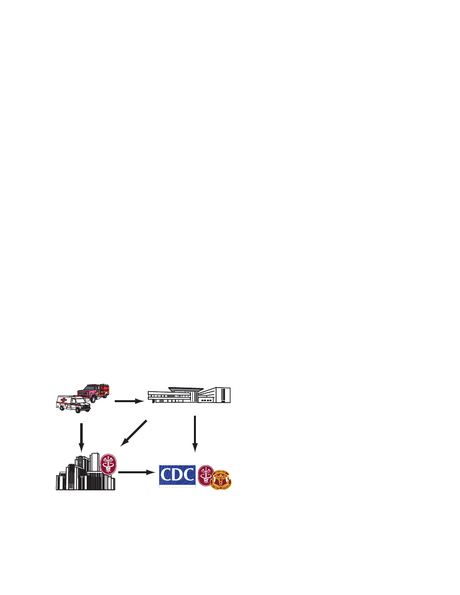
395
Laboratory Identification of Biological Threats
SAFER • HEALTHIER • PEOPLE
TM
First Responders
Level A Laboratories
Reference Laboratories
National Laboratories
Fig. 18-1. The network of military laboratories with connec-
tions to federal and state civilian response systems provides
unparalleled depth and resources to the biological threat
response.
personnel have specific training in handling patho-
genic agents and are directed by competent scientists.
Access to BSL-2 laboratories is restricted when work
is being conducted and safety precautions are taken
with contaminated sharp items. Procedures that may
create infectious aerosols are conducted only in bio-
logical safety cabinets or other physical containment
equipment. When samples must be processed on a
bench top, laboratory personnel must use other pri-
mary barrier equipment, such as plexiglass shields,
protective eyewear, lab coat and gloves, and work in
low-traffic areas with minimum air movement. BSL-3
conditions, which consist of additional environmental
controls (ie, negative pressure laboratories) and pro-
cedures, are intended for work involving indigenous
or exotic agents that may cause serious or potentially
lethal disease from inhalational exposure. Limited
prophylactic vaccines and therapeutics may be avail-
able to treat exposed personnel in case of an accident.
BSL-4 conditions are reserved for the most dangerous
biological agents for which specific medical interven-
tions are not available and an extreme risk for aerosol
exposure exists. BSL-4 requires the use of negative
pressure laboratories and one-piece, positive-pressure
personnel suits ventilated by a life support system.
Laboratory personnel should incorporate universal
bloodborne pathogen precautions and follow the
guidelines outlined in federal regulation 29 Code of
Federal Regulations (CFR) 1910.1030, “Occupational
Exposure to Blood-borne Pathogens.”
6
Specific pre-
cautions for each of the highest priority biological
threats can be found in the Basic Protocols for Level
A (Sentinel) Laboratories (http://www.bt.cdc.gov or
http://www.asm.org).
Biosurety
The 2001 anthrax letter attacks, which resulted in
22 cases of cutaneous or inhalational anthrax and
five deaths, raised the national concern about the
safety and security of laboratory stocks of biological
threats in government, commercial, and academic
laboratories.
7
As a result, the DHHS promulgated new
regulations (42 CFR, Part 73) that provided substantial
controls over access to biological select agents and
toxins (BSATs), required registration of facilities, and
established processes for screening and registering
laboratory personnel.
8
DHHS and the US Department
of Agriculture (USDA) identified over 80 biological
agents that required these regulatory controls (see
Exhibit 18-1). In addition to federal regulations, the
US Department of Defense (DoD) directed additional
controls for access to BSATs and required the establish-
ment of biosurety programs. These actions were taken
to foster public trust and assurance that BSATs are
handled safely and securely in military laboratories.
Among the services, the Army has established the most
comprehensive set of draft regulations (AR 50-XX) with
implementing memoranda.
At USAMRIID the framework for the military bio-
surety program was derived from the DoD’s experi-
ence with chemical and nuclear surety programs.
9-11
These surety programs incorporate reliability, safety,
and security controls to protect particular chemical and
nuclear weapons. The DoD biological surety program
applies many of the same controls as the chemical and
nuclear surety programs to medical biological defense
research and exceeds the standards of biosecurity pro-
grams in other federal and nonfederal laboratories.
Every military facility that stores and uses BSATs
must be registered not only with the CDC (see 42 CFR
Part 73) but also with the DoD.
8,9
In the case of Army
laboratories, registrations are completed through
the Assistant Secretary of the Army (Installation and
Environment). Army clinical laboratories, especially
those participating in the LRN triservice initiative,
are coordinated through the Army Medical Command
health policy and services. Not all clinical laboratories
need to be registered. However, unregistered laborato-
ries must follow the 42 CFR 73 “Clinical Laboratories
Exemption,” which states that clinical laboratories
identifying select agents have 7 days to forward or
destroy them. The transfer of BSAT cultures requires
the exchange of transfer documents (ie, CDC/APHIS
Form 2) between CDC-registered facilities.
Laboratory directors who supervise activities that
stock BSATs must be prepared to implement a variety of
stringent personnel, physical security, safety, and agent-
inventory guidelines. The law established penalties of

396
Medical Aspects of Biological Warfare
up to $250,000 (individual) or $500,000 (organization)
for each violation. Enhanced safety procedures are
required to work with or store BSATs. The DoD Bio-
logical Defense Safety Program is codified in Title 32
United States Code Part 627 and published as Army
Regulation 385-69. Guidelines for the safe handling
of BSATs can be found in CDC guidelines “Biosafety
in Microbiological and Biomedical Laboratories.”
5
Although many bacterial agents can be handled in
the BSL-2 clinical laboratory (Table 18-1), most work
TABLE 18-1
KEY IDENTITY MARKERS FOR SELECTED BIOLOGICAL SELECT AGENTS AND TOXINS
Biological Select
Biosafety
Agent and Toxin
Key Identity Markers
Level*
Confirmatory Methods
Anthrax
Gram-positive rod; spore-forming; aerobic; nonmotile;
22
•
Gamma phage sensitivity
catalase positive; large, gray-white to white;
large, gray-white to white;
•
Immunohistochemistry
nonhemolytic colonies on sheep blood agar plates
•
PCR
Botulism
Gram-positive rod; spore-forming; obligate anaerobe
2
•
Immunoassay
catalase negative; lipase production on egg yolk agar;
•
Mouse neutralization assay
150,000 dal protein toxin (types A,B,C,D,E,F,G); 2
•
PCR
subunits
Plague
Gram-negative coccobacilli often pleomorphic; nonspore
2
•
Immunofluorescence assay
forming; facultative anaerobe; nonmotile beaten copper
•
PCR
colonies (MacConkey’s agar)
Smallpox
Large double-stranded DNA virus; enveloped, brick-
4
•
PCR
shaped morphology; Guarnieri bodies (virus inclusions)
•
EM
under light microscopy
•
Immunohistochemistry
•
Immunoassay
Tularemia
Extremely small, pleomorphic, gram-negative coccobacilli;
2
•
PCR
nonspore forming; facultative intracellular parasite;
•
Immunoassay
nonmotile; catalase positive opalescent smooth colonies
on cysteine heart agar
Ebola
Linear, negative-sense single-stranded RNA virus;
4
•
PCR
enveloped; filamentous or pleomorphic, with extensive
•
EM
branching, or U-shaped, 6-shaped, or circular forms;
•
Immunoassay
limited cytopathic effect in Vero cells
•
Immunohistochemistry
Marburg
Morphologically identical to Ebola virus
4
•
PCR
•
EM
•
Immunoassay
•
Immunohistochemistry
Viral encephalitides Linear positive-sense single-stranded RNA virus;
3
•
PCR
enveloped, spherical virions with distinct glycoprotein
•
EM
spikes; cytopathic effect in Vero cells
•
Immunoassay
•
Immunohistochemistry
Ricin toxin
60,000–65,000 dal protein toxin; 2 subunits castor bean
2
•
Immunoassay
origin
Data sources: (1) Burnett JC, Henchal EA, Schmaljohn AL, Bavari S. The evolving field of biodefense: therapeutic developments and diag-
nostics. Nat Rev Drug Discov. 2005;4:281–297. (2) Henchal EA, Teska JD, Ludwig GV, Shoemaker DR, Ezzell JW. Current laboratory methods
for biological threat agent identification. Clin Lab Med. 2001;21:661–678.
*BSL-2 bacterial agents must be handled at BSL-3 with additional precautions or in a biological safety cabinet if laboratory procedures
might generate aerosols.
EM: electron microscopy
PCR: polymerase chain reaction

397
Laboratory Identification of Biological Threats
requires at least a class II biological safety cabinet or
hood and BSL-3 practices if there is a potential to create
aerosols.
5
Biosurety guidelines require that personnel
complete biological safety training before having ac-
cess to BSATs. A key goal of the guidelines is to prevent
access to BSATs by unauthorized personnel. In addition
to locked doors and freezers, continuous monitoring
of areas where BSATs are held is required. Moreover,
the capability to respond to the loss of agent must be
incorporated into a response plan. Physical security of
a facility by armed guards who can respond in minutes
is a component of Army regulations.
Perhaps the most controversial of the DoD and
Army guidelines is the requirement for a personnel
reliability program, which requires that reviewing offi-
cials (usually the military unit commander, laboratory
director, or otherwise delegated officer) aided by cer-
tifying officials (or employee supervisors) review the
suitability of every staff member with access to BSATs
with regard to behavioral tendencies, characteristics,
medical history, financial history, work habits, at-
titude, training, and more. Additionally, employees
are actively screened for illegal drug use through
urinalysis and alcohol abuse by observation. The
biosurety personnel reliability program incorporates
the requirements of the chemical and nuclear surety
programs, which were not incorporated into federal
law (except for the need for national agency and credit
checks). The DoD views the personnel reliability
program as essential because threat assessments have
identified the lone disgruntled insider as the most
serious threat to the biodefense program. On-site
and off-site contractors who support DoD programs
must implement the same safeguards under the cur-
rent policies. These regulations may seem excessive
because many BSATs can be obtained from natural
sources; however, the DoD and the Army provided
these guidelines to minimize risks associated with
the release of a high-consequence pathogen from
military facilities.
IDENTIFICATION APPROACHES
Specimen Collection and Processing
Clinical specimens can be divided into three differ-
ent categories based on the suspected disease course:
(1) early postexposure, (2) clinical, and (3) convales-
cent.
12
The most common specimens collected include
nasal and throat swabs, induced respiratory secretions,
blood cultures, serum, sputum, urine, stool, skin scrap-
ings, lesion aspirates, and biopsy materials.
2
Nasal
swab samples should not be used for making decisions
about individual medical care; however, they should
support the rapid identification of a biological threat
(post-attack) and subsequent epidemiological sur-
veys.
13,14
After overt attacks with a suspected biological
agent, baseline serum samples should be collected on
all exposed personnel. In the case of suspicious deaths,
pathology samples should be taken at autopsy to assist
in outbreak investigations. Specimens and cultures
containing possible select biological agents should
be handled in accordance with established biosafety
precautions. Specimens should be sent rapidly (within
24 hours) to the analytical laboratory on wet ice at 2°C
to 8°C. Blood cultures should be collected before the
administration of antibiotics and shipped to the labora-
tory within 24 hours at room temperature (21°C–23°C).
Blood culture bottles incubated in continuous moni-
toring instrumentation should be received and placed
within 8 hours of collection. Overseas (OCONUS) labo-
ratories should not attempt to ship clinical specimens
to CONUS reference laboratories using only wet ice.
Shipments requiring more than 24 hours should be
frozen on dry ice or liquid nitrogen. Specific shipping
guidance should be obtained from the supporting
laboratory before shipment. Specimens for complex
analysis, such as gene amplification methods, should
not be treated with permanent fixatives (eg, formalin
or formaldehyde). International, US, and commercial
regulations mandate the proper packing, documenta-
tion, and safe shipment of dangerous goods to protect
the public, airline workers, couriers, and other persons
who work for commercial shippers and who handle
the dangerous goods within the many segments of
the shipping process. In addition, proper packing and
shipping of dangerous goods reduces the exposure of
the shipper to the risks of criminal and civil liabilities
associated with shipping dangerous goods, particu-
larly infectious substances. Specific specimen collec-
tion and handling guidelines for the highest priority
bioterrorism agents are available from CDC and the
American Society for Microbiology (see http://www.
bt.cdc.gov or http://www.asm.org).
Clinical Microbiological Methods
Laboratory methods for biological threat agent iden-
tification were previously reviewed in this chapter.
2,15
Specific LRN guidelines for identifying the highest
priority (category A) bioterrorism agents can be ob-
tained from the CDC (http:\www.bt.cdc.gov). The
physician’s clinical observations and direct smears of
clinical specimens should guide the analytical plan (see
Table 18-1).
2,15
Most aerobic bacterial threat agents can

398
Medical Aspects of Biological Warfare
be isolated by using four bacteriological media: (1) 5%
sheep blood agar (SBA), (2) MacConkey agar (MAC),
(3) chocolate agar (CHOC), and (4) cystine heart agar
(CHA) supplemented with 5% sheep blood. Nonselec-
tive SBA supports the growth of Bacillus anthracis, Bru-
cella, Burkholderia, and Yersinia pestis. MAC agar, which
is the preferred selective medium for gram-negative
Enterobacteriaceae, supports Burkholderia and Y pestis.
CHA is the preferred medium for Francisella tularensis,
but CHOC agar also suffices. A liquid medium, such
as thioglycollate broth or trypticase soy broth, can also
be used followed by subculturing to SBA or CHOC
when solid medium initially fails to produce growth.
The selection of culture medium can be modified
when the target microorganism is known or highly
suspected; however, in most cases, the use of multiple
media options is recommended. Liquid samples can
be directly inoculated onto solid agar and streaked to
obtain isolated colonies. Specific culture details for the
highest priority biological threats are available from
the CDC (www.bt.cdc.gov).
Antibiotic Susceptibility Testing
Screening for unique antibiotic resistance or sus-
ceptibility may be critical to recognizing organisms
that acquire natural or directed enhancements. Mul-
tiple drug-resistant Y pestis, Brucella abortus, and Burk-
holderia strains have been identified.
16-20
In addition
to classical Kirby-Bauer disk diffusion antibiotic sus-
ceptibility tests or minimum inhibitory concentration
determinations, a variety of commercial antibiotic
susceptibility testing devices for use by community
hospitals have been standardized to reduce the time
required to achieve results.
21-24
Unfortunately, these
more rapid tests may not always be optimum for
detecting emerging resistance. Although standard-
ization of protocols by the Clinical and Laboratory
Standards Institute has ensured reproducibility of
results, emerging technology for detecting resistance
markers is not available in most clinical laboratories.
In addition, detecting progressive stepwise resistance
is limited to known and standardized techniques.
25
Molecular methods that could enhance screening
for unique genetic markers of resistance have been
developed
26-30
; however, genetic analysis approaches
can be cumbersome when multiple loci are involved,
as in the case of resistance to antibiotics related to
tetracycline or penicillin.
29,30
DNA microarrays offer
the potential for simultaneous testing for specific an-
tibiotic resistance genes, loci, and markers.
28,29
Grimm
imm
et al differentiated 102 of 106 different TEM beta-lac-
tamase variant sequences by using DNA microarray
analysis.
29
However, a comprehensive database of
However, a comprehensive database of
resistance genetic determinants for many biological
threats is not available, and new loci may emerge.
In response to the problem of emerging enteric dis-
eases, an electronic network has been established to
detect outbreaks of selected foodborne illnesses by
using pulsed-field gel electrophoresis.
31,32
Fontana
et al demonstrated pulsed-field gel electrophoresis
combined with ribotyping (a molecular method
based on the analysis of restriction fragment length
polymorphisms of ribosomal RNA genes) as an ef-
fective approach for detecting multidrug-resistant
Salmonella.
32
Applying these methods to the broader
array of potential threats should be an intensive future
research effort.
Immunodiagnostic Methods
An integrated approach to agent detection and
identification, which is essential for a complete and
accurate disease diagnosis, provides the most reliable
laboratory data.
2
Immunodiagnostic techniques may
play a key role in diagnosing disease by detection of
agent-specific antigens and/or antibodies present in
clinical samples. The most significant problem associ-
ated with the development of an integrated diagnostic
system has been the inability of such technologies to
detect agents with sensitivities approaching those
of more sensitive nucleic-acid–detection technolo-
gies. These differences in assay sensitivity increase
the probability of obtaining disparate results, which
could complicate medical decisions. However, recent
advances in immunodiagnostic technologies provide
the basis for developing antigen- and antibody-detec-
tion platforms capable of meeting requirements for
sensitivity, specificity, assay speed, robustness, and
simplicity.
Detecting specific protein or other antigens or host-
produced antibodies directed against such antigens
constitutes one of the most widely used and successful
methods for identifying biological agents and diagnos-
ing the diseases they cause. Nearly all methods for de-
tecting antigens and antibodies rely on the production
of complexes made of one or more receptor molecules
and the entity being detected.
Traditionally, assays for detecting proteins and other
non-nucleic acid targets, including antigens, antibod-
ies, carbohydrates, and other organic molecules, were
conducted using antibodies produced in appropriate
host animals. As a result, these assays were generically
referred to as immunodiagnostic or immunodetection
methods. In reality, numerous other nonantibody mol-
ecules, including aptamers, peptides, and engineered
antibody fragments, are now being used in affinity-
based detection technologies.
33-42

399
Laboratory Identification of Biological Threats
Diagnosing disease by immunodiagnostic technolo-
gies is a multistep process involving formation of com-
plexes bound to a solid substrate. This process is like
making a sandwich: detecting the biological agent or
antibody depends on incorporating all the “sandwich”
components. Elimination of any one part of the sandwich
results in a negative response (Figure 18-2). The primary
ligands used in most immunoassays are polyclonal or
monoclonal antibodies or antibody fragments.
Binding one or more of the antibodies onto a solid
substrate is usually the first event of the assay reac-
tion cascade. Immunoassays can generally be termed
as either heterogeneous or homogeneous, depending
on the nature of the solid substrate. A heterogeneous
assay requires physical separation of bound from un-
bound reactants by using techniques such as washing
or centrifugation. These types of assays can remove
interfering substances and are, therefore, usually more
specific. However, heterogeneous assays require more
steps and increased manipulation that cumulatively
affect assay precision. A homogeneous assay requires
no physical separation but may require pretreatment
steps to remove interfering substances. Homogeneous
assays are usually faster and more conducive to auto-
mation because of their simplicity. However, the cost
of these assays is usually greater because of the types
of reagents and equipment required.
The final step in any immunoassay is the detection
of a signal generated by one or more assay components.
This detection step is typically accomplished by us-
ing antibodies bound to (or labeled with) inorganic
or organic molecules that produce a detectable signal
under specific chemical or environmental conditions.
The earliest labels used were molecules containing
radioactive isotopes; however, radioisotope labels have
generally been replaced with less cumbersome labels
such as enzymes. Enzymes are effective labels because
they catalyze chemical reactions, which can produce a
signal. Depending on the nature of the signal, the re-
actants may be detected visually, electronically, chemi-
cally, or physically. Because a single enzyme molecule
can catalyze many chemical reactions without being
consumed in the reaction, these labels are effective at
amplifying assay signals. Most common enzyme-sub-
strate reactions used in immunodiagnostics produce a
visual signal that can be detected with the naked eye
or by a spectrophotometer.
Fluorescent dyes and other organic and inorganic
molecules capable of generating luminescent signals
are also commonly used labels in immunoassays. As-
says using these molecules are often more sensitive
than enzyme immunoassays but require specialized
instrumentation and often suffer from high back-
ground contamination from the intrinsic fluorescent
and luminescent qualities of some proteins and light-
scattering effects. Signals in assays using these types
of labels are amplified by integrating light signals over
time and cyclic generation of photons. Other com-
monly used labels include gold, latex, and magnetic
or paramagnetic particles. Each of these labels, which
can be visualized by the naked eye or by instruments,
are stable under a variety of environmental condi-
tions. However, because these labels are essentially
inert, they do not produce an amplified signal. Signal
amplification is useful and desirable because it results
in increased assay sensitivity.
Advances in biomedical engineering, chemistry,
physics, and biology have led to an explosion of new
diagnostic platforms and assay systems that offer great
promise for improving diagnostic capabilities. The
following overview discusses technologies currently
used for identifying biological agents and also used
(or under development) for diagnosing the diseases
caused by these agents.
Enzyme-Linked Immunosorbent Assay
Since the 1970s the ELISA has remained a core
technology for diagnosing disease caused by a wide
variety of infectious and noninfectious agents. As a
result, the ELISA is perhaps the most widely used and
best understood immunoassay technology. Developed
in many formats, assays can be designed to detect
either antibodies produced in response to infection
or antigens associated with the agents themselves.
ELISAs that detect biological agents or agent-specific
antibodies are heterogeneous assays in which an agent-
specific antigen or host-derived antibody is captured
onto a plastic multi-well plate by an antibody or an-
tigen previously bound to the plate surface (capture
moiety). Bound antigen or antibody is then detected
using a secondary antibody (the detector antibody).
The detector antibody can be directly labeled with a
Antigen Detection
Antibody Detection
Signal-Generating Components
Secondary Detector
Antibody
Primary Detector
Antibody
Analyte of Interest
Capture
Antibody/Antigen
Solid Phase
Fig. 18-2. Standard Sandwich Immunoassay. Detecting the
biological agent or antibody depends on incorporating all
the “sandwich” components. Elimination of any one part of
the sandwich results in a negative response.

400
Medical Aspects of Biological Warfare
signal-generating molecule or it can be detected with
another antibody labeled with an enzyme. These
enzymes catalyze a chemical reaction with substrate,
which results in a colorimetric change. The intensity
of this color can be measured by a modified spectro-
photometer that determines the optical density of
the reaction by using a specific wavelength of light.
In many cases, the assay can be interpreted without
instrumentation by simply viewing the color that ap-
pears in the reaction vessel.
The major advantage of ELISAs is their ability to be
configured for a variety of uses and applications. Use
of ELISAs in field laboratory settings is possible but
does require certain fixed-site logistical needs, such as
controlled temperature incubators and refrigerators,
the power needed to run them, and other ancillary
equipment needs. In addition, ELISAs are commonly
used and understood by clinical laboratories and phy-
sicians, are amenable to high-throughput laboratory
use and automation, do not require highly purified
antibodies, and are relatively inexpensive to perform.
The major disadvantages are that they are labor inten-
sive, temperature dependent, have a narrow antigen
concentration dynamic range that makes quantification
difficult, and are relatively slow.
The DoD has successfully developed antigen-detec-
tion ELISAs for nearly 40 different biological agents
and antibody-detection ELISAs for nearly 90 different
agents. All of these assays were developed by using
the same solid phase buffers and other reagents, incu-
bation periods, incubation temperatures, and general
procedures (Table 18-2). Although there is significant
variation in assay limits of detection, ELISAs typically
are capable of detecting as little as 1 ng of antigen per
mL of sample.
Electrochemiluminescence
Among the most promising new immunodiagnostic
technologies is a method based on electrochemilumi-
nescence (ECL) detection. One ECL system makes use
of antigen-capture assays and a chemiluminescent
label (ruthenium [Ru]) and includes magnetic beads
to concentrate target agents. These beads are coated
with capture antibody, and in the presence of biologi-
cal agent, immune complexes are formed between the
agent and the labeled detector antibody. Because of
its small size (1,057 kDa), Ru can be easily conjugated
to any protein ligand by using standard chemistries
without affecting immunoreactivity or solubility of
the protein. The heart of the ECL analyzer is an elec-
trochemical flow cell with a photodetector placed just
above the electrode. A magnet positioned just below
the electrode captures the magnetic-bead-Ru-tagged
TABLE 18-2
COMPARISON OF IMMUNODIAGNOSTIC METHODS
Dissociation-
enhanced
lanthanide
fluorescence
Enzyme-Linked immunoassay
Immunosorbent time-resolved
Electrochemi-
Hand-Held
Assay
fluorescence
luminescence
Flow-Based
Assay
Assay Parameters
Incubation time
3.5 h
2.2 h
15 min
30 min
15 min
Number of steps
5
4
1
1
1
Detection method
Colorimetric
Fluorescence Chemiluminescence Fluorescence
Visual
Multiplexing
No
Potential
No
Yes
Potential
Key Performance Parameters
Intra-assay variation (%)
15–20
20–50
2–12
10–25
Undetermined
Limit of detection: Yersinia pestis
250,000
250
500
62,500
125,000
F1 (colony-forming units)
Limit of detection: Staphylococcal
0.63
0.04
0.05
3.13
6.25
enterotoxin B (ng)
Limit of detection: Venezuelan
1.25 x 10⁷
3.13 x 10⁶
1.0 x 10⁷
3.13 x 10⁸
6.25 x 10⁸
equine encephalitis virus (plaque-
forming units)

401
Laboratory Identification of Biological Threats
immune complex and holds it against the electrode.
The application of an electric field results in a rapid
electron transfer reaction between the substrate (tripro-
pylamine) and the Ru. Excitation with as little as 1.5 v
results in light emission, which in turn is detected. The
magnetic beads provide a greater surface area than
conventional surface-binding assays like the ELISA.
The reaction does not suffer from the surface steric
and diffusion limitations encountered in solid-phase
immunoassays; instead, it occurs in a turbulent bead
suspension, thus allowing for rapid-reaction kinetics
and short incubation time. Detection limits as low as
200 fmol/L with a linear dynamic range can span six
orders of magnitude.
43-44
A field-ready ECL system consists of an analyzer
and a personal computer with software. ECL systems
possess several advantages, including speed, sensitiv-
ity, accuracy, and precision over a wide dynamic range.
In a typical agent-detection assay, sample is added to
reagents consisting of capture antibody-coated para-
magnetic beads and a Ru-conjugated detector antibody.
Reagents can be lyophilized. After a short, 15-minute
incubation period, the analyzer draws the sample into
the flow cell, captures and washes the magnetic beads,
and measures the electrochemiluminescent signal (up
to 1 min per sample cleaning and reading time). The
system uses 96-well plates and is therefore able to
handle large sample throughput requirements.
The ECL system has been demonstrated to be effec-
tive for detecting staphylococcal enterotoxin B, ricin
toxin, botulinum toxin, F tularensis, Y pestis F1 antigen,
B anthracis protective antigen, and Venezuelan equine
encephalitis virus.
2,45,46
The ECL system, which has
been demonstrated in field settings, is used as one
part of an integrated diagnostic system in several
deployable and deployed laboratories. Critical assay
performance characteristics and detection limits from
three typical ECL agent-detection assays are shown
in Table 18-2.
Time-Resolved Fluorescence
Time-resolved fluorescence (TRF) is an immunodi-
agnostic technology with assays available for detecting
agent-specific antibodies, microorganisms, drugs, and
therapeutic agents.
47-49
In practice, TRF-based assays
are sandwich-type assays similar to those used for
ELISA. The solid phase is a micro-well plate coated
in some manner with specific capture antibody (simi-
lar to that used with colorimetric ELISA platforms).
However, instead of being labeled with enzymes, de-
tector antibodies are labeled with lanthanide chelates.
The technology takes advantage of the differential
fluorescence lifespan of lanthanide chelate labels
compared to background fluorescence. The labels
have an intense, long-lived fluorescence signal and
a large Stokes shift, which result in an assay with a
very high signal-to-noise ratio and high sensitivity.
50
Unlike ECL, TRF produces detectable fluorescence
through the excitation of the lanthanide chelate by a
specific wavelength of light. Fluorescence is initiated
in TRF with a pulse of excitation energy, repeatedly
and reproducibly. In 1 second, the fluorescent material
can be pulse-excited 1,000 times with an accumulation
of the generated signal. One TRF format is dissocia-
tion-enhanced lanthanide fluorescence immunoassay
(DELFIA) in which dissociation of the complex-bound
chelate caused by adding a low-pH enhancement solu-
tion forms long-lasting fluorescent micelles. Detection
limits as low as 10
-17
moles of europium per well with
a dynamic range of at least four orders of magnitude
have been demonstrated.
The strength of DELFIA assays derives from their
sensitivity, similarity to the commonly used ELISA
techniques, and potential for multiplexing. Four dif-
ferent lanthanides are available (europium, samarium,
terbium, and dysprosium), and each has its own
unique narrow emission spectrum.
51
Both immunoas-
says and nucleic acid detection assays are compatible
with this platform. Like the ECL assays, DELFIA as-
says require purified high-quality antibodies. Critical
assay performance characteristics and assay limits of
detection from three typical DELFIA agent detection
assays are shown in Table 18-2. Although a field-ready
version of this instrument is not available, the system
is common to clinical laboratories and is used by the
CDC-sponsored LRN.
Flow Cytometry
Flow cytometry, the measurement of physical and
chemical characteristics of small particles, has many
current research and healthcare applications and is
commonplace in most large clinical laboratories. Ap-
plications include cytokine detection, cell differentia-
tion, chromosome analysis, cell sorting and typing,
bacterial counting, hematology, DNA content, and
drug discovery. The technique involves placing bio-
logical samples (ie, cells or other particles) into a liquid
suspension. A fluorescent dye, the choice of which is
based on its ability to bind to the particles of interest, is
added to the solution. The suspension is made to flow
in a stream past a laser beam. The light is scattered,
showing distribution and intensity characteristic of the
particular sample. A wavelength of the light is selected
that causes the dye, bound to the particle of interest,
to fluoresce, and a computer counts or analyzes the
fluorescent sample as it passes through the laser beam.

402
Medical Aspects of Biological Warfare
Using the same excitation source, the fluorescence may
be split into different color components so that several
different fluorophores can be measured simultaneous-
ly and the signals interpreted by specialized software.
A number of multiplexed flow cytometry assays have
been demonstrated.
52
Particles can also be sorted from
the stream and diverted into separate containers by
applying a charge to the particles of interest.
One commercially available platform is a rapid
assay system that reportedly can perform up to
100 tests simultaneously on a single sample. This
system incorporates three familiar technologies: (1)
bioassays, (2) microspheres, and (3) fluorescence.
The system consists of a flow cytometer with a
specific digital signal processing board and control
software. Assays occur in solution, thus allowing
for rapid reaction kinetics and shorter incubation
times. Capture antibodies or ligands are bound to
microspheres labeled with two spectrally distinct
fluorochromes. By adjusting the ratio of each fluoro-
chrome, microspheres can be distinguished based on
their spectral address. Bioassays are conducted on the
surfaces of these microspheres. Detector antibodies
are labeled with any of a number of different green
fluorescent dyes. This detector-bound fluorochrome
measures the extent of interaction that occurs at the
microsphere surface, ie, it detects antigen in a typi-
cal antigen-detection assay. The instrument uses two
lasers: one for detecting the microsphere itself, and
the other for the detector. Microspheres, which are
analyzed individually as they pass by two separate
laser beams, are classified based on their spectral
address and are measured in real time. Thousands
(20,000) of microspheres are processed per second,
resulting in an assay system theoretically capable of
analyzing up to 100 different reactions on a single
sample in just seconds. The manufacturer reports
assay sensitivities in the femtomole level, a dynamic
range of three to four orders of magnitude, and highly
consistent and reproducible results.
53
Because the
intensity of the fluorescent label is read only at the
surface of each microsphere, any unbound reporter
molecules remaining in solution do not affect the
assay, making homogeneous assay formats possible.
The system, which can be automated, can use tubes
as well as 96- and 384-well plates. Many multiplexed
assay kits are commercially available from a number
of manufacturers for various cytokines, phosphopro-
teins, and hormones.
Critical assay performance characteristics and
limits of detection from three typical flow-based
agent-detection assays are shown in Table 18-2. No
field-ready versions of these instruments are avail-
able, however, limiting the practical use of this plat-
form in deployment situations, and no commercial or
DoD sources for biothreat agent assays are available
for this platform.
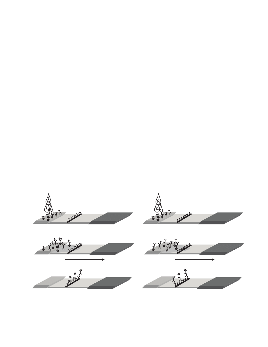
Lateral Flow Assays
Commercially produced lateral flow assays, which
have been on the market for many years, are so simple
to use and interpret that some types are approved for
over-the-counter use by the US Food and Drug Admin-
istration. Lateral flow assays are typically designed on
natural or synthetic membranes contained within a
plastic or cardboard housing. A capture antibody (for
antigen detection) or antigen (for antibody detection) is
bound to the membrane, and a second antibody labeled
with a visible marker element is placed on a sample ap-
plication pad. As the sample flows across the membrane,
antigen or antibody present in the sample binds to the
labeled antibody and is captured as the complex passes
the bound antibody or antigen (Figure 18-3). Colloidal
gold, carbon, paramagnetic, or colored latex beads are
commonly used particles that create a visible line in the
capture zone of the assay membrane.
One of the greatest advantages of lateral flow as-
says is their lack of reliance on instrumentation and
the associated logistical needs. However, this lack of
instrumentation decreases the utility of the tests be-
cause results cannot be quantified. To respond to this
deficiency, several technologies are being developed
to make these assays more quantitative (they also
increase the assays’ sensitivity). One technology al-
lows for quantitative interpretation of the lateral flow
assay.
54
Another method for quantitative detection of
antibody/antigen complex formation in lateral flow
assays uses up-converting phosphors.
55,56
Paramag-
netic particles have similarly been used in assays and
instruments capable of detecting changes in magnetic
flux within the capture zone, improving sensitivity
by as much as several orders of magnitude over more
traditional lateral flow assays.
Lateral flow assays are commonly used by the DoD
for detecting biological threat agents. In addition,
several companies have begun to market a variety of
threat agent tests for use by first responders. However,
independent evaluation of these assays has not typi-
cally been performed, so data acquired from the use
of these assays must be interpreted carefully. Another
common disadvantage of lateral flow assays is their
inability to run a full spectrum of control assays on a
single strip assay. Only flow controls are included with
most lateral flow assays. These controls show that the
conditions were correct for reagent flow across the
membrane but do not indicate the ability of the assay
to appropriately capture antigen.
403
Laboratory Identification of Biological Threats
Molecular Detection Methods
Polymerase Chain Reaction
Originally conceived in 1983 by Kary Mullis at the
Cetus Corporation,
57
polymerase chain reaction (PCR)
became a reality only 2 years later with the publication
by Saiki et al of its first practical application.
58
This first
description of PCR by Mullis et al marked a milestone
in biotechnology and the beginning of the field now
known as molecular diagnostics. PCR is a simple, in-vi-
tro chemical reaction that permits the synthesis of almost
limitless quantities of a targeted nucleic acid sequence.
At its simplest, the PCR consists of target DNA (also
called template DNA), two oligonucleotide primers
that flank the target DNA sequence to be amplified,
a heat-stable DNA polymerase, a defined solution of
salts, and an equimolar mixture of deoxyribonucleotide
triphosphates (dNTPs). The mixture is then subjected
to repeated cycles of defined temperature changes that
help to facilitate denaturation of the template DNA,
annealing of the primers to the target DNA, and exten-
sion of the primers so that the target DNA sequence
is replicated. A typical PCR protocol comprises 30
to 50 thermal cycles. Each time a cycle is completed,
there is a theoretical doubling of the target sequence.
Therefore, under ideal conditions, a single copy of a
nucleic acid target can be multiplied over a billion-fold
after 30 cycles. The whole procedure is carried out in a
programmable thermal cycler that precisely controls the
temperature at which the steps occur, the length of time
the reaction is held at the different temperatures, and
the number of cycles. The PCR products are typically
visualized as bands on an agarose gel after electropho-
resis and staining with a DNA intercalating dye such
as ethidium bromide or Sybr green.
In multiplex PCR, two or more sets of primers spe-
cific for different targets are included in the same reac-
tion mixture, allowing for multiple target sequences
to be amplified simultaneously.
59
The primers used in
multiplexed reactions must be carefully designed to
have similar annealing temperatures and lack comple-
mentarity. Multiplex PCR assays have played a larger
role in human and cancer genetics than in the detec-
tion of infectious organisms, where they have proven
more complicated to develop and often result in lower
sensitivity than PCR assays using single primer sets.
Reverse Transcriptase-PCR
The PCR method described previously was designed
to amplify DNA. However, many important human
diseases are caused by viruses with an RNA genome.
Therefore, reverse transcriptase PCR (RT-PCR) was
developed to amplify specific RNA targets. In this pro-
cess, extracted RNA is first converted to complementary
Sample Flow
a
b
c
Sample Flow
a
b
c
a
b
Fig. 18-3. Lateral flow assay format: A capture antibody (for antigen detection [a]) or antigen (for antibody detection [b])
is bound to the membrane, and a second antibody labeled with a visible marker element is placed on a sample application
pad. As the sample flows across the membrane, antigen or antibody present in the sample binds to the labeled antibody and
is captured as the complex passes the bound antibody or antigen.

404
Medical Aspects of Biological Warfare
DNA (cDNA) by reverse transcription, and then the
cDNA is amplified by PCR. As originally described,
reverse transcription of RNA into cDNA was carried
out using retroviral RT enzymes from either avian my-
eloblastosis virus or Moloney murine leukemia virus.
These enzymes are heat-labile and cannot be used at
temperatures above about 42°C, which presents prob-
lems in terms of both nonspecific primer annealing and
inefficient primer extension resulting from the potential
formation of RNA secondary structures. These problems
have largely been overcome by the development of a
thermostable DNA polymerase derived from Thermus
thermophilus, which, under the right conditions, can
act as both a reverse transcriptase and a DNA poly-
merase.
60,61
These and other similar enzymes can amplify
RNA targets without the need for a separate RT step.
Thus, this so-called “one-step” RT-PCR eliminates the
need for the cumbersome, time consuming, and con-
tamination-prone transfer of RT products to a separate
PCR tube. Commercial RT-PCR assays are available for
detecting a few important RNA viruses such as hepa-
titis C virus and human immunodeficiency virus, with
numerous others published in the scientific literature
as in-house or “home-brew” assays.
Real-Time PCR
By far the most important development in rapid
identification of biological agents has been the de-
velopment of “real-time” PCR methods. Although
traditional PCR was a powerful analytical tool that
launched a revolution in molecular biology, it was
difficult to use in clinical and field laboratories. As
originally conceived, gene amplification assays could
take more than 5 to 6 hours to complete, not including
the sample processing required before amplification.
The improvement of assay throughput came with the
development of assay chemistries that allowed the
PCR reaction to be monitored during the exponential
amplification phase on fast thermocyclers. Lee et al and
Livak et al demonstrated assays based on the detec-
tion and quantification of fluorescent reporters that
increased in direct proportion to the amount of PCR
product in a reaction.
62,63
By recording the amount of
fluorescence emission at each cycle, it is possible to
monitor the PCR reaction during the exponential phase,
in which the first significant increase in the amount of
PCR product correlates to the initial amount of target
template. The higher the starting copy number of the
nucleic acid target, the sooner a significant increase
in fluorescence is observed. A significant increase in
fluorescence above the baseline value measured during
cycles 3 through 15 indicates the detection of accumu-
lated PCR product. There are three main probe-based
fluorescence-monitoring systems for DNA amplifica-
tion: (1) hydrolysis probes, (2) hybridization probes,
and (3) DNA-binding agents. Hydrolysis probes most
exemplified by TaqMan (Applied Biosystems, Foster
City, Calif) chemistries have been the most successful
for rapidly identifying biological threats. Probe hydro-
lysis assays use the fluorogenic 5’ exonuclease activity
of Taq polymerase.
Fast thermocycling was achieved first by using
small volume assays in sealed capillary tubes placed
in convection ovens and later by solid-state electronic
modules.
64,65
Optimal assay development coupled to
instrument improvements has allowed the identifi-
cation of selected biological agents within 20 to 40
minutes after specimen processing. Over 50 assays
against 26 infectious agents have been developed us-
ing these approaches by the DoD, the CDC, and the US
Department of Energy.
2
Commercially available rapid
thermocycling instruments that can detect the fluores-
cent signals are now available from several sources,
including Applied Biosystems (Foster City, Calif),
Roche Diagnostics (Indianapolis, Ind), Idaho Technolo-
gies (Salt Lake City, Utah), Cepheid (Sunnyvale, Calif),
and Bio-Rad (Hercules, Calif). The Idaho Technolo-
gies Ruggedized Advanced Pathogen Identification
Device (RAPID) instrument has been incorporated
into the first generation of the JBAIDS for use in field
medical laboratories. By using new sample-processing
techniques, the presumptive identification of most bio-
logical agents can be completed in 3 hours or less with
rapid fluorescent-probe–based methods, compared
to approximately 6 hours with older PCR methods.
Other assay formats, such as fluorescent resonance
energy transfer, have allowed the resolution of closely
related species and mutation detection by character-
izing the melting point of the detection probe.
66,67
The
demonstration of integrated sample preparation and
gene amplification cartridges (such as Genexpert; Ce-
pheid, Sunnyvale, Calif) has the potential to improve
the reliability of PCR identification of biothreats by
decreasing the need for extensive operator training
and assay contamination.
68
Integrated cartridge gene
amplification systems have been incorporated into the
biohazard detection systems deployed to protect the
US Postal Service.
69
TIGER
A significant obstacle for detecting future bio-
threats is the requirement of many technologies,
such as immunoassays and most gene amplification
methods, to have identified target biomarkers ahead
of time. A unique coupling of broadly targeted gene
amplification with mass-based detection of amplified
products may allow for early recognition of replicat-
ing etiological agents without any preknowledge of

405
Laboratory Identification of Biological Threats
known, newly emergent, and bioengineered agents in
a single test (http://www.ibisrna.com/; valid August
8, 2004). This rapid, robust, and culture-free system
could have been used to identify agents such as
SARS-related coronaviruses, before their recognition
and characterization by traditional methods.
71
Robust
and portable TIGER systems are being developed for
civilian and military applications.
TABLE 18-3
BIOTERRORISM INCIDENTS, 1984–2004
Biological Agent
Description
Salmonella typhimurium Rajneeshee cult, The Dalles,
Oregon, 1984
1
Ricin toxin
Patriots Council, Minnesota;
Canada, 1991–1997
2,3
Bacillus anthracis
Aum Shinrikyo cult, Tokyo,
Japan, 1995
4
Shigella dysenteriae
Clinical lab, 1996
5
Various
Hoax incidents, Nevada, 1997–1998
6
B anthracis
Letters, Palm Beach, Florida;
civilian news operations in New
York City and in the Hart Senate
Office Building, Washington, DC;
also US postal facilities in the na-
tional capital area and in Trenton,
NJ; 2001
7
Ricin toxin
Manchester, England, 2002
3
;
Possible Chechen separatist plan
to attack the Russian embassy,
London, England, 2003
Ricin toxin
Dirksen Senate Office Build-
ing, Mailroom serving Senate
Majority Leader Bill Frist’s office,
Washington, DC, 2004
3
Data sources: (1) Torok TJ, Tauxe RV, Wise RP, et al. A large commu-
nity outbreak of salmonellosis caused by intentional contamination
of restaurant salad bars. JAMA. 1997;278:389–395. (2) Mirarchi FL,
Allswede M. CBRNE–ricin. eMedicine [serial online]. Available at:
http://www.emedicine.com/emerg/topic889.htm. Accessed March
16, 2005. (3) Shea D, Gottron F. Ricin: technical background and potential
role in terrorism. Washington, DC: Congressional Printing Office;
February 4, 2004. Congressional Research Service Report RS21383.
(4) Keim P, Smith KL, Keys C, Takahashi H, Kurata T, Kaufmann
A. Molecular investigation of the Aum Shinrikyo anthrax release in
Kameido, Japan. J Clin Microbiol. 2001;39:4566–4567. (5) Kolavic SA,
Kimura A, Simons SL, Slutsker L, Barth S, Haley CE. An outbreak
of Shigella dysenteriae type 2 among laboratory workers due to
intentional food contamination. JAMA. 1997;278:396–398. (6) Tucker
JB. Historical trends related to bioterrorism: an empirical analysis.
Emerg Infect Dis. 1999;5:498–504. (7) Bush LM, Abrams BH, Beall A,
Johnson CC. Index case of fatal inhalational anthrax due to bioter-
rorism in the United States. N Engl J Med. 2001;345:1607–1610.
the targets. Sampath and Ecker have described the
amplification of variable gene regions flanked by con-
served sequences, followed by electrospray ionization
mass spectrometry and base composition analysis
of the products.
70,71
This method, known as TIGER
(triangulation identification for genetic evaluation
of risks), provides for a high-throughput, multiple
detection and identification system for nearly all
EMERGING THREATS
The emergence of new biological threats is a
particular challenge for the military clinical or field
laboratory. For the past 50 years, the biological de-
fense research program has focused on known or
hypothesized collections of biological threats in the
biological weapons program of the United States
(ended in 1969) or of the former Soviet Union.
72,73
However, several critical events have broadened the
scope of the biological threat since 1984. First was
the recognition after 1984 that nonstate actors might
use biological agents in terrorist scenarios to advance
political, religious, or social agendas (Table 18-3).
74-80
These demonstrations suggest a more dangerous
future because individuals or groups without any na-
tional allegiance use biological threats in small-scale
scenarios outside of battlefield boundaries. Second,
the discovery of an emerging biological weapons
program in Iraq after the Persian Gulf War included
several unexpected new threats, including aflatoxins,
Shigella, and camelpox virus, in conjunction with
historical biological threats, such as anthrax, ricin
toxin, cholera, Clostridium perfringens and C botuli-
num neurotoxins.
81
This discovery suggested that
any etiological agent or combinations of biological
agents, beyond those identified previously as opti-
mal for past biological weapons of mass destruction,
could be used by US adversaries to create fear and
confusion. Third, the maturation and proliferation
of biotechnology have resulted in several laboratory
demonstrations of genetically engineered threats with
new, potentially lethal characteristics.
81-85
Jackson et
al demonstrated the virulence of orthopoxviruses en-
hanced by the insertion of immunoregulatory genes,
such as interleukin-4.
82
In other work, Athamna et
al demonstrated the intentional selection of antibi-
otic-resistant B anthracis.
83
Borzenkov et al modified
Francisella, Brucella, and Yersinia species by inserting
beta-endorphin genes.
84,85
As a result of the prolifera-
tion of these biotechniques, public health officials can
no longer depend on an adversary choosing any of
the 15 to 20 biological threats of past generations, but
now must prepare for a future of an infinite number
of threats, some of which may have been genetically
engineered to enhance virulence or avoid detection.

406
Medical Aspects of Biological Warfare
These new threats will require the development of
identification and diagnostic systems that can be
flexibly used to allow early recognition of a unique
biothreat, representing one of the next major research
and development challenges of the DoD and the Na-
tional Institutes of Health.
BIOFORENSICS
Military clinical and field laboratories are not re-
sponsible for forensics protocols, which are required
to support biocrime investigations and identify the
origins of a biological threat. However, law enforce-
ment personnel and military unit commanders may
request the support of clinical laboratory experts and
microbiologists to protect the nation’s health and safety
immediately after an attack. When allowed by com-
mand policy, military laboratories may assist in the
evaluation of suspicious materials and rule out hoax
materials if they use approved agent-identification
protocols. Laboratories should not attempt to perform
independent forensic analyses unless requested and
supervised by appropriate law enforcement authori-
ties. In CONUS, the intentional release of a biological
threat is a crime and therefore is investigated by lo-
cal and federal law enforcement agencies. OCONUS
laboratories should coordinate closely with theater
command staff and regional reference centers before
conducting any analyses. At the national level, the US
Department of Homeland Security National Bioforen-
sic Analysis Center is responsible for providing highly
regulated evaluations of biological threat materials
from civilian and military sources. The Center also is
responsible for establishing standards and coordinat-
ing analyses performed in supporting laboratories.
Although many clinical laboratories may be familiar
with epidemiological investigations, bioforensic activi-
ties require a strict chain-of-custody and documenta-
tion process. Standards for analysis have been estab-
lished by the American Society of Crime Laboratory
Directors (see http://www.fbi.gov/hq/lab/codis/
forensic.htm; accessed September 23, 2005). Related
guidance can be found in International Organization
for Standardization 17025 (Guide 25).
86
All laboratory
activities must be directed to preserving the original
evidence. Only validated analysis methods, in which
the performance variables such as sensitivity, specific-
ity, precision, robustness, and reliability have been sci-
entifically peer reviewed, should be used. Laboratory
protocols used in the CDC-sponsored LRN have been
accepted by law enforcement officials for the analysis
of evidentiary materials.
The biological and ecological complexities of most
biothreat agents present forensic microbiologists with a
number of significant analytical and interpretive chal-
lenges. Several available methods would be useful in
characterizing biocrime evidence. Classical phenotypic
assays for physiological properties are among the most
basic. Other methods include
•
sequencing of DNA/RNA in samples and
genomic sequencing of culture isolates;
•
determination of phylogenetic patterns of
single nucleotide polymorphisms from se-
quence data;
•
association of microorganism genotypes with
phenotypes;
•
use of pathogenicity arrays (including 16S
rRNA probes) to detect artificially constructed
hybrid microorganisms; and
•
use of screening tests for detection of antimi-
crobial resistance markers.
Use of multiple test methods is desirable to avoid
misidentification of agents caused by induced or en-
gineered mutations. To this end, portions of samples
should be saved for additional investigation or confir-
matory testing. Blind, barcoded sample replicates (eg,
10% of the replicates) are recommended.
87
Although the number of bioterrorism incidents has
been small, integrated forensic and epidemiological
approaches have assisted in past investigations. For
example, a combination of epidemiological methods,
classical phenotyping, and restriction endonuclease
digest of marker plasmids contributed to the identifi-
cation of a large community outbreak of salmonellosis
caused by intentional contamination of restaurant
salad bars.
74
The introduction of pulse field analysis
of DNA from culture isolates helped to determine the
magnitude and source of an outbreak of Salmonella
dysenteriae type 2 among laboratory workers resulting
from intentional food contamination.
76
Differentiation of B anthracis strains has been prob-
lematic because phenotypic and genetic markers are
shared among the members of the B cereus family.
88
Worldwide clone-based diversity patterns have been
demonstrated for B anthracis.
89
With the identifica-
tion of variable number tandem repeats, identifying
strains (unique genotypes) by multiple locus variable
number tandem repeats analysis is now possible.
Keim et al have suggested that there are about six
major worldwide clonal lineages and nearly 100
unique types.
89,90
Using these methods on B anthracis

407
Laboratory Identification of Biological Threats
spores that were aerosolized over Kameido, Japan, by
the Aum Shinrikyo cult were identified as consistent
with strain Sterne 34F2, which was used in Japan
for protecting animals against anthrax.
79
Molecular
subtyping of B anthracis played an important role in
differentiating and identifying strains during the 2001
bioterrorism-associated outbreak.
91
Because phylo-
genetic reconstruction using molecular data is often
subject to inaccurate conclusions about phylogenetic
relationships among operational taxonomic units, the
analysis of single nucleotide polymorphisms, which
exhibit extremely low mutation rates, may be more
valuable for phylogenetic analyses. Using a remark-
able set of 990 single nucleotide polymorphisms, Pear-
son et al demonstrated that nonhomoplastic, whole
genome single nucleotide polymorphism characters
allowed branch points and clade membership for B
anthracis laboratory reference strains to be estimated
with great precision, providing greater insight into
epidemiological, ecological, and forensic questions.
92
These investigators determined the ancestral root
of B anthracis, showing that it lies closer to a newly
described “C” phylogenetic branch than to either
of two previously described “A” or “B” branches.
Similar analytical methods are evolving for character-
izing strains of Y pestis and F tularensis.
93,94
Continued
maturation of genetic fingerprinting methods in the
forensic environment can significantly deter biocrime
and biological warfare in the future and result in more
rapid identification of perpetrators.
FUTURE APPROACHES
Early Recognition of the Host Response
The host responds to microbial invasion immu-
nologically and also responds to pathological factors
expressed by the foreign organism or toxin. Identifying
early changes in the host gene response may provide
an immediate indication of exposure to an agent and
subsequently lead to early identification of the specific
agent, before the onset of disease. Several biological
agents and toxins directly affect components important
for innate immunity, such as macrophage or dendritic
cell functions or immunomodulator expression. Stud-
ies suggest that anthrax lethal factor may induce apop-
tosis in peripheral blood mononuclear cells, inhibit
production of proinflammatory cytokines in peripheral
blood mononuclear cells, and impair dendritic cells.
95,96
Poxviruses may possess several mechanisms to inhibit
innate immunity.
97
Gibb et al reported that alveolar
macrophages infected with Ebola virus demonstrated
transient increases in cytokine and chemokine mRNA
levels that were markedly reduced after 2 hours
postexposure.
98
Others have shown that Ebola virus
infections are characterized by dysregulation of normal
host immune responses.
99
However, directly detecting
these effects, especially inhibition of cytokine expres-
sion, is technically difficult to measure in potentially
exposed populations.
New approaches that evaluate the regulation of
host genes in microarrays may allow for early disease
recognition.
100,101
A complicated picture is emerging
that goes beyond dysregulation of genes related to
innate immunity. Relman et al suggested that there
are genome-wide responses to pathogenic agents.
102
Mendis et al identified cDNA fragments that were
differentially expressed after 16 hours of in-vitro expo-
sure of human peripheral blood mononuclear cells to
staphylococcal enterotoxin B.
103
By using custom cDNA
microarrays and RT-PCR analysis, these investigators
found a unique set of genes associated with staphylo-
coccal enterotoxin B exposure. By 16 hours, there was
a convergence of some gene expression responses, and
many of those genes code for proteins such as protein-
ases, transcription factors, vascular tone regulators,
and respiratory distress. Additional studies are needed
to characterize normal baseline parameters from a
diverse group of individuals undergoing common
physiological responses to the environment, as well
as responses to the highest priority biological agents
and toxins in appropriate animal models. Approaches
that integrate detection of early host responses with
the sensitive detection of biological agent markers
can decrease morbidity and mortality by encouraging
optimal therapeutic intervention.
Joint Biological Agent Identification and Diagnostic
System
An integrated diagnostic approach is required to
recognize the biological threats of the future.
2
No
single technology is sufficient to definitively identify
any biological threat; thus, diagnostic systems must
be able to detect multiple biological markers. Future
systems must use a combination of immunological,
gene amplification, and classical identification meth-
ods to identify important virulence factors, genus and
species markers, common pathogenic markers, and
antibiotic markers (Figure 18-4). The DoD is devel-
oping the JBAIDS as a flexible diagnostic platform
that can incorporate a variety of new technologies.
104
JBAIDS will be a comprehensive integrated diagnostic

408
Medical Aspects of Biological Warfare
platform capable of reliably identifying multiple bio-
logical threat agents and endemic infectious diseases.
An acquisition strategy has been developed that will
allow the integration of identification technologies
into a single platform. Initial systems will include
gene and antigen-detection systems linked to an inter-
active information-management framework. JBAIDS
will support reliable, fast, and specific identification
of biological agents from a variety of clinical and
environmental sources and samples. JBAIDS will en-
hance healthcare by guiding the choice of appropriate
treatments, effective preventive measures, and pro-
phylaxis at the earliest stage of disease. In addition,
JBAIDS will identify and quantify biological agents
that could affect military readiness and effectiveness.
Reliability, technological maturity, and supportability
are the primary criteria used for selecting technolo-
gies included in JBAIDS.
SUMMARY
Biomarkers
Host response markers
Common pathogenic markers
and antibiotic resistance
Genus and species markers
Specific virulence markers
Avoid Technological Surprise!
Depth
& Diver
sit
y
Fig. 18-4. Diagnostic systems must be able to detect multiple
biological markers. No single technology is sufficient to de-
finitively identify any biological threat. Future systems must
use a combination of immunological, gene amplification,
and classical identification methods to identify important
virulence factors, genus and species markers, common
pathogenic markers, and antibiotic markers.
Protection of service members and their families
from the effects of attack by biological agents requires
the combined resources of the US military healthcare
system and coordination with civilian public health
officials. Military clinical and field laboratories serve
as unique sentinels in CONUS and OCONUS areas for
biological threats and emerging infectious diseases.
Field laboratories in forward areas, which are equipped
with the basic tools necessary to rule out endemic infec-
tious diseases, can be augmented with the capability
to identify the most likely biological warfare agents.
CONUS military laboratories conform to standards
and protocols established for the CDC-sponsored
LRN for the identification of biological threats. This
response is supplemented by the comprehensive
capabilities of the national laboratories, such as the
CDC and USAMRIID, and military reference centers.
Classical microbiology methods will remain as part of
the core capability, which is being expanded to include
integrated rapid immunodiagnostics and gene analysis
technologies. The laboratory response for biological
threats must be flexible to accommodate emerging and
“nonclassical” agents. Future research will continue to
develop real-time, simple, reliable, and robust methods
that will be useable throughout the military healthcare
and surveillance system.
REFERENCES
1. Rotz LD, Khan AS, Lillibridge SR, Ostroff SM, Hughes JM. Public health assessment of potential biological terrorism
agents. Emerg Infect Dis. 2002;8:225–230.
2. Henchal EA, Teska JD, Ludwig GV, Shoemaker DR, Ezzell JW. Current laboratory methods for biological threat agent
identification. Clin Lab Med. 2001;21:661–678.
3. Gilchrist MJ. A national laboratory network for bioterrorism: evolution from a prototype network of laboratories
performing routine surveillance. Mil Med. 2000;165(7 Suppl 2):28–31.
4. US Department of the Army. Combat Health Support in Stability Operations and Support Operations. Washington, DC:
DA; 1997. Medical Field Manual 8-42.
5. US Department of Health and Human Services. In Richmond J, McKinney R, eds. Biosafety in Microbiological and Bio-
medical Laboratories. 4th ed. Washington, DC: US Government Printing Office; 1999.
6. Occupational exposure to bloodborne pathogens; needlesticks and other sharps injuries, 29 CFR, Part 1910.1030 (2001).

409
Laboratory Identification of Biological Threats
7. Jernigan DB, Raghunathan PL, Bell BP, et al. Investigation of bioterrorism-related anthrax, United States, 2001: epide-
miologic findings. Emerg Infect Dis. 2002;8:1019–1028.
8. Possession, use and transfer of select agents and toxins, 42 CFR, Part 73 (2002).
9. Carr K, Henchal EA, Wilhelmsen C, Carr B. Implementation of biosurety systems in a Department of Defense medical
research laboratory. Biosecur Bioterror. 2004;2:7–16.
10. US Department of Defense. Nuclear Weapons Personnel Reliability Program. Washington, DC: DoD; 2001. DoD Directive
5210.42.
11. US Department of Defense. Chemical Agent Security Program. Washington, DC: DoD; 1986. DoD Directive 5210.65.
12. Woods JB, ed. USAMRIID’s Medical Management of Biological Casualties Handbook. 6th ed. Fort Detrick, Md: US Depart-
ment of the Army; 2005.
13. Hail AS, Rossi CA, Ludwig GV, Ivins BE, Tammariello RF, Henchal EA. Comparison of noninvasive sampling sites for
early detection of Bacillus anthracis spores from rhesus monkeys after aerosol exposure. Mil Med. 1999;164:833–837.
14. Centers for Disease Control and Prevention. Update: investigation of bioterrorism-related anthrax and interim guide-
lines for exposure management and antimicrobial therapy, October 2001. JAMA. 2001;286:2226–2232.
15. Burnett JC, Henchal EA, Schmaljohn AL, Bavari S. The evolving field of biodefense: therapeutic developments and
diagnostics. Nat Rev Drug Discov. 2005;4:281–297.
16. Galimand M, Guiyoule A, Gerbaud G, et al. Multidrug resistance in Yersinia pestis mediated by a transferable plasmid.
N Engl J Med. 1997;337:677–680.
17. Abaev IV, Astashkin EI, Pachkunov DM, Stagis NI, Shitov VT, Svetoch EA. Pseudomonas mallei and Pseudomonas pseu-
domallei: introduction and maintenance of natural and recombinant plasmid replicons [in Russian]. Mol Gen Mikrobiol
Virusol. January–March 1995:28–36.
18. Abaev IV, Akimova LA, Shitov VT, Volozhantsev NV, Svetoch EA. Transformation of pathogenic pseudomonas by
plasmid DNA [in Russian]. Mol Gen Mikrobiol Virusol. 1992:17–20.
19. Gorelov VN, Gubina EA, Grekova NA, Skavronskaia AG. The possibility of creating a vaccinal strain of Brucella abortus
19-BA with multiple antibiotic resistance [in Russian]. Zh Mikrobiol Epidemiol Immunobiol. Sep 1991:2–4.
20. Verevkin VV, Volozhantsev NV, Miakinina VP, Svetoch EA. Effect of TRA-system of plasmids RP4 and R68.45 on
pseudomonas mallei virulence [in Russian]. Vestn Ross Akad Med Nauk. 1997:37–40.
21. Guinet RM, Mazoyer MA. Laser nephelometric semi-automated system for rapid bacterial susceptibility testing. J
Antimicrob Chemother. 1983;12:257–263.
22. Kayser F, Machka K, Wieczorek L, Banauch D, Bablok W, Braveny I. Evaluation of the Micur microdilution systems
for antibiotic susceptibility testing of gram-negative and gram-positive bacteria. Eur J Clin Microbiol. 1982;1:361–366.
23. Horstkotte MA, Knobloch JK, Rohde H, Dobinsky S, Mack D. Evaluation of the BD PHOENIX automated microbiology
system for detection of methicillin resistance in coagulase-negative staphylococci. J Clin Microbiol. 2004;42:5041–5046.
24. Krisher KK, Linscott A. Comparison of three commercial MIC systems, E test, fastidious antimicrobial susceptibility
panel, and FOX fastidious panel, for confirmation of penicillin and cephalosporin resistance in Streptococcus pneumoniae.
J Clin Microbiol. 1994;32:2242–2245.
25. Antibiotic resistance: solutions to a growing public health threat: Hearing before the US Senate Committee on Health,
Education, Labor, and Pensions, Subcommittee on Public Health, Association of Public Health Laboratories, 106th
Congress. February 25, 1999. Testimony of Mary JR Gilchrist.

410
Medical Aspects of Biological Warfare
26. Lindler LE, Fan W, Jahan N. Detection of ciprofloxacin-resistant Yersinia pestis by fluorogenic PCR using the LightCy-
cler. J Clin Microbiol. 2001;39:3649–3655.
27. Marianelli C, Ciuchini F, Tarantino M, Pasquali P, Adone R. Genetic bases of the rifampin resistance phenotype in
Brucella spp. J Clin Microbiol. 2004;42:5439–5443.
28. Strizhkov BN, Drobyshev AL, Mikhailovich VM, Mirzabekov AD. PCR amplification on a microarray of gel-im-
mobilized oligonucleotides: detection of bacterial toxin- and drug-resistant genes and their mutations. Biotechniques.
2000;29:844–848,850–852,854.
29. Grimm V, Ezaki S, Susa M, Knabbe C, Schmid RD, Bachmann TT. Use of DNA microarrays for rapid genotyping of
TEM beta-lactamases that confer resistance. J Clin Microbiol. 2004;42:3766–3774.
30. Levy SB, McMurry LM, Barbosa TM, et al. Nomenclature for new tetracycline resistance determinants. Antimicrob
Agents Chemother. 1999;43:1523–1524.
31. Graves LM, Swaminathan B. PulseNet standardized protocol for subtyping Listeria monocytogenes by macrorestric-
tion and pulsed-field gel electrophoresis. Int J Food Microbiol. 2001;65:55–62.
32. Fontana J, Stout A, Bolstorff B, Timperi R. Automated ribotyping and pulsed-field gel electrophoresis for rapid iden-
tification of multidrug-resistant Salmonella serotype newport. Emerg Infect Dis. 2003;9:496–499.
33. Tuerk C, Gold L. Systematic evolution of ligands by exponential enrichment: RNA ligands to bacteriophage T4 DNA
polymerase. Science. 1990;249:505–510.
34. Brody EN, Gold L. Aptamers as therapeutic and diagnostic agents. J Biotechnol. 2000;74:5–13.
35. Ringquist S, Parma D. Anti-L-selectin oligonucleotide ligands recognize CD62L-positive leukocytes: binding affinity
and specificity of univalent and bivalent ligands. Cytometry. 1998;33:394–405.
36. Kurz M, Gu K, Al-Gawari A, Lohse PA. cDNA—protein fusions: covalent protein—gene conjugates for the in vitro
selection of peptides and proteins. Chembiochem. 2001;2:666–672.
37. Kurz M, Gu K, Lohse PA. Psoralen photo-crosslinked mRNA-puromycin conjugates: a novel template for the rapid
and facile preparation of mRNA-protein fusions. Nucleic Acids Res. 2000;28:E83.
38. Kreider BL. PROfusion: genetically tagged proteins for functional proteomics and beyond. Med Res Rev. 2000;20:212–215.
39. McPherson M, Yang Y, Hammond PW, Kreider BL. Drug receptor identification from multiple tissues using cellular-
derived mRNA display libraries. Chem Biol. 2002;9:691–698.
40. Huse WD, Sastry L, Iverson SA, et al. Generation of a large combinatorial library of the immunoglobulin repertoire
in phage lambda. Biotechnology. 1992;24:517–523.
41. Persson MA, Caothien RH, Burton DR. Generation of diverse high-affinity human monoclonal antibodies by repertoire
cloning. Proc Natl Acad Sci U S A. 1991;88:2432–2436.
42. Burton DR, Barbas CF, Persson MA, Koenig S, Chanock RM, Lerner RA. A large array of human monoclonal antibod-
ies to type 1 human immunodeficiency virus from combinatorial libraries of asymptomatic seropositive individuals.
Proc Natl Acad Sci U S A. 1991;88:10134–10137.
43. Yang H, Leland JK, Yost D, Massey RJ. Electrochemiluminescence: a new diagnostic and research tool. ECL detection
technology promises scientists new “yardsticks” for quantification. Biotechnology (N Y). 1994;12:193–194.
44. Carlowicz M. Electrochemiluminescence could spark an assay revolution. Clin Lab News. 1995;21:1–3.
45. Kijek TM, Rossi CA, Moss D, Parker RW, Henchal EA. Rapid and sensitive immunomagnetic-electrochemiluminescent
detection of staphylococcal enterotoxin B. J Immunol Methods. 2000;236:9–17.

411
Laboratory Identification of Biological Threats
46. Higgins JA, Ibrahim MS, Knauert FK, et al. Sensitive and rapid identification of biological threat agents. Ann N Y Acad
Sci. 1999;894:130–148.
47. Barnard G, Helmick B, Madden S, Gilbourne C, Patel R. The measurement of prion protein in bovine brain tissue us-
ing differential extraction and DELFIA as a diagnostic test for BSE. Luminescence. 2000; 15:357–362.
48. Crooks SR, Ross P, Thompson CS, Haggan SA, Elliott CT. Detection of unwanted residues of ivermectin in bovine
milk by dissociation-enhanced lanthanide fluoroimmunoassay. Luminescence. 2000;15:371–376.
49. Hierholzer JC, Johansson KH, Anderson LJ, Tsou CJ, Halonen PE. Comparison of monoclonal time-resolved fluoroim-
munoassay with monoclonal capture-biotinylated detector enzyme immunoassay for adenovirus antigen detection. J
Clin Microbiol. 1987;25:1662–1667.
50. Hemmila I, Dakubu S, Mukkala VM, Siitari H, Lovgren T. Europium as a label in time-resolved immunofluorometric
assays. Anal Biochem. 1984;137:335–343.
51. Merio L, Pettersson K, Lovgren T. Monoclonal antibody-based dual-label time-resolved fluorometric assays in a sim-
plified one-step format. Clin Chem. 1996;42:1513–1517.
52. Carson RT, Vignali DA. Simultaneous quantitation of 15 cytokines using a multiplexed flow cytometric assay. J Im-
munol Methods. 1999;227:41–52.
53. Fulton RJ, McDade RL, Smith PL, Kienker LJ, Kettman JR Jr. Advanced multiplexed analysis with the FlowMetrix
system. Clin Chem. 1997;43:1749–1756.
54. Harris P, Stephenson J. Automating POC instrumentation: a systems view. IVD Technology. 2000;6:45–49.
55. Niedbala RS, Feindt H, Kardos K, et al. Detection of analytes by immunoassay using up-converting phosphor technol-
ogy. Anal Biochem. 2001;293:22–30.
56. Corstjens P, Zuiderwijk M, Brink A, et al. Use of up-converting phosphor reporters in lateral-flow assays to detect
specific nucleic acid sequences: a rapid, sensitive DNA test to identify human papillomavirus type 16 infection. Clin
Chem. 2001;47:1885–1893.
57. Mullis KB. The unusual origin of the polymerase chain reaction. Sci Am. 1990;262:56–61,64–65.
58. Saiki RK, Scharf S, Faloona F, et al. Enzymatic amplification of beta-globin genomic sequences and restriction site
analysis for diagnosis of sickle cell anemia. Biotechnology. 1992;24:476–480.
59. Chamberlain JS, Gibbs RA, Ranier JE, Nguyen PN, Caskey CT. Deletion screening of the Duchenne muscular dystrophy
locus via multiplex DNA amplification. Nucleic Acids Res. 1988;16:11141–11156.
60. Myers TW, Gelfand DH. Reverse transcription and DNA amplification by a Thermus thermophilus DNA polymerase.
Biochemistry. 1991;30:7661–7666.
61. Young KK, Resnick RM, Myers TW. Detection of hepatitis C virus RNA by a combined reverse transcription-polymerase
chain reaction assay. J Clin Microbiol. 1993;31:882–886.
62. Lee LG, Connell CR, Bloch W. Allelic discrimination by nick-translation PCR with fluorogenic probes. Nucleic Acids
Res. 1993;11;21:3761–3766.
63. Livak KJ, Flood SJ, Marmaro J, Giusti W, Deetz K. Oligonucleotides with fluorescent dyes at opposite ends provide
a quenched probe system useful for detecting PCR product and nucleic acid hybridization. PCR Methods Appl.
1995;4:357–362.
64. Wittwer CT, Herrmann MG, Moss AA, Rasmussen RP. Continuous fluorescence monitoring of rapid cycle DNA am-
plification. Biotechniques. 1997;22:130–131, 134–138.

412
Medical Aspects of Biological Warfare
65. Northrup MA, Benett B, Hadley D, et al. A miniature analytical instrument for nucleic acids based on micromachined
silicon reaction chambers. Anal Chem. 1998;70:918–922.
66. Hiyoshi M, Hosoi S. Assay of DNA denaturation by polymerase chain reaction-driven fluorescent label incorporation
and fluorescence resonance energy transfer. Anal Biochem. 1994;221:306–311.
67. Panning M, Asper M, Kramme S, Schmitz H, Drosten C. Rapid detection and differentiation of human pathogenic
orthopox viruses by a fluorescence resonance energy transfer real-time PCR assay. Clin Chem. 2004;50:702–708.
68. Taylor MT, Belgrader P, Joshi R, Kintz GA, Northrup MA. Fully automated sample preparation for pathogen detection
performed in a microfluidic cassette. In: Ramsey M, van den Berg A, eds. Micro Total Analysis Systems 2001. Dordrecht,
Netherlands: Kluwer Academic Publishers; 2001.
69. Meehan PJ, Rosenstein NE, Gillen M, et al. Responding to detection of aerosolized Bacillus anthracis by autonomous
detection systems in the workplace. MMWR Recomm Rep. 2004;53:1–12.
70. Sampath R, Hofstadler SA, Blyn LB, et al. Rapid identification of emerging pathogens: coronavirus. Emerg Infect Dis.
2005;11:373–379.
71. Sampath R, Ecker DJ. Novel biosensor for infectious disease diagnostics. In: Knobler S, Mahmoud A, Lemon S, Mack
A, Sivitz L, Oberholtzer K, eds. Learning from SARS: preparing for the next disease outbreak. In: Diagnostics, Thera-
peutics, and Other Technologies to Control SARS. Washington, DC: The National Academies Press; 2004: 181–185.
72. Christopher GW, Cieslak TJ, Pavlin JA, Eitzen EM Jr. Biological warfare: a historical perspective. JAMA. 1997;278:412–417.
73. US Department of Defense, Office of the Secretary. Proliferation: Threat and Response. Washington, DC: US Government
Printing Office; 2001.
74. Torok TJ, Tauxe RV, Wise RP, et al. A large community outbreak of salmonellosis caused by intentional contamination
of restaurant salad bars. JAMA. 1997;278:389–395.
75. Tucker JB. Historical trends related to bioterrorism: an empirical analysis. Emerg Infect Dis. 1999;5:498–504.
76. Kolavic SA, Kimura A, Simons SL, Slutsker L, Barth S, Haley CE. An outbreak of Shigella dysenteriae type 2 among
laboratory workers due to intentional food contamination. JAMA. 1997;278:396–398.
77. Mirarchi FL, Allswede M. CBRNE–ricin. eMedicine [serial online]. Available at: http://www.emedicine.com/emerg/
topic889.htm. Accessed March 16, 2005.
78. Shea D, Gottron F. Ricin: technical background and potential role in terrorism. Washington, DC: Congressional Printing
Office; February 4, 2004. Congressional Research Service Report RS21383.
79. Keim P, Smith KL, Keys C, Takahashi H, Kurata T, Kaufmann A. Molecular investigation of the Aum Shinrikyo anthrax
release in Kameido, Japan. J Clin Microbiol. 2001;39:4566–4567.
80. Bush LM, Abrams BH, Beall A, Johnson CC. Index case of fatal inhalational anthrax due to bioterrorism in the United
States. N Engl J Med. 2001;345:1607–1610.
81. Zilinskas RA. Iraq’s biological weapons. The past as future? JAMA. 1997;278:418–424.
82. Jackson RJ, Ramsay AJ, Christensen CD, Beaton S, Hall DF, Ramshaw IA. Expression of mouse interleukin-4 by a re-
combinant ectromelia virus suppresses cytolytic lymphocyte responses and overcomes genetic resistance to mousepox.
J Virol. 2001;75:1205–1210.
83. Athamna A, Athamna M, Abu-Rashed N, Medlej B, Bast DJ, Rubinstein E. Selection of Bacillus anthracis isolates resistant
to antibiotics. J Antimicrob Chemother. 2004;54:424–428.

413
Laboratory Identification of Biological Threats
84. Borzenkov VM, Pomerantsev AP, Pomerantseva OM, Ashmarin IP. Study of nonpathogenic strains of Francisella, Bru-
cella and Yersinia as producers of recombinant beta-endorphin [in Russian]. Biull Eksp Biol Med. 1994;117:612–615.
85. Borzenkov VM, Pomerantsev AP, Ashmarin IP. The additive synthesis of a regulatory peptide in vivo: the admin-
istration of a vaccinal Francisella tularensis strain that produces beta-endorphin [in Russian]. Biull Eksp Biol Med.
1993;116:151–153.
86. International Standards Organization/International Electrotechnical Commission. General Requirements for the Com-
petence of Testing and Calibration Laboratories. New York, NY: American National Standards Institute; 1999. ISO/IEC
Standard 17025.
87. Budowle B, Schutzer SE, Einseln A, et al. Public health: building microbial forensics as a response to bioterrorism.
Science. 2003;301:1852–1853.
88. Helgason E, Okstad OA, Caugant DA, et al. Bacillus anthracis, Bacillus cereus, and Bacillus thuringiensis—one species
on the basis of genetic evidence. Appl Environ Microbiol. 2000;66:2627–2630.
89. Keim P, Price LB, Klevytska AM, et al. Multiple-locus variable-number tandem repeat analysis reveals genetic relation-
ships within Bacillus anthracis. J Bacteriol. 2000;182:2928–2936.
90. Keim P, Van Ert MN, Pearson T, Vogler AJ, Huynh LY, Wagner DM. Anthrax molecular epidemiology and forensics:
using the appropriate marker for different evolutionary scales. Infect Genet Evol. 2004;4:205–213.
91. Hoffmaster AR, Fitzgerald CC, Ribot E, Mayer LW, Popovic T. Molecular subtyping of Bacillus anthracis and the 2001
bioterrorism-associated anthrax outbreak, United States. Emerg Infect Dis. 2002;8:1111–1116.
92. Pearson T, Busch JD, Ravel J, et al. Phylogenetic discovery bias in Bacillus anthracis using single-nucleotide polymor-
phisms from whole-genome sequencing. Proc Natl Acad Sci U S A. 2004;101:13536–13541.
93. Klevytska AM, Price LB, Schupp JM, Worsham PL, Wong J, Keim P. Identification and characterization of variable-
number tandem repeats in the Yersinia pestis genome. J Clin Microbiol. 2001;39:3179–3185.
94. Farlow J, Smith KL, Wong J, Abrams M, Lytle M, Keim P. Francisella tularensis strain typing using multiple-locus, vari-
able-number tandem repeat analysis. J Clin Microbiol. 2001;39:3186–3192.
95. Popov SG, Villasmil R, Bernardi J, et al. Effect of Bacillus anthracis lethal toxin on human peripheral blood mononuclear
cells. FEBS Lett. 2002;527:211–215.
96. Agrawal A, Lingappa J, Leppla SH, et al. Impairment of dendritic cells and adaptive immunity by anthrax lethal toxin.
Nature. 2003;424:329–334.
97. Seet BT, Johnston JB, Brunetti CR, et al. Poxviruses and immune evasion. Annu Rev Immunol. 2003;21:377–423.
98. Gibb TR, Norwood DA Jr, Woollen N, Henchal EA. Viral replication and host gene expression in alveolar macrophages
infected with Ebola virus (Zaire strain). Clin Diagn Lab Immunol. 2002;9:19–27.
99. Geisbert TW, Young HA, Jahrling PB, Davis KJ, Kagan E, Hensley LE. Mechanisms underlying coagulation abnormali-
ties in ebola hemorrhagic fever: overexpression of tissue factor in primate monocytes/macrophages is a key event. J
Infect Dis. 2003;188:1618–1629.
100. Whitney AR, Diehn M, Popper SJ, et al. Individuality and variation in gene expression patterns in human blood. Proc
Natl Acad Sci U S A. 2003;100:1896–1901.
101. Relman DA. Shedding light on microbial detection. N Engl J Med. 2003;349:2162–2163.
102. Relman DA. Genome-wide responses of a pathogenic bacterium to its host. J Clin Invest. 2002;110:1071–1073.

414
Medical Aspects of Biological Warfare
103. Mendis C, Das R, Hammamieh R, et al. Transcriptional response signature of human lymphoid cells to staphylococcal
enterotoxin B. Genes Immun. 2005;6:84–94.
104. Niemeyer D, Baker B, Belrose B, et al. Improving laboratory capabilities for biological agent identification. J Homeland
Security [serial online]. March 2004. Available at: http://www.homelandsecurity.org/journal/Articles/Niemeyer_Im-
proving_Lab.HTML. Accessed January 31, 2007.
Wyszukiwarka
Podobne podstrony:
Bezp Państwa T 2, W 10, BW
45 06 BW Hydraulika stosowana
51 07 BW Gospodarka wodna
factoring00wellrich bw
Cersanit wanna, Resources, Budownictwo, BUDOWNICTWO OGÓLNE, Budownictwo Ogólne I i II, Budownictwo o
Akumulator do BOMAG BW BW??L
Ch18 Stress Calculations
hartalgebracou0wellrich bw
44 06 BW Budowle wodne
BW ch14
elementarypracti00doddrich bw
bw 1
Ch18 Assemble Complex Models
elementsofalgebr00lillrich bw
BW ch23
IncentiveBee BW
Ch18 pg593 612
więcej podobnych podstron