Biodegradation of Low-Density Polyethylene (LDPE) by
Mixed Culture of
Lysinibacillus xylanilyticus
and
Aspergillus niger
in Soil
Atefeh Esmaeili
1
*
, Ahmad Ali Pourbabaee
1
, Hossein Ali Alikhani
1
, Farzin Shabani
2
, Ensieh Esmaeili
3
1 Soil Science Department, Faculty of Agricultural Engineering and Technology, University College of Agriculture and Natural Resources, University of Tehran, Karaj, Iran,
2 Ecosystem Management, School of Environmental and Rural Science, University of New England, Armidale, Australia, 3 Department of the Environment, Tehran, Iran
Abstract
In this study, two strains of Aspergillus sp. and Lysinibacillus sp. with remarkable abilities to degrade low-density
polyethylene (LDPE) were isolated from landfill soils in Tehran using enrichment culture and screening procedures. The
biodegradation process was performed for 126 days in soil using UV- and non-UV-irradiated pure LDPE films without pro-
oxidant additives in the presence and absence of mixed cultures of selected microorganisms. The process was monitored by
measuring the microbial population, the biomass carbon, pH and respiration in the soil, and the mechanical properties of
the films. The carbon dioxide measurements in the soil showed that the biodegradation in the un-inoculated treatments
were slow and were about 7.6% and 8.6% of the mineralisation measured for the non-UV-irradiated and UV-irradiated LDPE,
respectively, after 126 days. In contrast, in the presence of the selected microorganisms, biodegradation was much more
efficient and the percentages of biodegradation were 29.5% and 15.8% for the UV-irradiated and non-UV-irradiated films,
respectively. The percentage decrease in the carbonyl index was higher for the UV-irradiated LDPE when the
biodegradation was performed in soil inoculated with the selected microorganisms. The percentage elongation of the
films decreased during the biodegradation process. The Fourier transform infra-red (FT-IR), x-ray diffraction (XRD) and
scanning electron microscopy (SEM) were used to determine structural, morphological and surface changes on
polyethylene. These analyses showed that the selected microorganisms could modify and colonise both types of
polyethylene. This study also confirmed the ability of these isolates to utilise virgin polyethylene without pro-oxidant
additives and oxidation pretreatment, as the carbon source.
Citation: Esmaeili A, Pourbabaee AA, Alikhani HA, Shabani F, Esmaeili E (2013) Biodegradation of Low-Density Polyethylene (LDPE) by Mixed Culture of
Lysinibacillus xylanilyticus and Aspergillus niger in Soil. PLoS ONE 8(9): e71720. doi:10.1371/journal.pone.0071720
Editor: Stephen J. Johnson, University of Kansas, United States of America
Received June 2, 2013; Accepted July 9, 2013; Published September 23, 2013
Copyright: ß 2013 Esmaeili et al. This is an open-access article distributed under the terms of the Creative Commons Attribution License, which permits
unrestricted use, distribution, and reproduction in any medium, provided the original author and source are credited.
Funding: The authors have no support or funding to report.
Competing Interests: The authors have declared that no competing interests exist.
* E-mail: aesmaeili@ut.ac.ir
Introduction
Synthetic plastics, such as polyethylene, are used extensively in
packaging and other industrial and agricultural applications.
These plastics are characteristically inert and are resistant to
microbial attack, leading to their accumulation in the environ-
ment. Recently, the biodegradation of plastic waste and the use of
microorganisms to degrade the polymers have gained notable
importance because of the inefficiency of the chemical and
physical disposal methods used for these pollutants, and the
environmental problems they cause. Microorganisms play a
significant role in the biological decomposition of material [1].
However, the high molecular weight, 3
-dimensional structure,
hydrophobic nature and lack of functional groups in the LDPE
interfere with microbial attack. The generation of biodegradable
PE (polyethylene) requires modifying the properties that are
responsible for the PE resistance to degradation. The UV
irradiation (photo
-oxidation) and, thermal and chemical oxidation
of PE prior to its exposure to a biotic environment enhances
biodegradation [2]. These pretreatments increase surface hydro-
philicity of the polymer by the formation of additional groups such
as carbonyl groups that can be utilised by microorganisms [3,4,5].
Various methods are available to estimate the biodegradability of
plastics. It is desirable to estimate the biodegradability of plastic
wastes under natural condition such as soil [6]. A standard test to
determine the biodegradation of plastic materials when exposed to
soil was developed by the ASTM [7]. The microbial degradation
process of polymers is initiated by the secretion of enzymes which
cause a chain cleavage of the polymer into monomers. Metabolism
of the split portions leads to progressive enzymatic dissimilation of
the macromolecules from the chain
-ends; eventually, the chain
fragments become short enough to be consumed by microorgan-
isms [8]. In most studies, fungi have been investigated for the
biodegradation of LDPE because these organisms produce
degrading enzymes [1] and, extracellular polymers, such as
polysaccharides, which can help to colonise the polymer surface
[9], and the distribution and penetration ability of the fungal
hyphae is an advantage. Some studies have investigated the PE
biodegradation process using fungal isolates, such as Phanerochaete
chrysosporium [6], Aspergillus niger [9,10], and other strains of the
Aspergillus genus including A. terreus, A. fumigatus [11] and A. flavus
[12]. There are reports in the literature confirming the ability of
bacteria to degrade PE. Sivan et al. [13] isolated a biofilm
-
producing strain of Rhodococcus ruber (C208) that degraded PE at a
rate of 0.86% per week. Hadad et al. [14] isolated a thermophilic
PLOS ONE | www.plosone.org
1
September 2013 | Volume 8 | Issue 9 | e71720
bacterial strain (707), identified as Brevibacillus borstelensis, which
utilised standard and photo-oxidised PE. The ability of Bacillus
species to utilise PE, with and without pro
-oxidant additives, was
also evaluated [15]. In this study, selected microorganisms were
isolated from a typical aged landfill and were identified as
Aspergillus niger (designated F1) and Lysinibacillus xylanilyticus XDB9
(T) strain S7
–10F. The ability of these isolates to degrade LDPE
films in soil was investigated.
Materials and Methods
1. Materials
An Iranian petrochemical company provided the low-density
polyethylene granules (LF0200, with a density of 0.920 gr.cm
23
)
and the ethylene oligomer (C
20
–C
40
). The LDPE films (20
m
m
thick) were made from the LDPE granules using a blowing film
extruder.
2. Enrichment culture and isolation of microorganisms
The enrichment procedure was performed to isolate microor-
ganisms that utilise PE as the sole source of carbon. Different soil
samples (11 in total) were collected randomly from landfills in
which PE wastes had been buried for different periods. In the
remainder of this paper, no specific permissions were required for
soil sampling or the described field studies done by the first author
of this study. Also, it should be confirmed that the study area was
not privately-owned and the field studies did not involve
endangered or protected species.
The following 3 methods for the enrichment culture were
performed using LDPE films and powder:
1. Soil samples, 10 g each, were placed in test tubes containing
4 ml of synthetic mineral medium containing (grams per litre):
NH
4
NO
3
, 1.0; MgSO
4
.7H
2
O, 0.2; K
2
HPO
4
, 1.0;
CaCl
2
.2H
2
O, 0.1, KCl, 0.15 and approximately 300 mg of
polyethylene film. The test tubes were incubated for 20 weeks
at 30
uC [2].
2. Each soil sample (10 g) was placed in 250 ml Erlenmeyer flasks
containing 50 ml of synthetic mineral medium, and 1 g of PE
powder (LF0200) was added to each flask as the sole source of
carbon. The cultures were incubated on a rotary shaker
(120 rpm) at 30
uC for 12 weeks.
3. This method is the same as method 2 except the flasks were
incubated without shaking at 30
uC for 12 weeks.
After termination of the enrichment procedure, the initial
isolation of microorganisms was performed in solid media
(synthetic mineral medium
-agar) containing linear paraffin as the
sole source of carbon, and the microorganisms were selected
through growth comparisons. Next, the screening of the selected
microorganisms was performed by comparing their growth ability
in solid media containing 2% liquid ethylene oligomer as the sole
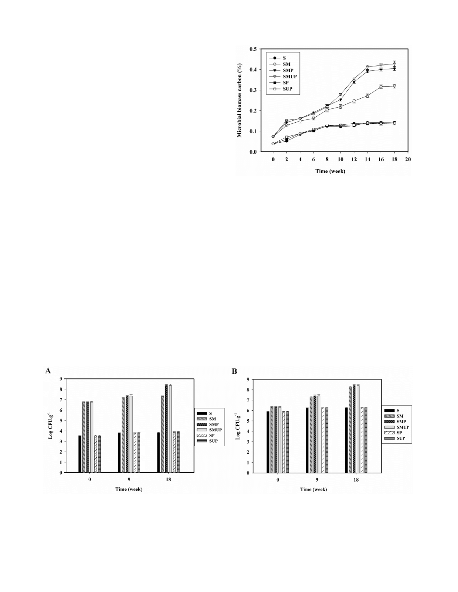
Figure 1. CFU count for fungal and bacterial isolates in various treatments containing LDPE films incubated in the soil for 126 days.
(A) CFU count for fungal isolates in various treatments containing UV- and non-UV-irradiated pure LDPE films without pro-oxidant additives
incubated in the soil for 126 days. (B) CFU count for bacterial isolates in various treatments containing UV- and non-UV-irradiated pure LDPE films
without pro-oxidant additives incubated in the soil for 126 days. Each data point represents the average of three replicates 6 SD. (S: Soil; SM: Soil
+
Selected Microorganisms; SMP: Soil
+ Selected Microorganisms + non-UV-irradiated PE; SMUP: Soil + Selected Microorganisms + UV-irradiated PE; SP:
Soil
+ non-UV-irradiated PE; SUP: Soil + UV-irradiated PE).
doi:10.1371/journal.pone.0071720.g001
Figure 2. Soil microbial biomass carbon for different treat-
ments containing UV- and non-UV-irradiated pure LDPE films
incubated in the soil for 126 days. Each data point represents the
average of three replicates 6 SD. (S: Soil; SM: Soil
+ Selected
Microorganisms; SMP: Soil
+ Selected Microorganisms + non-UV-
irradiated PE; SMUP: Soil
+ Selected Microorganisms + UV-irradiated
PE; SP: Soil
+ non-UV-irradiated PE; SUP: Soil + UV-irradiated PE).
doi:10.1371/journal.pone.0071720.g002
Biodegradation of Low Density Polyethylene in Soil
PLOS ONE | www.plosone.org
2
September 2013 | Volume 8 | Issue 9 | e71720
source of carbon. The microorganisms that were selected were
cultured in liquid mineral medium (synthetic mineral medium)
containing different concentrations of liquid ethylene oligomer (3,
4 and 5%). The microorganisms with the ability to grow in the
presence of 5% ethylene oligomer were transferred to synthetic
mineral medium containing 0.1% PE powder as the sole source of
carbon for the final screening step.
3. Identification of isolated microorganisms
The taxonomic identification of the bacterial isolate, including
biochemical characterisation and PCR amplification of the 16S
rDNA, was performed at the Iranian Biological Resource Center
(IBRC). The partial nucleated sequence of the 16S rDNA from
isolate S7
–10F was determined by the Macrogen Co. in South
Korea (using ABI system 3730 XL) and was deposited in the NCBI
database under Genbank Accession No: JF838304.
The identification of the fungal isolate was performed by
recognising the diagnostic morphological features of genera using
macroscopic and microscopic examinations [16]. In addition, the
molecular identification methods using the PCR to amplify a
segment of the rRNA operon encompassing the 5.8S rRNA gene
and the flanking internal transcribed spacers (ITS) is now in
progress at the Iranian Biological Resource Center (IBRC).
4. Evaluation of LDPE degradation in soil
4-1. Ultraviolet irradiation of polyethylene.
The LDPE
films were irradiated for 25 days under UV light (two 55 W lamps
(Osram) made in Germany) in a laminar flow cabinet and were cut
into pieces measuring approximately 363 cm and 1561.5 cm for
use in the biodegradation assays. For the mechanical properties
analysis, the LDPE films were cut into strips with dimensions of
1561.5 cm.
4-2. Soil preparation and inoculation.
The biodegradation
assay was performed according to ASTM D5988
–03 [7], a
respirometric test based on the measurement of CO
2
evolution.
The soil was collected from farmlands and contained very low
amounts of carbon compounds (organic carbon). The texture of
the soil was loam and was composed of 35.8% sand, 26.6% clay
and 37.6% silt. The organic content of the soil was 1.58% and the
pH was 7.5. This pH was found to be near optimal for
hydrocarbon biodegradation and it was assumed that this pH
would also favour the biodegradation of plastic materials [17]. The
water
-holding capacity of the soil was determined and used to
adjust the water content of the soil to 50% of the holding capacity
[17,7,18]. The soil was sieved (,2 mm) and stored at 4
uC sealed
in a plastic container.
To prepare the inoculums, the fungal isolate was cultured on
MEA (malt extract agar) plates and was incubated at 30
uC until
complete growth was obtained for the next stage. Next, 5 plugs
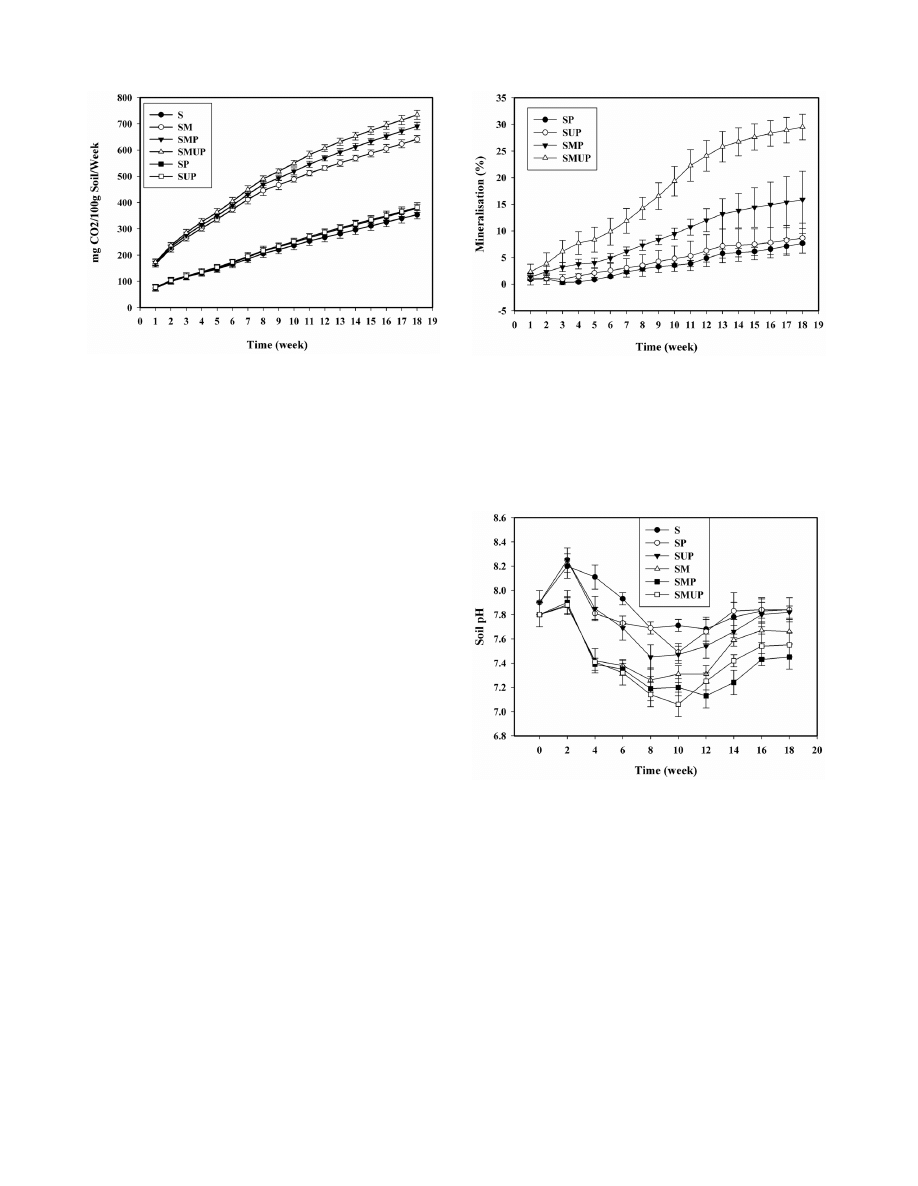
Figure 3. The cumulative CO2 evolution of UV- and non-UV-
irradiated pure LDPE films incubated in the soil with various
treatments for 126 days. Each data point represents the average of
three replicates 6 SD. (S: Soil; SM: Soil
+ Selected Microorganisms; SMP:
Soil
+ Selected Microorganisms + non-UV-irradiated PE; SMUP: Soil+
Selected Microorganisms
+ UV-irradiated PE; SP: Soil + non-UV-
irradiated PE; SUP: Soil
+ UV-irradiated PE).
doi:10.1371/journal.pone.0071720.g003
Figure 4. Mineralisation profile of UV- and non-UV-irradiated
pure LDPE films incubated in the soil with various treatments
for 126 days. Each data point represents the average of three
replicates 6 SD. (SP: Soil
+ non-UV-irradiated PE; SUP: Soil + UV-
irradiated PE; SMP: Soil
+ Selected Microorganisms + non-UV-irradiated
PE; SMUP: Soil
+ Selected Microorganisms + UV-irradiated PE).
doi:10.1371/journal.pone.0071720.g004
Figure 5. pH changes in inoculated, un-inoculated and blank
soil samples for UV- and non-UV-irradiated pure LDPE films.
Each data point represents the average of three replicates 6 SD. (S: Soil;
SM: Soil
+ Selected Microorganisms; SMP: Soil + Selected Microorgan-
isms
+ non-UV-irradiated PE; SMUP: Soil+ Selected Microorganisms +
UV-irradiated PE; SP: Soil
+ non-UV-irradiated PE; SUP: Soil + UV-
irradiated PE).
doi:10.1371/journal.pone.0071720.g005
Biodegradation of Low Density Polyethylene in Soil
PLOS ONE | www.plosone.org
3
September 2013 | Volume 8 | Issue 9 | e71720

(161 cm) of the fungus from the MEA plates were transferred into
50 ml Erlenmeyer flasks containing 15 ml of culture medium
containing the following: (grams per litre): glucose, 10; malt
extract, 10; peptone, 2; yeast extract, 2; asparagine, 1; K
2
HPO
4
, 2;
MgSO
4
.7H
2
O, 1; and Thiamine
-HCL, 0.001. The flasks were
incubated at 30
uC until sufficient biomass was obtained. The soil
(100 g) was placed in the bottom of 2
-litre desiccator jars. The
LDPE pieces (0.1 g) were mixed with the soil and the desiccators
were inoculated with the mixture of the fungus (5 ml of the fungus
inoculums) and the bacterium (5 ml of a mid
-exponential-phase
culture of the bacterium grown in NB (nutrient broth) medium
containing 1.5610
8
colony
-forming units). The desiccators were
incubated in a sterilised chamber at 30
uC for 126 days.
The reduced water content of the soil was supplemented once
per week with a mineral solution (pH 6.5) containing (grams per
litre) KH
2
PO
4
, 0.7; K
2
HPO
4
, 0.7; MgSO
4
.7H
2
O, 0.7; NH
4
NO
3
,
1.0; NaCl, 0.005; MnSO
4
.7H
2
O, 0.001; ZnSO
4
.7H
2
O, 0.002;
and FeSO
4
, 0.002 [6].
In this study, two main treatments were performed: soil +
selected microorganisms + UV
-irradiated LDPE films (SMUP)
and soil + selected microorganisms + non
-UV-irradiated LDPE
films (SMP). In each treatment, three blanks were included: soil
(S), soil with the selected microorganisms (SM) and soil with UV
-
or non
-UV-irradiated LDPE films (SUP and SP, respectively).
Each treatment was performed in triplicate.
After 126 days, the process was terminated, and the LDPE
pieces were washed in distilled water, were dried and were
analysed for biodegradation.
4-3. Soil analyses.
Microbial count: The microbial
population in the treatments was measured periodically (every
6 weeks) using the dilution plate method [19]. The bacteria and
fungi were distinguished using different agar media containing
(grams per litre) the following: for fungi, malt extract, 20; glucose,
20; peptone, 1; and for bacteria, K
2
HPO
4
, 1.0; MgSO
4
.7H
2
O,
0.02, CaCl
2
, 0.1; NaCl, 0.1; FeCl
3
, trace, KNO
3
, 0.5; asparagine,
0.5; mannitol, 1; and yeast extract, 0.25 [6]. Sterile physiological
serum was used to dilute the soil samples by 10
21
to 10
28
. For
each of the 10
23
, 10
24
, 10
25
and 10
26
dilutions, 3 plates were
inoculated. The plates were incubated at 28
uC. The colonies were
counted after an appropriate incubation time.
Microbial biomass carbon: The microbial biomass carbon
was measured every two weeks using the chloroform fumigation/
direct extraction method for all treatments [20].
Carbon dioxide evolution: To trap the evolved CO
2
, each
desiccator was equipped with a beaker containing 20 ml of a
0.2 mol/L NaOH (Merck) solution, which was titrated with a
0.2 mol/L HCL (Merck) solution. The desiccators were main-
tained at 30
uC and were opened at appropriate intervals to allow
aeration and titration of the NaOH solution. Prior to the titration,
2 ml of a 0.5 mol/L BaCl
2
solution was added to the NaOH
solutions. Desiccators (3 in total) containing only the absorbing
solution and no soil were also included as technical controls [7].
The percentage of biodegradation of the samples (mineralisa-
tion) was calculated taking into account the theoretical amount of
carbon dioxide ([CO
2
]
Theor
) of the samples; and the percentage
biodegradation: ([CO
2
]
T
*100/([CO
2
]
Theor
).
Soil pH measurement: The pH of the soil was determined in
a 5:1 (distilled water: soil) slurry using a glass combination
electrode calibrated with standard buffers, following the guidelines
given in the Test Method ASTM D 1293
–99, a standard test
method for pH of water [21,7].
4-4. LDPE analyses.
Mechanical properties: The tensile
strength (percentage elongation) was determined using a tensile
tester (Gotech, model U 60) at room temperature and 50 mm/
min with a 5
-cm gap. The samples were equilibrated to 50%
relative humidity for at least 40 h before the analysis [22].
Fourier transform infra-red (FT-IR) analysis: The
structural change in the LDPE surface was investigated using
the EQUINOX 55 FT
-IR spectrometer. For each LDPE film, a
spectrum was taken from 400 to 4000 wavenumbers.cm
21
. The
carbonyl and double bond indices were calculated based on the
relative intensities of the carbonyl band at 1,715 cm
21
and the
double bond band at 1,650 cm
21
to that of the methylene
scissoring band at 1,460 cm
21
[23].
X-ray diffraction (XRD) analysis: The X-ray diffraction
patterns of the films were measured with a X-ray diffractometer
(D5000, Siemens Diffractometer) which is operated fully automat-
ically using Cu Ka radiation (l = 1.5418 A
u). The scattered
radiation was registered in the angular interval (2fi) from 2
uto
40
u. A current of 30 mA and a voltage of 40 kV were used. All
Table 1. Changes in the percentage elongation of non-UV-irradiated PE films before and after 63 and 126 days of biodegradation
in soil in various treatments.
Treatments
Time 0
Week 9
Week 18
e
r
± SD
e
r
± SD
(D%)
e
r
± SD
(D%)
SP
299.566.2 a
297.669.7 a
0.6 (2)
272.967 b
8.9 (2)
SMP
299.566.2
247.3613.3 c
17.4 (2)
155.5611.8 d
48 (2)
e
r
elongation at break (%), (D %) difference between percentage elongation of films before and after biodegradation process (shown as a percentage). Values
accompanied by a similar letter are not significantly different according to Duncan’s multiple- range test (P = 0.05). Each value represents the average of four replicates
6
SD. (SP: Soil
+ non-UV-irradiated PE; SMP: Soil + Selected Microorganisms + non-UV-irradiated PE).
doi:10.1371/journal.pone.0071720.t001
Table 2. Changes in the percentage elongation of UV-
irradiated PE films before and after 63 and 126 days of
biodegradation in soil in various treatments.
Treatments Time 0
Week 9
Week 18
e
r
± SD
e
r
± SD
(D%)
e
r
± SD
(D%)
SUP
12.661.2 a
10.361.2 b
17.8 (2)
*
2
SMUP
12. 661.2
2.160.4 c
83 (2)
*
2
e
r
elongation at break (%), * fragile specimens, (D %) difference between
percentage elongation of films before and after biodegradation process (shown
as a percentage). Values accompanied by a similar letter are not significantly
different according to Duncan’s multiple range test (P = 0.05). Each value
represents the average of four replicates 6 SD. (SUP: Soil
+ UV-irradiated PE;
SMUP: Soil
+ Selected Microorganisms + UV-irradiated PE).
doi:10.1371/journal.pone.0071720.t002
Biodegradation of Low Density Polyethylene in Soil
PLOS ONE | www.plosone.org
4
September 2013 | Volume 8 | Issue 9 | e71720
diffraction patterns were examined at room temperature and
under constant operating conditions.
Scanning electron microscopy (SEM): The polyethylene
samples were removed from the soil and were dried in a desiccator
for 24 h under vacuum. The samples were vapour
-fixed at room
temperature for three days in a sealable glass container containing
two beakers, one containing 10 ml of 25% glutaraldehyde in H
2
O
and the other containing 5 ml of 5% OsO
4
in 0.1 M phosphate
buffer at pH 7.0. After fixation, the container was aerated for 20 h
[13]. The samples were gold
-coated using BAL-TEC-SCDOOS
and were examined using a Philips
-X LP30 scanning electron
microscope.
Results and Discussion
1. Isolation and identification of the isolates
In the initial step of the isolation, 144 isolates were selected by
comparing their ability to growth in solid mineral medium
containing linear paraffin. In addition, the screening of these
isolates was performed by comparing their growth ability in solid
media containing synthetic mineral medium supplemented with
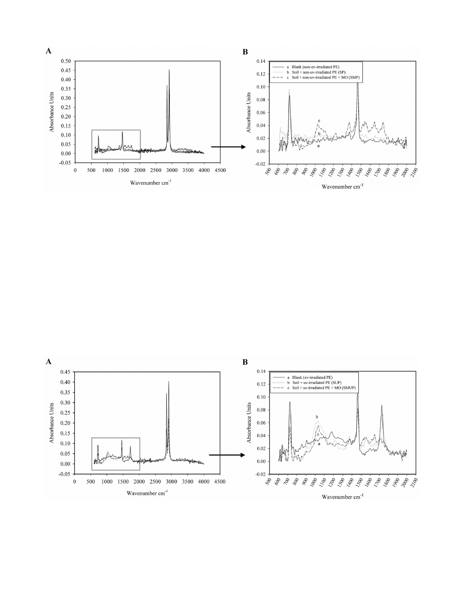
Figure 6. FT-IR spectra of non-UV-irradiated pure LDPE films before and after incubation in soil in various treatments. (A) FT-IR
spectra of non-UV-irradiated pure LDPE films without pro-oxidant additives before and after 126 days of incubation in soil in the presence and
absence of the selected microorganisms from 500–4000 cm-1. (B) The changes in the bands between 500 and 2,000 cm-1 of the FT-IR spectra of non-
UV-irradiated pure LDPE films without pro-oxidant additives before and after 126 days of incubation in soil with different treatments: (a) blank (no UV
irradiation, no incubation); (b) non-UV-irradiated LDPE after incubation in soil in the absence of the selected microorganisms (SP treatment); (c) non-
UV-irradiated LDPE after incubation in soil in the presence of the selected microorganisms (SMP treatment).
doi:10.1371/journal.pone.0071720.g006
Figure 7. FT-IR spectra of UV-irradiated pure LDPE films before and after incubation in soil in various treatments. (A) FT-IR spectra of
UV-irradiated pure LDPE films without pro-oxidant additives before and after 126 days of incubation in soil in the presence and absence of the
selected microorganisms from 500–4000 cm-1. (B) The changes in the bands between 500 and 2,000 cm-1 of the FT-IR spectra of UV-irradiated pure
LDPE films without pro-oxidant additives before and after 126 days of incubation in soil with different treatments: (a) blank (after 25 days’ UV
irradiation, no incubation); (b) UV-irradiated LDPE after incubation in soil in the absence of the selected microorganisms (SUP treatment); (c) UV-
irradiated LDPE after incubation in soil in the presence of the selected microorganisms (SMUP treatment).
doi:10.1371/journal.pone.0071720.g007
Biodegradation of Low Density Polyethylene in Soil
PLOS ONE | www.plosone.org
5
September 2013 | Volume 8 | Issue 9 | e71720
2% ethylene oligomer, resulting in the selection of 53 isolates. In
total, five gram
-positive and spore-forming Bacilli and five fungal
isolates were screened based on their growth ability in a liquid
mineral medium containing 5% ethylene oligomer. Of these
isolates, one bacterial and one fungal isolate were selected as the
final strains by comparing the growth ability in mineral medium
containing PE powder as the sole source of carbon. The
morphological characterisation of the fungus, including the colour
of the colonies cultured on agar and the dimensions of the
conidiophores and conidia, indicated that this isolate resembled
Aspergillus niger. This strain was designated strain F1 in this study.
The taxonomic identification of the bacterial isolate (S7
–10F),
in accordance with Bergey’s Manual of Systematic Bacteriology
[24] and 16S rDNA sequencing indicated a 99.4% resemblance to
Lysinibacillus xylanilyticus.
2. Soil microbial count
The fungal and bacterial population was measured separately in
the beginning, middle and end of the incubation period (Fig. 1A,
B). As shown in Fig. 1, the microbial population increased from
the beginning to the end of the incubation period for both the
fungi and the bacteria, and this increment was much higher in the
treatments inoculated with the selected microorganisms (SMUP,
SMP and SM). These data demonstrate that soil-indigenous
microorganisms cannot utilise PE as the sole carbon source (S, SP
and SUP treatments). The initial fungal population of the soil was
lower than the bacterial population; therefore, the difference
between the fungal population in the treatments with and without
the selected microorganisms, at time zero and during the
incubation, was because of the growth of the selected fungal
isolate, strain F1 (Fig. 1A). The bacterial population demonstrated
a similar result. The difference between the un
-inoculated
treatments and the treatments inoculated with the selected
bacterial isolate during the process indicated the ability of strain
S7
–10F to utilise both types of PE (UV-irradiated and non-UV-
irradiated LDPE) as the source of carbon (Fig. 1B). For the SMUP
and SMP treatments, the bacterial and fungal populations
exhibited the highest number of live colonies in the 126 days,
Table 3. Carbonyl and double bond indices values determined using FTIR from LDPE films before and after 126 days incubation in
soil in various treatments.
Treatments
Time 0
Week 18
(D%)
CI
DBI
CI
DBI
CI
DBI
SP
0.151
0.154
0.187
0.216
23.8 (
+)
40.2 (
+)
SUP
0.747
0.168
0.705
0.280
5.6 (2)
66.7 (
+)
SMP
0.151
0.154
0.401
0.401
165. 6 (
+)
160.4 (
+)
SMUP
0.747
0.168
0.433
0.507
42 (2)
201. 8 (
+)
CI carbonyl index, DBI double bond index, (D%) difference between carbonyl and double bond indices of films before and after 126 days of biodegradation (shown as a
percentage). (SP: Soil
+ non-UV-irradiated PE; SUP: Soil + UV-irradiated PE; SMP: Soil + Selected Microorganisms + non-UV-irradiated PE; SMUP: Soil + Selected Micro-
organisms
+ UV-irradiated PE).
doi:10.1371/journal.pone.0071720.t003
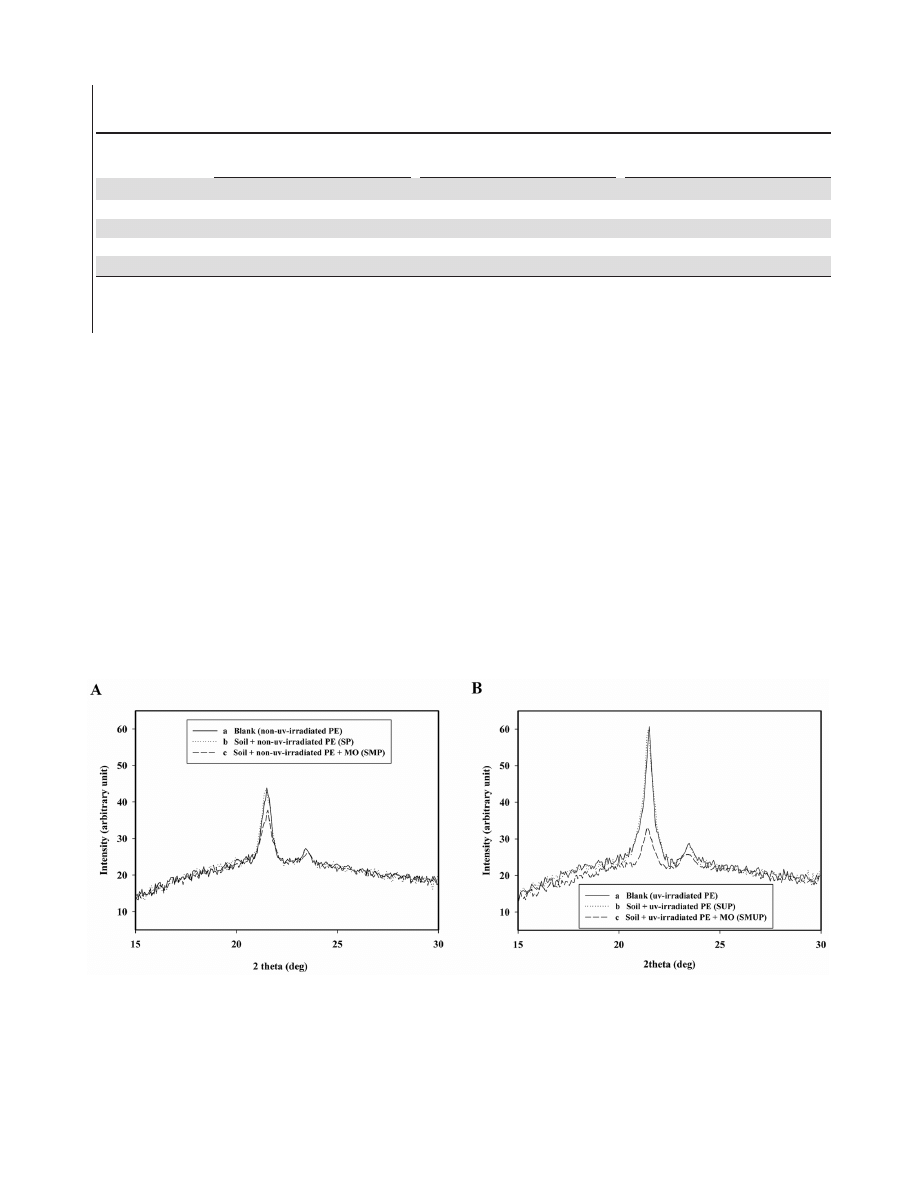
Figure 8. XRD spectra of non-UV and UV-irradiated pure LDPE films before and after incubation in soil with different treatments. (A)
XRD spectra of non-UV-irradiated pure LDPE films without pro-oxidant additives before and after 126 days of incubation in soil with different
treatments: (a) blank (no UV irradiation, no incubation); (b) non-UV-irradiated LDPE after incubation in soil in the absence of the selected
microorganisms (SP treatment); (c) non-UV-irradiated LDPE after incubation in soil in the presence of the selected microorganisms (SMP treatment).
(B) XRD spectra of UV-irradiated pure LDPE films without pro-oxidant additives before and after 126 days of incubation in soil with different
treatments: (a) blank (after 25 days’ UV irradiation, no incubation); (b) UV-irradiated LDPE after incubation in soil in the absence of the selected
microorganisms (SUP treatment); (c) UV-irradiated LDPE after incubation in soil in the presence of the selected microorganisms (SMUP treatment).
doi:10.1371/journal.pone.0071720.g008
Biodegradation of Low Density Polyethylene in Soil
PLOS ONE | www.plosone.org
6
September 2013 | Volume 8 | Issue 9 | e71720
indicating the utilisation of UV
-irradiated and non-UV-irradiated
LDPE by the selected microorganisms in these treatments,
respectively.
3. Soil microbial biomass carbon
The soil microbial biomass carbon (MBC) was measured every
two weeks. The MBC results exhibited a similar pattern to the
CFU data. The MBC increased more rapidly in the inoculated
treatments (SM, SMP and SMUP, especially in the treatments
containing PE as the source of carbon) than in the un
-inoculated
treatments (SUP and SP) (Fig. 2).
4. CO
2
evolution
Currently, the laboratory tests used to determine the biodeg-
radation of plastics are based on the measurement of carbon
dioxide evolution or oxygen consumption when the original
polymer is exposed to controlled environmental conditions (e.g.,
soil, compost, active sludge, etc.). Generally, biodegradation is
measured as the degree of mineralisation, namely the conversion
into CO
2
. This method is considered the optimum approach for
confirming the total biodegradability, i.e., the total conversion of
organic carbon into inorganic carbon [25]. The amount of CO
2
evolved was determined by the titration of the remaining NaOH.
The results are presented in Fig. 3. There were no significant
differences in CO
2
evolution between the S, SP and SUP
treatments. These treatments showed a slight and gradual increase
in CO
2
generation during biological degradation compared to the
inoculated treatments (SMUP, SMP and SM). Of the treatments
that were inoculated with the selected microorganisms, the SMUP
treatment demonstrated the highest amount of CO
2
production.
The CO
2
levels reached 734 mg CO
2
.g soil
21
after 126 days,
which was due to the utilisation of carbonyl groups of the UV-
irradiated films by the S7
–10F and F1 strains. In addition,
significant differences were observed between the SMP and SM
treatments, indicating the ability of the selected microorganisms to
utilise the non
-UV-irradiated LDPE as the carbon source. The
mineralisation profiles of the samples are shown in Fig. 4. The
percentage biodegradation was higher in the inoculated treatments
(SMP and SMUP) than in the un
-inoculated treatments (SP and
SUP). The mineralisation of the UV
-irradiated LDPE films in the
inoculated treatment (SMUP, 29.5%), when compared with the
corresponding un
-inoculated treatment (SUP, 8.6%), was remark-
ably elevated. This difference clearly demonstrates that the
selected bacterial and fungal isolates utilised the oxidation
products in the pre
-oxidised films. Similar results were obtained
with similar treatments containing non
-UV-irradiated films (SMP
and SP). The mineralisation values for pure PE without oxidation
pre
-treatment in the SMP and SP treatments were 15.8% and 7.
6% respectively. The pronounced difference between these two
treatments indicates that the selected microorganisms are not only
able to utilise pre
-oxidised PE as a carbon source, as described, but
can also utilise pure LDPE without oxidation pre
-treatment.
The negligible difference in the CO
2
production and mineral-
isation values between the SP and SUP treatments, and the very
high level of CO
2
generation in the SMUP and SMP treatments,
clearly demonstrates the important role of the selected fungal and
bacterial isolates in the biodegradation of UV
-irradiated and non-
UV
-irradiated LDPE compared with the indigenous soil microbial
population. Most related studies have investigated the biodegra-
dation of abiotically aged LDPE containing pro
-oxidant additives
or PE modified with starch. The percentage mineralisation for the
pre
-oxidised LDPE and LDPE containing pro-oxidant additives
was 16% and 24% after 317 days incubation in compost, and 9%
and 12% after 317 days incubation in soil, respectively [26].
The mineralisation of thermally oxidised biodegradable LDPE
after two years of incubation in soil was reported as approximately
91% [27].
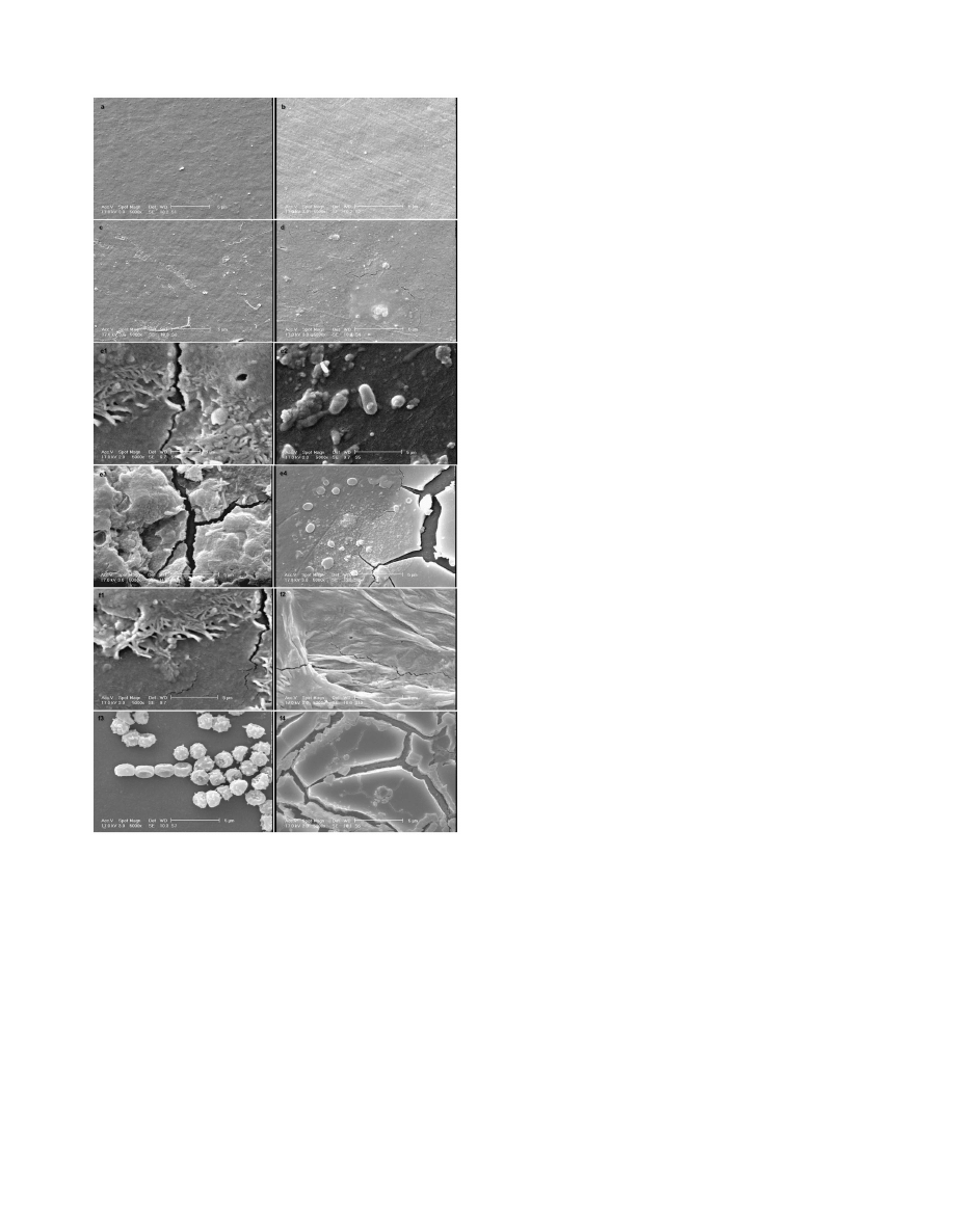
Figure 9. SEM micrograph of pure LDPE films before and after
126 days of incubation in soil with different treatments. (a)
Blank (no UV irradiation, no incubation). (b) UV-irradiated LDPE film
without incubation. (c) non-UV-irradiated LDPE film incubated in soil in
the absence of the selected microorganisms (SP). (d) UV-irradiated LDPE
film incubated in soil in the absence of the selected microorganisms
(SUP). (e) non-UV-irradiated LDPE film incubated in soil in the presence
of the selected microorganisms (SMP): (e1) penetration of hyphae into
the LDPE matrix; (e2) formation of bacterial biofilm on the surface of
LDPE; (e3 and e4) formation of pits and cavities on the surface of LDPE.
(f) UV-irradiated LDPE film incubated in soil in the presence of the
selected microorganisms (SMUP): (f1 and f2) penetration of hyphae into
the LDPE matrix; (f3) formation of bacterial biofilm on the surface of
LDPE; and (f4) formation of pits and cavities on the surface of LDPE.
doi:10.1371/journal.pone.0071720.g009
Biodegradation of Low Density Polyethylene in Soil
PLOS ONE | www.plosone.org
7
September 2013 | Volume 8 | Issue 9 | e71720

Our results demonstrated higher mineralisation rates for pure
LDPE without any pro
-oxidant additives, with and without
oxidation pretreatment, compared to the mineralisation rates
reported by Ojeda et al. [28] for traditional LDPE without pro
-
oxidant additives exposed to sun light for 7 and 30 days. The study
also reported approximately 1% mineralisation for these films after
90 days incubation in compost. Abrusci et al. [15] reported a
pronounced difference between pure PE (2
–2.5% biodegradation
after 90 days) and the corresponding material containing pro
-
oxidant additives (7
–10% biodegradation after 90 days) incubated
with a mixture of Bacillus species.
5. Soil pH measurement
The pH value is a key factor for the survival and activity of
microorganisms. Generally, the pH should be between 6 and 8 [7].
The periodic measurements of the soil pH are presented in Fig. 5.
The initial increase in the pH values may be because of the
ammonification of nitrogen components [11]. The decrease and
increase in pH was also reported in a study by Jakubowicz et al.
[27] in which the biodegradation of thermally oxidised biode-
gradable LDPE in soil for a period of 606 days was evaluated.
6. Mechanical properties of polyethylene films
The mechanical properties of the non
-UV- and UV-irradiated
films are shown respectively in Tables 1 and 2, during and after
the biodegradation process. There was no significant difference
(P = 0.05) in percentage elongation between time zero and after
63 days’ incubation of non-UV-irradiation films in the absence of
the selected microorganisms (Table 1, treatment SP). The
elongation at break (%) of the non
-UV-irradiated LDPE films
decreased 8.9% in the un
-inoculated treatment (SP) after
126 days’ incubation, whereas the percentage elongation of these
films decreased 17.4% and 48% in the inoculated treatment (SMP)
after 63 and 126 days’ incubation, respectively (Table 1). Reduc-
tions in elongation of 17.8% and 83% were recorded after 63 days
for the UV
-irradiated LDPE films in the un-inoculated (SUP) and
inoculated (SMUP) treatments, respectively (Table 2).
The long polymer chains were likely cut into shorter pieces
because of the action of enzymes secreted by the microorganisms.
Because the films became fragile and lighter in weight indicates the
preliminary stages of microbial decomposition, consisting of a
reduction in molecular weight [29].
The UV irradiation of the films caused a reduction in the
percentage elongation of 95. 8%. This result is in agreement with a
study by Lee et al. [30] reporting an increase in the percentage
elongation of degradable films after 2 weeks of UV irradiation,
and a marked decrease in this parameter after 4 and 8 weeks of
treatment. The percentage elongation of 8
-week UV-irradiated
degradable films was near to zero. Two-week UV
-irradiated
degradable films demonstrated a reduction in the percentage
elongation after 4 weeks’ of incubation in culture media contain-
ing ligninolytic microorganisms. A reduction in the percentage
elongation of the LDPE film after thermal oxidation was reported
by Jakubowicz et al. [27]. In addition, Orhan and Buyukungor
[6], Jakubowicz et al. [31] and Nowak et al. [29] reported a
reduction in the percentage elongation of polyethylene films after
the biodegradation process.
7. FT-IR analysis
In the biodegradation of polyethylene, the initial abiotic step
involves the oxidation of the polymer chain leading to the
formation of carbonyl groups. These groups eventually form
carboxylic groups, which subsequently undergo b
-oxidation [23]
and are completely degraded via the citric acid cycle resulting in
the formation of CO
2
and H
2
O. b
-oxidation and the citric acid
cycle are catalysed by microorganisms. Monitoring the formation
and disappearance of carbonyl and double bond bands using FT-
IR is necessary to elucidate the mechanism of the biodegradation
process. Figure 6 shows the FT-IR spectra of non
-UV-irradiated
pure LDPE films without pro
-oxidant additives before and after
the 126 days of incubation in soil in the presence and absence of
the selected microorganisms. FT-IR spectra of non
-UV-irradiated
LDPE films before and after 126 days of incubation in soil in the
presence and absence of the selected microorganisms from 500–
4000 cm
21
is shown in Fig. 6A. The changes in the bands between
500 and 2,000 cm
21
are magnified in Fig. 6B. The FT-IR spectra
of the LDPE film without oxidation pretreatment is shown in
Fig. 6B.a. The spectrum of the non
-UV-irradiated LDPE film,
which is introduced into soil in the absence of the selected
microorganisms (SP treatment), is shown in Fig. Fig. 6B.b.
Compared with the corresponding control, significant changes in
the spectra of the non
-UV-irradiated LDPE film after 126 days
incubation were not observed (Fig. 6B.a, no UV irradiation, no
incubation). The spectrum of this film, incubated in soil that was
inoculated with the selected microorganisms, shows the appear-
ance of several new bands (Fig. 6B.c, SMP treatment). The
carbonyl absorption bands can be observed in the range of
1,71021,750 cm
21
because of the formation of ketone or
aldehyde C = O groups by the action of the selected microorgan-
isms. The absorbance in the region of 1,541 cm
21
is associated
with the carboxylate group [32]. Additionally, new absorption
bands between 1,000 and 1,700 cm
21
(1,029 and 1,371 cm
21
) of
the spectra are possibly due to the oxidised fractions, such as
moieties containing –OH groups, resulting from biodegradation
by the selected microorganisms [33]. The selected bacterial and
fungal isolates were capable of utilising long, hydrophobic
polyethylene chains. In our study, the selected microorganisms
from the landfill source oxidised even virgin LDPE without
oxidation pretreatment and pro-oxidant additives. These results
are in contrast to many reports in which the microorganisms could
assimilate only the products of pre
-oxidised PE [34,11]. Because
the initial breakage of PE chains is the longest and most difficult
step in its degradation, long incubation times produce significant
quantities of carbonyl groups to continue the decomposition
process [29]. Figure 7 shows the FT-IR spectra of the UV
-
irradiated pure LDPE films without pro
-oxidant additives before
and after the 126 days of incubation in soil in the presence and
absence of the selected microorganisms. FT-IR spectra of the UV-
irradiated LDPE films before and after 126 days of incubation in
soil in the presence and absence of the selected microorganisms
from 500–4000 cm
21
is shown in Fig. 7A. The changes in the
bands between 500 and 2,000 cm
21
are magnified in Fig. 7B. The
FT-IR spectra of the LDPE film after 25 days of UV irradiation
(Fig. 7B.a) shows the appearance of a peak in the range of 1,710
–
1,750 cm
21
due to the formation of carbonyl groups (abiotic
oxidation). The intensity of the bands in the 1,178 cm
21
region is
increased and is related to carbonyl groups [10]. Figure 7B.b
shows the FT-IR spectrum of the UV
-irradiated LDPE film
incubated in un
-inoculated soil (SUP treatment). The new
absorption bands between 1,000- and 1,700 cm
21
(1028 and
1373 cm
21
) are also because of the oxidised fractions, such as
moieties containing –OH groups resulting from the action of the
indigenous soil microorganisms [33]. The comparison of the
spectra from the SP and SUP treatments (Figs. 6B.b and 7B.b)
shows clearly that the indigenous soil microorganisms could not
utilise the virgin PE with a hydrophobic nature but were capable
of attaching to and partially oxidising the UV
-irradiated LDPE
film.
Biodegradation of Low Density Polyethylene in Soil
PLOS ONE | www.plosone.org
8
September 2013 | Volume 8 | Issue 9 | e71720

The spectrum of the UV
-irradiated LDPE film which is
introduced into soil in the presence of the selected microorgan-
isms (SMUP treatment), is shown in Fig. 7B.c. Compared with
the corresponding control, the intensity of the carbonyl band at
1,710
–1,750 cm
21
was significantly decreased during the process
with the selected microorganisms. The intensity of the bands in
the 1,000
–1,700 cm
21
range (1,071, 1,541 and 1,649 cm
21
) is
also attributed to the oxidised fractions because of the action of
the selected microorganisms. Part of the decreased absorption at
1,714 cm
21
is compensated by the appearance of carboxylates at
1,541 cm
21
(Fig. 7B.c, SMUP treatment) [32]. The indigenous
soil microorganisms reduced the carbonyl index (CI) 5.6%
(Table 3 and SUP treatment in Fig. 7B.b), whereas selected
microorganisms reduced the CI 42% (Table 3 and SMUP
treatment in Fig. 7B.c).
An increase in the double bond index (DBI) was observed in all
treatments and was especially significant in the SMP and SMUP
treatments (Table 3). The decrease in the CI in the SMUP and
SUP treatments and the increase in the DBI in all samples,
especially the SMP and SMUP treatments, can be explained using
the proposed mechanism for PE biodegradation. According to this
mechanism, formed carbonyl groups along the polymeric chain,
resulting from the action of abiotic factors, can be attacked
microbially (CI decrease), and lead to the release of unsaturated
chains (DBI increase) [9]. The oxidised group is transformed to a
carboxylic acid and is metabolised via b
-oxidation.
Manzur et al. [10] reported that the segments formed by the
rupturing of the chains because of the biological treatment could
cause the formation of the vinyl group and the increase in the DBI.
In addition, the increase in the DBI can be attributed to biotic
dehydrogenation [35].
8. XRD analysis
The XRD spectra of the non
-UV- and UV-irradiated pure
LDPE films is shown in Fig. 8, before and after 126 days of
incubation in soil in the presence and absence of the selected
microorganisms. As shown in this figure, the XRD spectra show
distinguished peaks at 21.4 and 23.5 of the angular position 2fi.
The intensity of the peaks of UV-irradiated films is higher than
that of non
-UV-irradiated one (Fig. 8A.a and Fig. 8B.a). This
difference clearly demonstrated that oxidation pretreatment
increased the degree of polyethylene crystalinity.
The intensity of the peaks was significantly decreased after
126 days of incubation in soil in the presence of the selected
microorganisms (Fig. 8A.c, SMP treatment and Fig. 8B.c, SMUP
treatment). There were no significant differences in degree of
crystalinity between corresponding controls and the films incu-
bated in soil in the absence of the selected microorganisms
(Fig. 8A.b, SP treatment and 8A.a, Fig. 8B.b, SUP treatment and
8B.a). These results indicated that crystalinity and the crystal sizes
for non-UV-irradiated and UV-irradiated films decreased during
process with the selected microorganisms [36].
9. Scanning electron microscopy analysis
The SEM analysis was performed to monitor the changes in the
surface of the films. The adhesion of the microorganisms to the
polymeric surface is fundamental for biodegradation to take place
[9]. Figure 9 shows the SEM micrographs of the PE surfaces
before and after 126 days of incubation with the different
treatments. Before processing, the samples had a smooth surface
with no defects (Figs. 9a and 9b). In addition, no special features
were detected in the SEM micrograph of the films introduced into
the soil without the selected microorganisms (Figs. 9c and 9d).
However, after incubation with the selected microorganisms,
surface erosion and the formation of pits and cavities on the
surface of the samples can be observed (Figs. 9e and 9f). The
presence of pits and cavities may be because of the absence of a
uniform distribution of short branches or photodegradable
products in the polymer matrix [10], suggesting that the fungus
(strain F1) penetrated into the LDPE matrix during degradation.
This penetration of hyphae into the matrix, and the formation of a
bacterial biofilm of the strain S7
–10F on the surface of the films
were observed in both the (UV- and non
-UV-irradiated PE)
incubated in the soil with the selected microorganisms (SMP and
SMUP treatments).
The LDPE degradation by Aspergillus niger and Aspergillus
fumigatus is consistent with the results obtained previously
[9,10,11]. Aspergillus terreus also participated in the degradation
of modified and unmodified PE [12]. Moreover, there are
examples in the literature confirming the ability of the genus
Bacillus to degrade PE. Bacillus pumilus and B. halodenitrificans
were able to degrade an abiotically aged LDPE containing
pro
-oxidant within 120 days [37]. Bacillus sphericus and B. cereus
have also been shown to degrade LDPE and HDPE unmodified
and modified with starch [34]. The biodegradation of photo
-
degraded LDPE containing pro
-oxidant additives by a mixture
of Bacillus strains (B. megaterium, B. subtilis and B. cereus) was
evaluated within 90 days, and biofilm formation developed only
in the photo
-degraded material after one week of the bacterial
treatment [15].
Conclusion
In this study, the biodegradation of pure LDPE films without
pro
-oxidant additives, with and without photo-oxidation pre-
treatment, was evaluated in soil in the presence and absence of a
mixed culture of selected landfill
-source microorganisms (Asper-
gillus niger designated strain F1 and Lysinibacillus xylanilyticus XDB9
(T) designated strain S7
–10F). The data obtained from the
respiration and microbial population measurements showed
significant differences between the inoculated and un
-inoculated
treatments with the selected microorganisms. The carbon dioxide
measurements showed that the biodegradation in the un
-
inoculated treatments were slow and were about 7.6% and
8.6% of mineralisation for the non
-UV-irradiated and UV-
irradiated LDPE respectively after 126 days. In contrast, in the
presence of the selected microorganisms, the biodegradation was
much more efficient and the percentages of biodegradation were
29.5% and 15.8% for the UV
-irradiated and non-UV-irradiated
films, respectively. The percentage decrease in the CI was higher
for the UV
-irradiated LDPE when the biodegradation was
performed in soil inoculated with the selected fungal and bacterial
isolates. The FT-IR, XRD and SEM analyses demonstrated the
ability of the selected microorganisms to modify and colonise
both types of PE as the carbon source, and demonstrated the
important role of these isolates in the PE biodegradation process.
The oxidation pretreatment facilitated the biodegradation of PE;
however, contrary to other reports, our study confirmed the
ability of the selected fungal and bacterial isolates to utilise virgin
PE without pro
-oxidant and oxidation pretreatments. The results
of this study show that the selected microorganisms (strains S7
–
10F and F1) exhibit great potential for LDPE biodegradation
under natural conditions, such as those found in soil. In the near
future, these microorganisms can be used to reduce the quantity
of solid waste, which is rapidly accumulating in the natural
environment.
Biodegradation of Low Density Polyethylene in Soil
PLOS ONE | www.plosone.org
9
September 2013 | Volume 8 | Issue 9 | e71720

Acknowledgments
The authors would like to thank Maleki Shamsabadi and Tehrani for
providing petrochemical materials needed for the experiments.
Author Contributions
Conceived and designed the experiments: AE AAP HAA. Performed the
experiments: AE. Analyzed the data: AE AAP EE. Contributed reagents/
materials/analysis tools: AE AAP HAA FS EE. Wrote the paper: AE.
References
1. Shah AA, Hasan F, Hameed A, Ahmed S (2008) Biological degradation of
plastics: a comprehensive review. Biotechnol Adv 26: 246–265.
2. Gilan I, Hadar Y, Sivan A (2004) Colonization and biofilm formation and
biodegradation of polyethylene by a strain of Rhodococcus rubber. Appl Microbiol
Biotechnol 65: 97–104.
3. Albertsson AC (1978) Biodegradation of synthetic polymers. 2. Limited
microbial conversation of C-14 in polyethylene to (CO-2)-C-14 by some soil
fungi. J Appl Polym Sci 22: 3419–3433.
4. Albertsson AC (1980) The shape of the biodegradation curve for low and high
density polyethylene in prolonged series of experiments. Eur Polym J 16: 623–630.
5. Cornell JH, Kaplan AM, Rogers MR (1984) Biodegradation of photooxidized
polyethylenes. J Appl Polym Sci 29: 2581–2597.
6. Orhan Y, Buyukgungor H (2000) Enhancement of biodegradability of disposable
polyethylene in controlled biological soil. Int Biodeterior Biodegradation 45:
49–55.
7. ASTM D 5988 (2003) Standard test method for determining aerobic
biodegradation in soil of plastic material or residual plastic materials after
composting.
8. Lau AK, Cheuk WW, Lo KV (2009) Degradation of greenhouse twines derived
from natural fibers and biodegradable polymer during composting. J Environ
Manage 90: 668–671.
9. Volke-Sepulveda T, Saucedo-Castanede G, Gutierrez-Rojas M, Manzur A,
Favela-Torres E (2002) Thermally treated low density polyethylene biodegra-
dation by Penicillium pinoohilum and Aspergillus niger. J Appl Polym Sci 83: 305–314.
10. Manzur A, Limon-Gonzalez M, Favela-Torres E (2004) Biodegradation of
physiochemically treated LDPE by a consortium of filamentous fungi. J Appl
Polym Sci 92: 265–271.
11. Sahebnazar Z, Shojaosadati SA, Mohammad-Taheri M, Nosrati M (2010)
Biodegradation of low-density polyethylene (LDPE) by isolated fungi in solid
waste medium. Waste Manage 30: 396–401.
12. El-Shafei HA, Nadia H, El-Nasser A, Kanosh AL, Ali AM (1998)
Biodegradation of disposable polyethylene by fungi and Streptomyces species.
Polym Degrad Stab 62: 361–365.
13. Sivan A, Szanto M, Pavlov V (2006) Biofilm development of the polyethylene-
degrading bacterium Rhodococcus ruber. Appl Microbiol Biotechnol 73: 346–352.
14. Hadad D, Geresh S, Sivan A (2005) Biodegradation of polyethylene by the
thermophilic bacterium Brevibacillus borstelensis. J Appl Microbiol 98: 1093–1100.
15. Abrusci C, Pablos JL, Corrales T, Lopez-Marin J, Marin L, et al. (2011)
Biodegradation of photo-degraded mulching films based on polyethylene and
stearates of calcium and iron as pro-oxidant additives. Int Biodeterior
Biodegradation 65: 451–459.
16. Watanabe T (2002) Pictorial Atlas of Soil and Seed Fungi. CRC PRESS.
17. Yabannavar AV, Bartha R (1994) Methods for assessment of biodegradability of
plastic films in soil. Appl Environ Microbiol 60: 3608–3614.
18. ISO17556 (2003) Plastics-Determination of the Ultimate Aerobic Biodegrad-
ability in Soils by Measuring the Oxygen Demand in a Respirometer or the
Amount of Carbon Dioxide Evolved.
19. Page AL, Miller RH, Keeney DR (2008) Methods of soil analysis. Part 2:
Chemical and Microbiological Properties. Soil Science Society of American.
Wisconsin. pp. 139–141.
20. Jenkinson DS, Brookes PC, Powelson DS (2004) Measuring soil microbial
biomass. Soil Biol Biochem 36: 5–7.
21. ASTM D1293 (1999) Standard test methods for pH of water.
22. ASTM D 882 (2003) Standard test method for tensile properties of thin plastic
sheeting.
23. Albertsson AC, Andersson SO, Karlsson S (1987) The mechanism of
biodegradation of polyethylene. Polym Degrad Stab 18: 73–87.
24. Claus D, Berkel R (1986) Genus Bacillus. In: Williams ST, Sharp ME, Holt JC,
editors. Bergey’s Manual of Systematic Bacteriology. Baltimor MD. Williams
and Wilkins. pp. 1105–1139.
25. Siotto M, Sezenna E, Saponaro S, Degli Innocenti F, Tosin M, et al. (2012)
Kinetics of monomer biodegradation in soil. J Environ Manage 93: 31–37.
26. Fontanella S, Bonhomme S, Kounty M, Husarova L, Brusson J-M, et al. (2010)
Comparison of biodegradability of various polyethylene films containing pro-
oxidant additives. Polym Degrad Stab 95: 1011–1021.
27. Jakubowicz I, Yarahmadi N, Arthurson V (2011) Kinetics of abiotic and biotic
degradability of low-density polyethylene containing pro-degradant additives
and its effect on the growth of microbial communities. Polym Degrad Stab 96:
919–928.
28. Ojeda TFM, Dalmolin E, Forte MMC, Jacques RJS, Bento FM, et al. (2009).
Abiotic and biotic degradation of oxo-biodegradable polyethylenes. Polym
Degrad Stab 94: 965–970.
29. Nowak B, Pajak J, Drozd-Btarkowicz M, Rymarz G (2011) Microorganisms
participating the biodegradation of modified polyethylene films in different soils
under laboratory conditions. Int Biodeterior Biodegradation xxx: 1–11.
30. Lee B, Pometto AL, Fratzke A, Bailey TB (1991) Biodegradation of dagradable
plastic polyethylene by Phanerochate and Streptomyces species. Appl Environ
Microbiol 57: 678–685.
31. Jakubowicz I, Yarahmadi N, Petersen H (2006) Evaluation of the rate of abiotic
degradation of biodegradable polyethylene in various environments. Polym
Degrad Stab 91: 1556–1562.
32. Weiland M, Daro A, David C (1995) Biodegradation of thermally oxidized
polyethylene. Polym Degrad Stab 48: 275–289.
33. Corti A, Muniyasami S, Vitali M, Imam SH, Chiellini E (2010) Oxidation and
biodegradation of polyethylene films containing pro-oxidant additives: Syner-
gistic effects of sunlight exposure, thermal aging and fungal biodegradation.
Polym Degrad Stab 95: 1106–1114.
34. Sudhakar M, Doble M, Sriyutha Murthy P, Venkatesan R (2008) Marine
microbe-mediated biodegradation of low- and high-density polyethylene. Int
Biodeterior Biodegradation 61: 203–213.
35. Chiellini E, Corti A, Swift G (2003) Biodegradation of thermally-oxidized
fragmented low density polyethylene. Polym Degrad Stab 81: 341–351.
36. El-Rehim HAA, Hegazy ESA, Ali AM, Rabie AM (2004) Synergistic effect of
combining UV-sunlight-soil burial treatment on the biodegradation rate of
LDPE/starch blends. J Photochem Photobiol 163: 547–556.
37. Roy PK, Titus S, Surekha P, Tulsi E, Deshmukh C, et al. (2008) Degradation of
abiotically aged LDPE films containing pro-oxidant by bacterial consortium.
Polym Degrad Stab 93:1917–1922.
Biodegradation of Low Density Polyethylene in Soil
PLOS ONE | www.plosone.org
10
September 2013 | Volume 8 | Issue 9 | e71720
Wyszukiwarka
Podobne podstrony:
Degradable Polymers and Plastics in Landfill Sites
Empire of the Petal Throne Generating Pe Choi Characters in Gardasiyal
Degradable Polymers and Plastics in Landfill Sites
Screening for distinct xylan degrading enzymes in complex shake flask
Protein Degradation in the Large Intestine Relevance to Colorectal Cancer
PE Miner Mining Structural Information to Detect Malicious Executables in Realtime
Education in Poland
Participation in international trade
in w4
Metaphor Examples in Literature
Die Baudenkmale in Deutschland
Sp asp proc kom cz VII 2010
PE Nr 04 97
PE Nr 06 94
więcej podobnych podstron