
215
Smallpox and Related Orthopoxviruses
Chapter 11
SMALLPOX AND RELATED
ORTHOPOXVIRUSES
PETER B. JAHRLING, P
h
D*; JOHN W. HUGGINS, P
h
D
†
; M. SOFI IBRAHIM, MS
c
, P
h
D
‡
; JAMES V. LAWLER, MD
§
;
and
JAMES W. MARTIN, MD, FACP
¥
INTRODUCTION
AGENT CHARACTERISTICS
Classification
Morphology
Phylogenetic Relationships
Replication
Pathogenesis
ORTHOPOXVIRUSES AS BIOLOGICAL WARFARE AND BIOTERRORISM
THREATS
CLINICAL ASPECTS OF ORTHOPOXVIRUS INFECTIONS
Smallpox
Monkeypox
Other Orthopoxviruses Infecting Humans
DIAGNOSIS
Clinical Diagnosis
Laboratory Diagnosis
Phenotypic Diagnosis
Immunodiagnosis
Nucleic Acid Diagnosis
MEDICAL MANAGEMENT
Prophylaxis
Treatment
SUMMARY
*Director, National Institute of Allergies and Infectious Diseases, Integrated Research Facility, National Institutes of Health, 6700A Rockledge Drive,
Bethesda, Maryland 20897; formerly, Senior Research Scientist, US Army Medical Research Institute of Infectious Diseases, 1425 Porter Street, Fort
Detrick, Maryland
†
Chief, Viral Therapeutics Branch, US Army Medical Institute of Infectious Diseases, 1425 Porter Street, Fort Detrick, Maryland 21702
‡
Lieutenant Colonel, Medical Service Corps, US Army Reserve; Microbiologist, Division of Virology, US Army Medical Research Institute of Infectious
Diseases, 1425 Porter Street, Fort Detrick, Maryland 21702
§
Lieutenant Commander, Medical Corps, US Navy Reserve; Director for Biodefense Policy, Homeland Security Council, The White House, Washington,
DC 20502; formerly, Infectious Diseases Physician, US Army Medical Research Institute of Infectious Diseases, 1425 Porter Street, Fort Detrick,
Maryland
¥
Colonel, Medical Corps, US Army; Chief, Operational Medicine Department, Division of Medicine, US Army Medical Research Institute of Infectious
Diseases, 1425 Porter Street, Fort Detrick, Maryland 21702
216
Medical Aspects of Biological Warfare
INTRODUCTION
significant adverse events,
6
which are more serious in
persons who are immunocompromised, and prerelease
vaccination is contraindicated for a significant portion
of the population.
Recent revelations that the former Soviet Union
produced ton quantities of smallpox virus as a strategic
weapon
3
and conducted open-air testing of aerosolized
variola on Vozrozhdeniye Island in the Aral Sea have in-
creased the plausibility of variola being used as a bioter-
rorism agent.
7
Considerable investment is being made in
biopreparedness measures against smallpox and related
orthopoxviruses, including emergency response plans
for mass immunization and quarantine,
8
as well as de-
velopment of improved countermeasures such as new
vaccines and antiviral drugs.
9
These countermeasures
are also needed to respond to the public health threat
of the closely related monkeypox virus, which occurs
naturally in western and central Africa and produces
a disease in humans that closely resembles smallpox.
Alibek claimed that monkeypox virus was weaponized
by the former Soviet Union.
10
Monkeypox virus was
imported inadvertently into the United States in 2003
via a shipment of rodents originating in Ghana, where,
in contrast to the significant morbidity and mortality
seen in the Democratic Republic of Congo, little mor-
bidity was associated with infection. Over 50 human
infections were documented in the United States as a
result, demonstrating the public health importance of
this agent and the potential bioterrorist threat.
11,12
Variola, the virus that causes smallpox, is one of
the most significant bioterrorist threat agents. During
the 20th century, smallpox is estimated to have caused
over 500 million human deaths.
1
Yet the disease and
the naturally circulating virus itself were eradicated
by the World Health Organization’s (WHO) global
eradication campaign, which was declared a success
in 1980.
2
This program, which involved vaccinating
all humans in a ring surrounding every suspected
case of variola infection, was successful in part be-
cause smallpox is solely a human disease; there are
no animal reservoirs to reintroduce the virus into
the human population. The impact of a smallpox
virus attack in the human population would be even
more catastrophic now than during the 20th century,
because most vaccination programs were abandoned
worldwide in the 1970s, the prevalence of immunosup-
pressed individuals has grown, and mobility, including
intercontinental air travel, has accelerated the pace of
viral spread. Smallpox virus is stable, highly infectious
via the aerosol route, and highly transmissible from
infected to susceptible persons, and it has a relatively
long asymptomatic incubation period, making contact
tracing difficult.
3
Mathematical models of a variola
reintroduction into contemporary human populations
indicate dire consequences.
4
Public health experts have
argued that a significant portion of the population
should be prevaccinated to blunt the impact of such
an attack.
5
However, the vaccine is associated with
AGENT CHARACTERISTICS
Classification
Poxviruses infect most vertebrates and invertebrates,
causing
a variety of diseases of veterinary and medical
importance.
The poxvirus family is divided into two
main subfamilies: (1)
the Chordopoxvirinae, which infects
vertebrates; and (2) the Entomopoxvirinae,
which infects
insects. Subfamily Chordopoxvirinae is divided into eight
genera, one of which is Orthopoxvirus, which includes
the human pathogens variola (Figure 11-1), monkeypox
virus, and other species that infect humans such as cow-
pox and vaccinia viruses. Members of the Orthopoxvirus
genus are mostly zoonotic pathogens, and a few of these
viruses produce disease in humans (Table 11-1).
Morphology
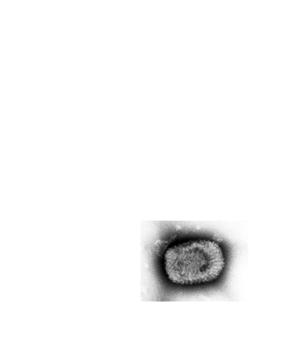
Orthopoxviruses are oval, brick-shaped particles
with a geometrically corrugated outer surface. Their
size ranges from 220 nm to 450 nm long and 140 nm
Fig. 11-1. A transmission electron micrograph of a tissue
section containing variola viruses.
Photograph: Courtesy of FA Murphy, University of Texas
Medical Branch, Galveston, Texas.
217
Smallpox and Related Orthopoxviruses
to 260 nm wide. The outer envelope consists of a lipo-
protein layer embedding surface tubules and enclosing
a core described as biconcave because of an electron
microscopy fixation artifact. The core contains the viral
DNA and core fibrils, and it is surrounded by the core
envelope and a tightly arranged layer of rod-shaped
structures known as the palisade layer. Between the
palisade layer and the outer envelope are two oval
masses known as the lateral bodies (Figure 11-2). Two
infectious forms of orthopoxviruses (described next)
result from the replication cycle.
Phylogenetic Relationships
The evolutionary relationships among the poxvi-
ruses have been facilitated by the recent availability
of complete DNA sequences for over 30 species. Phy-
logenetic analysis reveals that variola and camelpox
viruses are more closely related to each other than
any other members of the genus, and vaccinia is most
closely related to cowpox virus strain GRI-90.
13,14
Cowpox virus strain GRI-90 appears to be less closely
related to cowpox virus strain Brighton, indicating that
at least two separate species are included under the
name cowpox virus. Monkeypox virus does not group
closely with any other orthopoxvirus, which indicates
that it diverged from the rest of the genus members
long ago. Yet vaccination prevents monkeypox. Minor
modifications to the camelpox virus genome might
result in a virus with variola attributes. Virulence or
attenuation may hinge on a few genetic determinants.
For example, variola major (associated with a 30%
fatality rate) and variola minor ( < 1% fatality rate)
are greater than 98% identical over the length of the
185,000-kilobase (kb) genome.
As anticipated from the genomic homologies,
members of the Orthopoxvirus genus are antigenically
related. Serum absorption and monoclonal antibody
studies have identified cross-reacting and species-
specific neutralizing antigens.
15
Nine neutralizing
epitopes have been identified among the intracellular
Fig. 11-2. Thin section of smallpox virus growing in the cy-
toplasm of an infected chick embryo cell of infected person.
Intracellular mature virions (brick-shaped) and immature
virions (spherical) are visible. Magnification is approximately
x 25,000.
Photograph: Courtesy of FA Murphy, University of Texas
Medical Branch, Galveston, Texas.
TABLE 11-1
POXVIRUSES THAT CAUSE HUMAN DISEASE
Genus
Species
Animal Reservoir
Orthopoxvirus
Variola virus
None
Vaccinia virus
Unknown (none?)
Cowpox virus
Rodents
Monkeypox virus
Rodents
Parapoxvirus
Bovine popular stomatitis virus
Cattle
Orf virus
Sheep
Pseudocowpox virus
Cattle
Seal parapoxvirus
Seals
Parapoxvirus
Tanapox
Rodents (?)
Yabapox virus
Monkeys (?)
Molluscipoxvirus
Molluscum contagiosum virus
None
218
Medical Aspects of Biological Warfare
mature virion (IMV) particles of different species of
orthopoxviruses
16
; additional epitopes, believed to
be critical in protection against infection in vivo, ex-
ist on extracellular enveloped viral particles.
17,18
Viral
envelope proteins are important in protective antibody
responses: envelope antigens were absent from virion
suspensions used for inactivated smallpox vaccines
that proved to be ineffective.
19,20
Replication
Orthopoxvirus genomes are linear, double-stranded
DNA approximately 200 kb long. The genomes encode
about 176 to 266 proteins, including enzymes and fac-
tors that are necessary for self-replication and matura-
tion. The central region of the genome contains highly
conserved genes that are essential for viral replication,
and the terminal regions contain less conserved genes
that are important for virus-host interactions. The vi-
rus contains a number of virus-encoded enzymes, in
particular a DNA-dependent RNA polymerase that
transcribes the viral genome.
21
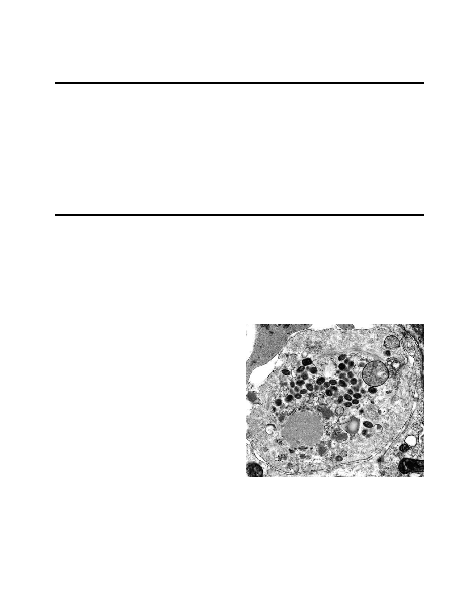
Replication occurs in
cytoplasmic factories referred to as B-type inclusions,
in which virions at various stages of assembly are seen.
Whether host cell nuclear factors are involved in viral
replication or maturation is unclear. Cells infected
with some poxviruses (eg, cowpox, avian poxviruses)
also contain electron-dense A-type inclusions, usually
containing mature virions; A-type inclusions are easily
seen by light microscopy (Figure 11-3).
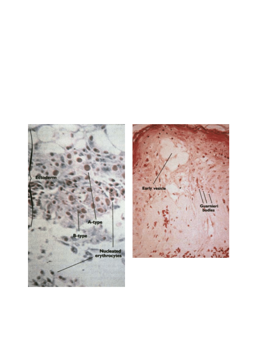
a
b
Fig. 11-3. Cytoplasmic inclusion bodies in cells infected with
orthopoxviruses. (a) B-type (pale-red, irregular) inclusion, or
Guarnieri, bodies, and A-type (large eosinophilic, with halo)
inclusion bodies in ectodermal cells of the chorioallantoic
membrane, in a pock produced by cowpox virus. A number
of nucleated erythrocytes are in the ectoderm and free in the
mesoderm, and the surface of the pock is ulcerated. Hematoxylin-eosin stain. (b) This section of the skin of a patient with
hemorrhagic-type smallpox shows Guarnieri bodies and free erythrocytes below an early vesicle. Hematoxylin-eosin stain.
Reproduced with permission from Fenner F, Henderson DA, Arita I, Jezek Z, Ladnyi ID. Smallpox and Its Eradication. Geneva,
Switzerland: World Health Organization; 1988: 85.

219
Smallpox and Related Orthopoxviruses
Viral replication begins with attachment of viral
particles to the host cell surface, most likely through
cell receptors, and involves expression of early, in-
termediate, and late genes.
21
Initial uncoating occurs
during entry, followed by synthesis of early mRNAs,
which are translated to facilitate further uncoating and
transcription of intermediate mRNAs. Intermediate
mRNAs, in turn, are translated to allow transcription
of the late mRNAs. The late mRNAs are translated into
structural and nonstructural proteins of the virions.
These proteins, along with DNA concatemers that
are formed during the early phase of replication, are
assembled into genomic DNA and packaged into im-
mature virions, which then evolve into brick-shaped
infectious IMVs. IMVs are infectious only when they
are released by cell lysis. IMV particles, which can
acquire a second membrane from an early endosomal
component to form the intracellular enveloped virion
(IEV), migrate to the cell surface via microtubules and
fuse with the cell membrane to form cell-associated
virions (CEVs). CEVs induce polymerization of actin
to form filaments that affect the direct transfer of CEVs
to adjacent cells. If CEVs become dissociated from the
cell membranes, they are called extracellular envel-
oped virions (EEVs). Although IMVs are produced
in greatest abundance in cell culture and are the most
stable to environmental degradation, CEVs and EEVs
probably play a more critical role in cell-to-cell spread
in the intact animal.
22
Many of the Orthopoxvirus gene products, known as
virokines and viroceptors, interact with and modulate
essential functions of the host cells and immune pro-
cesses.
21,23
The limited host range of variola may relate
to the unique association of viral gene products with
various host signaling pathways. Therefore, strategies
that block such key pathways in the replication and
maturation of poxviruses provide potential targets for
therapeutic intervention.
24
Pathogenesis
Most knowledge about smallpox pathogenesis is
inferred from animal studies of mousepox,
25,26
rab-
bitpox,
26
and monkeypox
27,28
in their respective hosts,
and from vaccinia in humans. Studies using primates
infected with variola
29
corroborate these findings and
lend further insight into human smallpox and monkey-
pox infections. In both natural and experimental infec-
tions, the virus is introduced via the respiratory tract,
where it first seeds the mucous membranes, including
membranes of the eye, and then passes into local lymph
nodes. The first round of replication occurs in the lymph
nodes, followed by a transient viremia, which seeds tis-
sues, especially those of the reticuloendothelial system,
including regional lymphatics, spleen, and tonsils. A
second, brief viremia transports the virus to the skin
and to visceral tissues immediately before the prodro-
mal phase. In humans, the prodrome is characterized
by an abrupt onset of headache, backache, and fever,
and usually sore throat resulting from viral replication
in the oral mucosa. Characteristic skin lesions develop
following viral invasion of the capillary epithelium of
the dermal layer. The virus may also be present in urine
and conjunctival secretions.
30
At death, most visceral
tissues contain massive virus concentrations.
In a review of all pathology reports published in
English over the past 200 years,
31
Martin suggested
that generally healthy patients who died of smallpox
usually died of renal failure, shock secondary to vol-
ume depletion, and difficulty with oxygenation and
ventilation as a result of viral pneumonia and airway
compromise, respectively. Degeneration of hepatocytes
might have caused a degree of compromise, but liver
failure was not usually the proximate cause of death.
Much of the pathogenesis of smallpox remains
a mystery because of the limited tools that were
available when it was an endemic disease. Detailed
analysis of the pathophysiology of the disease course
using the monkeypox and variola primate models and
in comparison with limited clinical and pathology
data from human smallpox victims suggests a role
for dysregulation of the immune response involv-
ing the production of proinflammatory cytokines,
lymphocyte apoptosis, and the development of co-
agulation abnormalities. High viral burdens, which
were identified in numerous target tissues in the
animal models, were probably associated with organ
dysfunction and multisystem failure. Immunohisto-
chemistry studies showing the distribution of viral
antigens as well as electron microscopy evidence of
the replicating virus correlated with pathology in the
lymphoid tissues, skin, oral mucosa, gastrointestinal
tract, reproductive system, and liver. Apoptosis was
a prominent observation in lymphoid tissues, with
a striking loss of T cells observed. The cause of this
widespread apoptosis remains unknown. However,
strong production of proinflammatory cytokines at
least in part likely contributed to the upregulation
of various proapoptotic genes. The strong upregula-
tion of cytokines may also have contributed to the
development of a hemorrhagic diathesis. The detec-
tion of D-dimers and other changes in hematologic
parameters in monkeys that developed classical or
hemorrhagic smallpox suggests that activation of the
coagulation cascade is a component of both disease
syndromes. In human populations, however, the oc-
currence of hemorrhagic smallpox was approximately
1% to 3% of the total cases observed.

220
Medical Aspects of Biological Warfare
From these recent studies of variola and monkeypox
virus infection in primates, the “toxemia” described by
clinicians for human smallpox
2
may be fundamentally
related to the processes underlying septic shock.
32
Common denominators include lymphocyte apopto-
sis; proinflammatory cytokines (exuberant production
of type I interferon [IFN], interleukin-6, tumor necrosis
factor-α, and IFN-γ measurable in plasma); and dis-
seminated intravascular coagulation. Aberrant acti-
vation of these pathways, which contributes to toxic
shock, is a hallmark of pathological activation of the
innate immune system.
To facilitate viral replication, orthopoxviruses gen-
erally modulate their host’s immune response to the
pathogen’s advantage. Poxviruses encode proteins that
target or interrupt the natural inflammatory response
and interfere with apoptosis, synthesis of steroids, and
initiation of the complement system. In general, these
proteins block either extracellular immune signals (by
mimicking or interfering with cytokine/chemokine
proteins and/or receptors), or they work intracellularly
by interfering with apoptosis, targeting by the immune
system, or intracellular immune cell signaling. A com-
bination of these mechanisms may allow the virus to
overcome immunological surveillance and establish
clinical disease in the host.
33
ORTHOPOXVIRUSES AS BIOLOGICAL WARFARE AND BIOTERRORISM THREATS
Using variola virus in warfare is an old concept. Brit-
ish colonial commanders used blankets from smallpox
victims as a biological weapon, distributing them among
Native Americans.
34-36
During the American Civil War,
allegations were made about the use of smallpox as a
biological weapon, although no definite evidence ex-
isted.
37,38
In the years leading up to and during World
War II, the Japanese military explored weaponization of
smallpox during the operations of Unit 731 in Mongolia
and China. More recently, the former Soviet Union de-
veloped smallpox as a strategic weapon and produced
ton quantities of liquid smallpox on a continuing basis
well into the 1980s.
10,39
The former Soviet Union also
conducted open air testing of weaponized smallpox
virus and demonstrated that infectious virus could drift
15 km downwind and infect humans.
7
Although declared stocks of smallpox virus exist
only at the two WHO repositories (the Centers for Dis-
ease Control and Prevention [CDC] in Atlanta, Georgia,
USA, and at the State Research Center of Virology and
Biotechnology/Vector in Koltsovo, Russia), it is of
concern that undeclared stocks may exist in military
sites within the former Soviet Union, or that they were
transferred from the Soviet program to programs in
Iraq, Iran, North Korea, or elsewhere.
39
The probability
that such stocks exist is impossible to assess, but the
catastrophic consequences of smallpox release in a
biological attack cannot be discounted.
4
Variola is a significant threat for use as a biological
weapon because of its stability, infectivity in aerosol
form, small infectious dose, severe disease manifesta-
tions, and interhuman transmissibility. Furthermore,
the anticipated morbidity and mortality for the general
population may be higher than historical averages
because of waning immunity following vaccinations
in the distant past and immunosuppression resulting
from HIV, cancer, organ transplants, and old age.
3
Oth-
er members of the Orthopoxvirus genus share many of
variola’s properties and are potential agents of a delib-
erate bioterrorist attack. Of the poxviruses other than
variola, monkeypox virus presents the greatest threat
for biological warfare or terrorism use. Monkeypox
can naturally produce severe disease in humans that
closely resembles smallpox, with mortality exceeding
15% in some outbreaks.
40
The disease is transmitted
from person to person, is highly transmissible by aero-
sol and, in at least some nonhuman primate models,
has an infectious dose as low as one tissue culture
infecting dose (TCID
50
).
27,41-43
Monkeypox virus, like
variola, is relatively stable and can resist desiccation
in both heat and cold.
44
The monkeypox virus also can
grow to high titers in cell culture systems, including the
chick chorioallantoic membrane of embryonated eggs,
a simple methodology described in older microbiol-
ogy texts using equipment and supplies available at
agricultural supply stores. A large dose of monkeypox
delivered by aerosol can produce a rapidly progressive
and overwhelming pneumonia in nonhuman primate
models.
28
Monkeypox virus may have already been
weaponized by the Soviet military.
10
Cowpox and buffalopox produce limited cutaneous
disease in humans in natural infection.
45
Buffalopox,
like cattlepox, may be essentially identical to vaccinia.
46
The effect of altering route of delivery, dose of virus,
or the actual viral agent itself on human disease mani-
festation is unclear. Several studies demonstrate that
orthopoxviruses produce different clinical syndromes
and immunological responses in animal models de-
pending on the route of infection.
28,47-51
Aerosol infec-
tion has the potential to produce more pronounced pul-
monary disease.
28,42,52
In addition, all orthopoxviruses
share a significant amount of homology with variola
and monkeypox.
14
If the critical virulence factors for
systemic human disease were found, then cowpox,

221
Smallpox and Related Orthopoxviruses
buffalopox, or other orthopoxviruses potentially could
be genetically modified to express these critical factors.
When designed as a weapon and delivered by aerosol,
these viruses could have significant impact in humans,
even without genetic modification.
Camelpox rarely, if ever, causes disease in humans.
However, because of Iraqi admissions of research with
camelpox as part of the country’s biological warfare
program, some concern exists over its potential use as
a biological weapon.
53
Camelpox virus is the closest
relative of variola virus; the major difference between
camelpox virus and variola strain Bangladesh-1975
genomes is four additional insertions, elongated
inverted terminal repeats, and a small area of gene
rearrangement present in camelpox virus.
13
As with
other orthopoxviruses, slight modifications in the
camelpox virus genome might dramatically change
its pathogenicity in humans. Although prohibited by
US law, genetic modification of camelpox would be
a likely starting point by any group that wanted to
construct variola based on published sequences. In
addition, it may soon be technically feasible to create
infectious variola using an oligonucleotide synthesizer,
analogous to the recent demonstration for creation of
the much simpler polio virus.
54
The possibility of genetically engineered ortho-
poxviruses remains unknown in biodefense research.
Studies have shown increased mousepox and vaccinia
virus virulence in mouse models by the incorporation
of cloned host cytokine genes into the virus genome.
55,56
Whether these results represent findings unique to
the virus-host model used or reflect a more general
premise of enhanced virulence is unclear.
57,58
The pos-
sibility of similar genetic engineering only increases
the threat of orthopoxviruses that are not significant
natural threats for human disease. Further research is
warranted to ensure that present and future counter-
measures are effective with modified viruses.
CLINICAL ASPECTS OF ORTHOPOXVIRUS INFECTIONS
Smallpox
Variola virus is stable and retains its infectivity for
long periods outside the host.
59
Variola virus is infec-
tious by aerosol,
3
but natural airborne spread other than
among close contacts is unusual.
60,61
Approximately
30% of susceptible contacts became infected during the
era of endemic smallpox,
62
and the WHO eradication
campaign was predicated upon the requirement of close
person-to-person proximity for reliable transmission
to occur. Nevertheless, two hospital outbreaks dem-
onstrated that the variola virus can be spread through
airborne dissemination in conditions of low relative
humidity.
63
The patients in these outbreaks were infec-
tious from the onset of their eruptive exanthem, most
commonly from days 3 through 6 after fever onset. If
the patient had a cough, then chances of infection were
greatly increased. Indirect transmission via contami-
nated bedding or other fomites was infrequent.
64
Some
people in close contact with patients harbored virus in
their throats without developing disease and may have
been a means of secondary transmission.
65,66
After exposure to aerosolized virus, variola trav-
els from the upper or the lower respiratory tract to
regional lymph nodes, where it replicates and gives
rise to viremia, which is followed by a rash.
67
The in-
cubation period of smallpox averages 12 days (range
9–14 days). Those in contact with infected patients are
quarantined for a minimum of 16 to 17 days follow-
ing exposure.
67
Following infection via the respiratory
route and replication in local lymph nodes, variola
virus disseminates systemically to other lymphoid
tissues, spleen, liver, bone marrow, and lung. During
this asymptomatic, prodromal period, variola virus
can be recovered from the blood, but the yield is lower
than later in the illness. Clinical manifestations begin
acutely with malaise, fever, rigors, vomiting, head-
ache, and backache; 15% of patients develop delirium.
Approximately 10% of light-skinned patients exhibit
an erythematous rash during this phase. After 2 to 3
more days, an enanthem appears concomitantly with
a discrete rash about the face, hands, and forearms.
Because of the lack of a keratin layer on mucous mem-
branes, lesions shed infected epithelial cells and give
rise to infectious oropharyngeal secretions in the first
few days of the eruptive illness, and occasionally 24
hours before eruption.
68
These respiratory secretions
are the most significant but not the sole means of virus
transmission. Following subsequent eruptions on the
lower extremities, the rash spreads centrally during
the next week to the trunk. Lesions quickly progress
from macules to papules and eventually to pustular
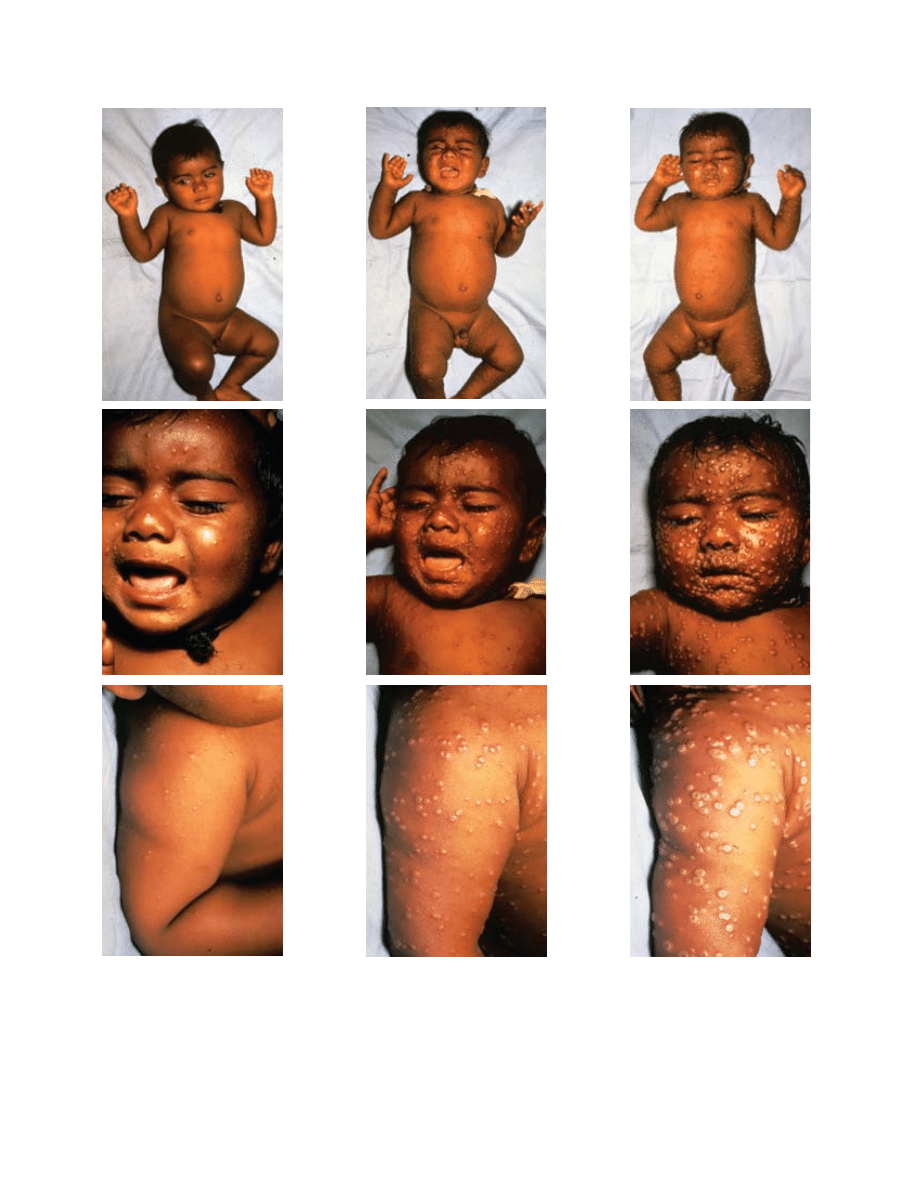
vesicles (Figure 11-4). Lesions are more abundant on
the extremities and face, and this centrifugal distribu-
tion is an important diagnostic feature. In contrast
to the lesions seen in varicella, smallpox lesions on
various segments of the body remain generally syn-
chronous in their stage of development. From 8 to 14
days after onset, the pustules form scabs, which leave
depressed depigmented scars on healing. Although
variola titers in the throat, conjunctiva, and urine di-
minish with time,
67
virus can readily be recovered from
222
Medical Aspects of Biological Warfare
Fig. 11-4. This series of photographs illustrates the evolution of skin lesions in an unvaccinated infant with the classic form
of variola major. (a) The third day of rash shows synchronous eruption of skin lesions; some are becoming vesiculated. (b)
On the fifth day of rash, almost all papules are vesicular or pustular. (c) On the seventh day of rash, many lesions are umbili-
cated, and all lesions are in the same general stage of development. Reproduced with permission from Fenner F, Henderson
DA, Arita I, Jezek Z, Ladnyi ID. Smallpox and Its Eradication. Geneva, Switzerland: World Health Organization; 1988: 10–14.
Photographs by I Arita.
b
a
c
223
Smallpox and Related Orthopoxviruses
scabs throughout convalescence.
69
Therefore, patients
should be isolated and considered infectious until all
scabs separate.
Two distinct forms of smallpox were recognized in
the last century of smallpox occurrence. Variola ma-
jor, the highly virulent, prototypical, and historically
significant form of the disease, remained prevalent
in Asia and parts of Africa during the 20th century.
Variola minor was distinguished by milder systemic
toxicity and more diminutive pox lesions.
2
However,
Dixon reported many cases that were indistinguishable
from variola major in his extensive comparison of le-
sion types.
70
Korte first described variola minor, found
in Africa, in 1904.
2
Chapin found a similar mild form
known as alastrim that occurred in North America as
early as 1896 and subsequently was exported to South
America, Europe, and Australia. Two distinct viral
strains of reduced virulence caused variola minor and
alastrim, and both typically caused 1% mortality in
unvaccinated victims.
2
The Rao classification specified five clinical pre-
sentations of variola.
71
Three quarters of variola major
cases were designated classic or ordinary type (see
Figure 11-4). After prodromal fever and constitutional
symptoms appeared, patients developed the typical
variola rash, centrifugal in distribution, with synchro-
nous progression from macules to papules, to vesicles
to pustules, and then to scabs. The fatality rate was
3% in vaccinated and 30% in unvaccinated patients.
Other clinical presentations of smallpox occurred
less frequently, probably because of the difference in
host immune response. Flat-type smallpox, noted in
2% to 5% of smallpox patients, was characterized by
both severe systemic toxicity and the slow evolution
of flat, soft, focal skin lesions that did not resemble
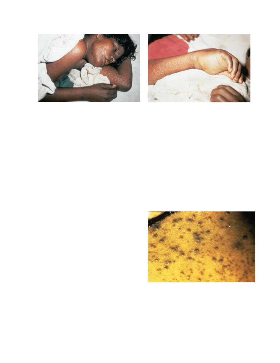
the classical variola exanthem (Figure 11-5). This syn-
drome caused 66% mortality in vaccinated patients
and 95% mortality in unvaccinated patients. Fewer
than 3% of smallpox patients developed hemorrhagic-
type smallpox, which was accompanied by extensive
petechiae (Figure 11-6), mucosal hemorrhage, and
intense toxemia; death usually occurred before typi-
cal pox lesions developed.
72
However, on occasions
hemorrhagic smallpox also occurred in the classic
type later in the disease. Both hemorrhagic-type and
flat-type smallpox may have indicated underlying im-
Fig. 11-5. Flat-type smallpox in an unvaccinated woman on the sixth day of rash. Extensive flat lesions (a and b) and systemic
toxicity with fatal outcome were typical. Reproduced with permission from Fenner F, Henderson DA, Arita I, Jezek Z, Ladnyi
ID. Smallpox and Its Eradication. Geneva, Switzerland: World Health Organization; 1988: 33. Photographs by F Dekking.
b
a
Fig. 11-6. Early hemorrhagic-type smallpox with cutaneous
signs of hemorrhagic diathesis. Death usually intervened
before the complete evolution of pox lesions. Reproduced
with permission from Herrlich A, Munz E, Rodenwaldt E.
Die pocken; Erreger, Epidemiologie und klinisches Bild. 2nd ed.
Stuttgart, Germany: Thieme; 1967. In: Fenner F, Henderson
DA, Arita I, Jezek Z, Ladnyi ID. Smallpox and Its Eradication.
Geneva, Switzerland: World Health Organization; 1988: 35.

224
Medical Aspects of Biological Warfare
munodeficiency; hemorrhagic forms occurred more
commonly in pregnant women and young children.
73
The modified type, which occurred typically but not
exclusively in previously vaccinated individuals,
was characterized by moderation of constitutional
symptoms, typically reduced numbers of lesions, and
rapid evolution of lesions, with scabs formed by the
9th day of the illness. The variola sine eruptione was
characterized by prodromal fever and constitutional
symptoms. These patients, most of whom had been
vaccinated, never developed a rash.
71
In actuality, the
manifestations of variola infection fall along a spec-
trum, and classification is primarily for the purpose
of prognosis.
Bacterial superinfection of pox lesions was rela-
tively common in the preantibiotic era, especially in
the absence of proper hygiene and medical care and
in tropical environments.
2
Arthritis and osteomyelitis
developed late in the disease in about 1% to 2% of
patients, occurred more frequently in children, and
often manifested as bilateral joint involvement, par-
ticularly of the elbows.
74
Viral inclusion bodies could
be demonstrated in the joint effusion and bone marrow
of the involved extremity. Cough and bronchitis were
occasionally reported as prominent manifestations of
smallpox, with implications for spread of contagion;
however, pneumonia was unusual.
2
Pulmonary edema
occurred frequently in hemorrhagic-type and flat-type
smallpox. Orchitis was noted in approximately 0.1%
of patients. Encephalitis developed in 1 in 500 cases of
variola major, compared with 1 in 2,000 cases of variola
minor. Keratitis and corneal ulcers were important
complications of smallpox, progressing to blindness
in slightly fewer than 1% of cases. Disease during
pregnancy precipitated high perinatal mortality, and
congenital infection was also recognized.
Partial immunity caused by vaccination resulted
in modified-type smallpox, in which sparse skin le-
sions evolved variably, often without pustules, and
quickly, with crusting occurring as early as the 7th
day of illness. When exposed to smallpox, some fully
immune individuals developed fever, sore throat, and
conjunctivitis (called contact fever), which lasted sev-
eral days but did not give rise to the toxicity or minor
skin lesions that signify variola sine eruptione. Persons
who recovered from smallpox possessed long-lasting
immunity, although a second attack may have occurred
in 1 in 1,000 persons after an intervening period of 15
to 20 years.
75
Both humoral and cellular responses are
important components of recovery from infection.
Neutralizing antibodies peak 2 to 3 weeks following
onset and last longer than 5 years,
76
up to several de-
cades in some individuals.
18
Monkeypox
The clinical features of human monkeypox are clas-
sically described as being similar to those of smallpox.
77
Disease begins with a 2- to 4-day disruptive phase with
high fever and prostration. The rash develops and
progresses synchronously over 2 to 4 weeks, evolving
from macules to papules, to vesicles and pustules, to
scabs. Lesions are usually umbilicated, have a centrifu-
gal distribution, and involve the palms and soles. Sore
throat and frank tonsillitis frequently occur during
the eruptive phase of human monkeypox.
77,78
Lymph-
adenopathy is a common finding that differentiates
monkeypox from smallpox. Lymphadenopathy, which
has been documented in up to 83% of unvaccinated
persons with monkeypox, arises most frequently early
in the course of infection, involving the submandibular
and cervical nodes and less frequently the axillary and
inguinal nodes.
Clinical manifestations of human monkeypox are
likely more diverse and not as stereotypical as those
of smallpox. Mild infections were frequent in the first
recognized African cases, with 14% of patients having
fewer than 25 lesions and no incapacity.
77
In a series
of 282 patients, the exanthema first appeared some-
where other than the face in 18% of the vaccinated
patients; 31% of vaccinated patients had pleomorphic
or “cropping” appearance of rash lesions, and 9.4%
had centripetal distribution.
79
All of these features are
inconsistent with a mimic of smallpox. Patients in the
recent US outbreak tended to have fewer mild lesions
than most African patients. Patients were hospitalized
in only 19 of 78 suspected cases in the United States,
and only 2 had significant illness requiring some
form of medical intervention.
80,81
None of the initial
cases was suspected as a smallpox-like disease. A sine
eruptione form of monkeypox has not been described,
but the number of serologically diagnosed infections
without consistent rash illness suggests that it is a pos-
sibility.
82
A hemorrhagic form of human monkeypox
has not been documented.
83,84
Complications of monkeypox are more common in
unvaccinated persons and children.
85
During intensive
surveillance in the Democratic Republic of the Congo
between 1980 and 1986, secondary bacterial superinfec-
tion of the skin was the most common complication
(19.2% of unvaccinated patients), followed by pul-
monary distress/pneumonia (11.6% of unvaccinated
patients), vomiting/diarrhea/dehydration (6.8% of
unvaccinated patients), and keratitis (4.4% of unvac-
cinated patients). With the exception of keratitis, the
incidence of these complications in vaccinated persons
was at least 3-fold less. Alopecia has been noted in

225
Smallpox and Related Orthopoxviruses
some cases.
86
Encephalitis was detected in at least
one monkeypox case in the Democratic Republic of
the Congo and in one of the cases in the US outbreak
of 2003.
79,81
As in smallpox, permanent pitted scars are
often left after scabs separate.
Severity of disease and death is related to age
and vaccination status, with younger unvaccinated
children faring worse.
77,86-88
The case fatality rate in
Africa varied in different outbreaks and periods of
increased surveillance. The fatality rate was 17% from
1970 through 1979, 10% from 1981 through 1986, and
1.5% from 1996 through 1997.
40
No fatalities occurred
among 78 suspected cases in the recent US outbreak.
80
The presence of comorbid illnesses, such as measles,
malaria, or diarrheal disease, may have a significant
impact on mortality in children.
85
Cause of death in
monkeypox is not universally clear, although 19 of 33
fatalities in one series of patients involved pulmonary
distress or bronchopneumonia, suggesting superim-
posed bacterial pneumonia.
Other Orthopoxviruses Infecting Humans
Cowpox is primarily a localized, cutaneous dis-
ease.
45
Baxby, Bennett, and Getty reviewed 54 cases
of cowpox infection with a detailed discussion of
clinical manifestations.
89
Disease usually consists
of single pock-like lesions on the hands or face,
although multiple lesions are seen in roughly one
quarter of cases. Typical lesions progress from mac-
ule to papule to vesicle to pustule to dark eschar,
with a hemorrhagic base being common in the late
vesicular stage. Progression from macule to eschar is
slow, often evolving over 2 to 3 weeks. Local edema,
induration, and inflammation are common and can be
pronounced. Lesions are painful and are accompanied
by regional lymphadenopathy. Complete healing and
scab separation usually occur within 6 to 8 weeks of
onset, but may take 12 weeks or longer. A majority
of patients experience some constitutional symptoms
before the eschar stage.
The majority of human cowpox infections are self-
limited and without complication. Ocular involve-
ment, including the cornea, can occur, but it usually
resolves without permanent damage. A few severe
generalized cowpox infections have been reported,
including one fatality.
89,90
Three of these four described
cases included a history of atopic dermatitis, indicat-
ing a risk of increased severity of disease analogous
to vaccinia.
Buffalopox infection in humans has not been ex-
tensively described. Limited data suggest that human
infection usually occurs on the hands and consists of
inflamed and painful pustular lesions progressing
through a Jennerian evolution.
91-93
Regional lymphade-
nopathy and fever can accompany local disease.
93
DIAGNOSIS
Clinical Diagnosis
The clinical presentation of smallpox is similar to
many vesicular and pustular rash illnesses, including
varicella, herpes simplex, drug reactions, and erythema
multiforme. Although the index of suspicion for an
eradicated disease may be low, the failure to recognize a
case of smallpox could result in the exposure of hospital
contacts and the seeding of an outbreak. The Smallpox
Diagnosis and Evaluation page on the CDC Web site
(http://www.bt.cdc.gov/agent/smallpox/diagnosis) is
an essential resource to assist a clinician in evaluating a
febrile patient presenting with a rash. This site contains
an algorithm to quickly determine the likelihood of clini-
cal smallpox and a standardized worksheet to classify
the risk of smallpox using the CDC criteria.
Laboratory Diagnosis
Collection of appropriate specimens is paramount
for accurate laboratory diagnosis of Orthopoxvirus
infection. For virological diagnosis, specimens from
skin lesions are most important, because when viremia
does occur in Orthopoxvirus infections, it is an early
phenomenon.
2
Ideally, cutaneous tissue and blood are
sent for diagnostic testing, with other samples being
sent at the request of public health officials or experts in
the field.
84
Detailed instructions for specimen collection
can be found in the Department of Defense Smallpox
Response Plan (http://www.bt.cdc.gov/agent/small-
pox/response-plan/index.asp) or on the CDC Web
site (http://www.cdc.gov/ncidod/monkeypox/di-
agspecimens.htm). Briefly, vesicles or pustules should
be unroofed, the detached vesicle skin sent in a dry
tube, and the base of the lesion scraped to make a
touch-prep on a glass slide. Biopsy specimens should
be split (if possible) and sent in formalin and in a dry
tube. If scabs are collected, two scabs should be sent
in a dry tube. Dacron or polyester swabs should be
used for oropharyngeal swabs and transported in dry
tubes. Blood should be collected in a marble-topped
or yellow-topped serum separator tube (which is
then centrifuged to separate serum) and in a purple-
topped anticoagulant tube for whole blood. Clinical

226
Medical Aspects of Biological Warfare
specimens potentially containing orthopoxviruses
other than variola virus, including monkeypox virus,
may be handled in a biosafety level 2 using biosafety
level 3 practices.
94
Many phenotypic and genotypical methods involv-
ing virological, immunological, and molecular ap-
proaches have been used to identify Orthopoxvirus.
Phenotypic Diagnosis
In the past, a presumptive diagnosis of orthopox-
viruses required a laboratory with capabilities and
expertise in viral diagnostics. Microscopists with
experience in poxvirus infections can often recognize
the characteristic inclusion bodies (Guarnieri bodies,
corresponding to B-type poxvirus inclusions [see Fig-
ure 11-3]) in tissue samples under light microscopy.
These cytoplasmic inclusions are hematoxylinophilic,
stain reddish purple with Giemsa stain, and contain
Feulgen-positive material.
95
Microscopy alone cannot
differentiate members of the Orthopoxvirus genus, yet
the epidemiological setting can suggest which species
is involved. The orthopoxviruses with pathogenic-
ity for humans (with the exception of molluscum
contagiosum) can be grown on the chorioallantoic
membranes of 12-day-old embryonated chicken eggs,
where they form characteristic pocks. These viruses
also grow readily in easily obtained cell cultures,
including VERO,
96
other monkey kidney cell lines,
A549, and others. Variola could characteristically
be differentiated from other viruses by a strict tem-
perature cut-off at 39
°
C. Methods for isolation and
identification of individual virus species have been
reviewed.
97,98
Electron microscopy reveals the unmis-
takable brick-like morphology of orthopoxviruses
in thin sections of infected materials. Immunogold
stains permit more precise identification to the spe-
cies level.
Immunodiagnosis
Serologic testing for anti-Orthopoxvirus antibodies is
an old technique, and various assays were used exten-
sively in the study of smallpox.
2
However, significant
serologic cross-reactivity exists between all the Ortho-
poxvirus species; therefore, species differentiation is not
possible with conventional serologic assays. Techniques
developed in the 1980s to detect monkeypox-specific
antibodies are complex and considered unreliable by
some experts.
82,99
Although complement-fixation tests
detect antibodies that disappear within 12 months of
infection, other traditional techniques, such as immuno-
fluorescence assay, radioimmunoassay, enzyme-linked
immunosorbent assay (ELISA), hemagglutination-inhi-
bition and neutralization assay, detect immunoglobulin
(IgG) antibodies that are persistent. Thus, differentiat-
ing antibodies due to acute infection from antibodies
resulting from prior vaccination can be difficult with
single specimens.
Immunofluorescence assays and ELISAs have been
used to detect IgM in acute infection directed against
cowpox and monkeypox, respectively.
90,99
Because IgM
seems to disappear within 6 months, IgM ELISAs can
be used to detect recent infections when virus detection
is not possible after lesions have healed and scabs have
separated. In the investigation of the 2003 US monkey-
pox outbreak, the CDC relied on anti-Orthopoxvirus
IgG and IgM ELISAs for serologic diagnosis.
81
More
recently, a combination of T-cell measurements and a
novel IgG ELISA was used to enhance epidemiological
follow-up studies to this outbreak.
100,101
Nucleic Acid Diagnosis
The molecular diagnostic approaches, including
DNA sequencing, polymerase chain reaction (PCR),
restriction fragment-length polymorphism (RFLP),
real-time PCR, and microarrays, are more sensitive and
specific than the conventional virological and immu-
nological approaches. Of these techniques, sequencing
provides the highest level of specificity for species or
strain identification, but current sequencing techniques
are not yet as practical as rapid diagnostic tools in
most laboratories. RFLP analysis
102,103
and microarray
genotyping
104
also provide high levels of specificity,
and when combined with PCR, these approaches
can offer high levels of sensitivity. Real-time PCR
methods provide exquisite levels of sensitivity and
specificity.
105
The basic concept behind real-time PCR
is the measurement, by fluorescence detection, of the
amount of nucleic acids produced during every cycle
of the PCR. Several detection chemistries, such as the
intercalating dyes (SYBR Green, Applied Biosystems,
Foster City, Calif), Hydrolysis probes (5’ nuclease or
Taqman, Minor Groove Binding Proteins [MGBP]),
Hybridization probes (Fluorescence Resonance Energy
Transfer [FRET]) and molecular beacons, are used.
There are several commercially available instruments
for real-time PCR, such as the ABI—7900 (Applied
Biosystems), Smart Cycler (Cepheid, Synntvale, Calif),
LightCycler (Roche Diagnostics Corporation, India-
napolis, Ind), MJ Opticon (Bio-Rad, Hercules, Calif),
RotorGene (Corbett Life Science, Sydney, Australia);
RAPID (Idaho Technology, Salt Lake City, Utah);
and others. When combined with portable analytical
platforms such as the Smart Cycler or LightCycler,
real-time PCR systems can be readily deployed to field
sites for rapid testing.

227
Smallpox and Related Orthopoxviruses
Successful performance of PCR-based diagnostics
requires extraction of DNA from body fluid and tissue
samples, careful design of oligonucleotide primers
and probes, and optimization of amplification and
detection conditions. There are numerous commercial
nucleic acid purification methods for various sample
types, which involve cell lysis and protein denatur-
ation followed by DNA precipitation or fractionation
by reversible binding to an affinity matrix. Selection of
appropriate primers, probes, and optimization of assay
conditions require knowledge of genome sequences
and molecular biology techniques.
One of the basic techniques used in PCR-based
diagnostics is gel analysis, in which PCR-amplified
regions of the genome are separated on agarose gels
by electrophoresis, and the amplicon sizes are used
to identify the sample. Several PCR gel-analysis
assays have been used to identify cowpox, mon-
keypox, vaccinia, and variola viruses from clinical
specimens.
98,106-108
Large fragment PCR-RFLP (LPCR-RFLP) analysis
requires amplifying large DNA fragments with high-
fidelity DNA polymerase enzymes. The amplified
LPCR products are purified on agarose gels and di-
gested with a restriction enzyme. The digested DNA
fragments are then electrophoresed on polyacrylamide
gels for a constant period at constant voltage and
stained with ethidium bromide. The restriction pattern
is then visualized and photographed with a digital
camera. The positions for all DNA fragments in each
restriction pattern are determined and digitized by
appropriate fingerprinting software. From this pattern,
a similarity coefficient is calculated for every pair of
restriction patterns and used as an index for species
differentiation.
Recently developed real-time PCR assays, which
can be performed in a few hours, can test clinical
specimens for all orthopoxviruses or for specific spe-
cies such as vaccinia, variola, or monkeypox.
105,109-111
Real-time PCR was one of the diagnostic techniques
used in the investigation of the 2003 US monkeypox
outbreak.
81
Because of its sensitivity, rapidity, and ease,
real-time PCR will likely become the primary method
of preliminary diagnosis of Orthopoxvirus infection,
with isolation and growth in a high-level containment
laboratory reserved for confirmation.
MEDICAL MANAGEMENT
Prophylaxis
Vaccination
History. Attempts to use infected material to induce
immunity to smallpox date to the first millennium;
the Chinese used scabs or pus collected from mild
smallpox cases to infect recipients usually via inser-
tion of bamboo splinters into the nasal mucosa. This
procedure produced disease in a controlled situation
that was typically milder than naturally occurring
disease and allowed for isolation or controlled expo-
sure of nonimmune individuals. The practice spread
to India and from there to Istanbul, where Europeans
encountered it in the early 18th century. In Europe the
inoculation of the skin with infected pock material
was later referred to as variolation to distinguish the
procedure from vaccination. Inducing immunity using
variola-contaminated materials had been known to the
British Royal Medical Society through Joseph Lister’s
reports from China as early as 1700, but the procedure
was not practiced until Lady Mary Wortley Montagu,
wife of the British ambassador to Turkey, introduced it
to British society. Lady Montagu, who had been badly
disfigured from smallpox, had her son inoculated in
Constantinople in 1717 and subsequently arranged for
surgeon Charles Maitland to inoculate her daughter in
1722. In the British American colonies, Cotton Mather
of Boston persuaded Dr Zabdiel Boylston to conduct
variolation on 224 people in 1721 after reading about
inoculation in a Royal Medical Society publication.
70
During a smallpox outbreak in Boston in 1752, over
2,000 persons underwent variolation, resulting in a
90% reduction in mortality among the population im-
munized. During the Revolutionary War, the Canadian
Campaign failed largely because the American rein-
forcements contracted smallpox. Continued problems
with recurring smallpox epidemics among recruits to
the Continental Army resulted in a directive in 1779
for variolation of all new recruits. General Washington,
who had undergone variolation himself as a young
man, was the first military commander to order im-
munization of his forces.
112
The practice of variolation, which was never widely
accepted, was outlawed at times because many of
those inoculated developed grave clinical illness.
Variolation often caused a 1% to 2% mortality rate,
and the individuals who died had the potential to
transmit natural smallpox. Edward Jenner overcame
problems of inoculation with variola by capitalizing
on the long-held observation that milkmaids had clear
complexions (without smallpox scars), presumably
because they had had cowpox, which causes milder
disease in humans. Folklore maintained that human
infection with cowpox conferred lifelong immunity to
smallpox. In 1796 Jenner scientifically demonstrated

228
Medical Aspects of Biological Warfare
that inoculation with material obtained from a milk-
maid’s cowpox lesions would result in immunity and
protection from infection with smallpox when intro-
duced by inoculation. Jenner published his findings in
1798, and in 1801 he reported that 100,000 persons had
been vaccinated in England. By the 1820s vaccination
had become widespread throughout Britain and much
of Europe. Although derivation of current vaccinia
strains is uncertain, it is not a form of cowpox, and
because Jenner lost his original material used for vac-
cination, the specific source of current vaccinia strains
remains unknown.
70
The United States began regulat-
ing production of the vaccine in 1925. Since then, the
New York City Board of Health strain of vaccinia has
been used as the primary US vaccine strain. The WHO
global vaccination program eventually led to smallpox
eradication, with the last serially transmitted smallpox
case reported in 1977. Routine vaccination of children
in the United States ceased in 1971, and vaccination
of hospital workers ceased in 1976. Vaccination of
military personnel was continued because of Cold War
concerns about its intentional use but eventually halted
in 1989. Because of the risk of bioterrorism, smallpox
vaccination in at-risk military personnel and civilian
healthcare workers was resumed in 2003.
113,114
During the WHO global eradication program, most
of the human population received vaccinia virus by
scarification. Although there were multiple manufac-
turers worldwide, and vaccine lots varied with respect
to potency and purity, almost all vaccinia administered
was derived from one of two lineages, the New York
Board of Health and Lister strains.
2
Live vaccinia
virus suspension was placed as a drop on the skin or
drawn up by capillary action between the tines of a
bifurcated needle; the nominal dose of live vaccinia
was about 10
5
virions. Usually, primary vaccination is
uneventful; following introduction into the skin, the
virus replicates in basal layer keratinocytes, spreads
cell-to-cell, and leads to discrete vesicle formation.
Within a week, the vesicle evolves into a pustule sur-
rounded by inflammatory tissue. This lesion scabs over
within 10 to 14 days; eventually, the scab is shed. Vac-
cinees in the global campaign often experienced ten-
der axillary lymph nodes, fever, and malaise for brief
periods. Occasionally, however, complications arose
with varying degrees of severity. Accidental transfer
of vaccinia from the inoculation site was common,
but of little consequence unless transferred to the eye.
Generalized vaccinia, which involved systemic spread
of the virus and eruption of multiple pocks at distant
sites, was more serious; in individuals with eczema or
atopic dermatitis, however, it sometimes led to exten-
sive inflammation and secondary bacterial infection.
More serious, life-threatening complications arose in
vaccinees with defects in cell-mediated immunity; the
vaccination site frequently enlarged to form an ulcer,
secondary ulcers appeared, and the infection cleared
slowly or not at all. The most serious event was post-
vaccinial encephalitis. Although rare, this condition
was frequently fatal. Death occurred in approximately
one in one million primary vaccinations.
115,116
Adverse
events may be more frequent and severe if mass immu-
nization were to be resumed in an unscreened general
population that now includes transplant recipients on
immunosuppressive drugs, HIV-infected individuals,
and geriatric patients.
Recent Vaccination Campaigns. The requirement
that any alternative vaccine must not be inferior to live
vaccinia sets a high standard. The successful immuni-
zation or “take rate” has been greater than 95%, both
historically and in a more recent series of over 450,000
military vaccinees.
113
In this recent series, one case of
encephalitis and 37 cases of myopericarditis were
documented in a prescreened, healthy, young adult
population. Although the incidence of myopericarditis
was below the historical average and the cases were
mild, this adverse event contributed to the general re-
luctance of the civilian healthcare population to accept
vaccination.
114
A potential replacement vaccinia was
prepared in massive quantities (> 300 million doses)
by selection of plaque-purified progeny virus from the
New York Board of Health strain, which was amplified
in VERO cell cultures. This vaccine is more purified
and free of adventitious agents in comparison with its
predecessor, which was prepared on calf skin. Phase I
safety and immunogenicity trials for ACAM 2000 in-
dicate greater than 95% take rates and adverse events
comparable to those of live vaccinia.
117
Historically, live
(replicating) vaccinia immunization has also been used
as postexposure prophylaxis and is believed effective
if administered within 4 days of exposure.
The recent immunization of modest numbers of
military and civilian individuals has provided an op-
portunity to study the nature of adverse events using
modern tools of immunology. A strong association
was established between adverse events and increased
systemic cytokines, in particular, IFN-γ, tumor ne-
crosis factor-α, interleukin-5, and interleukin-10.
118
Some researchers have speculated that cardiac events,
although rare, may be related to dramatic alterations
in cytokine profiles.
Protective immunity elicited by live vaccinia is
thought to depend on a combination of humoral and
cellular immune responses. Using a monkey model in
which animals are immunized with vaccinia and chal-
lenged with monkeypox, Edghill-Smith has shown that
vaccinia-specific B cells are critical for protection.
119
An-
tibody depletion of B cells, but not CD4
+
or CD8
+
T cells,
abrogated vaccinia-induced protection. Edghill-Smith
has also shown that simian-immunodeficiency-virus–

229
Smallpox and Related Orthopoxviruses
compromised monkeys could withstand vaccinia if it
was preceded by a dose of nonreplicating Modified
Vaccinia Ankara (MVA) strain vaccinia, but they were
not protected against monkeypox challenge when their
CD4
+
T-cell counts were less than 300 mm.
3.
MVA is an alternative vaccine that has promise as a
nonreplicating immunogen. MVA, which was used in
Germany in the later stages of global eradication, was
shown to be safe and immunogenic, but its protective
efficacy has not been established in humans. MVA was
generated by over 500 serial passages in chick embryo
fibroblasts, which resulted in multiple deletions and
mutations and an inability to replicate efficiently in
human and most other mammalian cells.
120
Ultrastruc-
tural examination of purified MVA reveals that most of
the particles are enveloped; the host restriction occurs
at a late stage of maturation. The presence of enveloped
particles is believed to be important to the elicitation
of protective immunity. Experimentally, MVA was
demonstrated to protect monkeys against a monkey-
pox virus challenge, after one or two doses of MVA
or MVA followed by Dryvax (Wyeth Laboratories,
Marietta, Pa).
121
Surprisingly, a single dose of MVA also
protected when challenge followed immunization by
as little as 10 days, although protection was not abso-
lute; a modest number of pocks and a low-level viremia
occurred in the MVA recipients following challenge.
Rhesus monkeys were used in a similar intravenous
challenge model to evaluate a DNA vaccine strategy,
a combination of four genes (L1R, A27L, A33R, and
B5R) with promising results.
122
The smallpox vaccine used in the United States is
Dried, Calf Lymph Type (Dryvax), a live-virus prepara-
tion of the New York Board of Health vaccinia strain
prepared from calf lymph. The calf lymph is purified,
concentrated, and lyophilized. The diluent for the
vaccine contains 50% glycerin and 0.25% phenol in
US Pharmacopeia sterile water, with no more than 200
bacterial organisms per milliliter in the reconstituted
product (Polymyxin B sulfate, dihydrostreptomycin
sulfate, chlortetracycline hydrochloride, and neomycin
sulfate are used in the processing of the vaccine, and
therefore small amounts of these antibiotics may be
present in the final product).
Vaccination is performed with a bifurcated needle
onto which the reconstituted vaccinia preparation
has been drawn, using three intradermal jabs for im-
munologically naïve individuals (new vaccinees) or
15 jabs for prevaccinated individuals, with enough
strength to produce a visible trace of bleeding. The
resulting vaccination lesion is then kept covered with
a nonadherent and nonimpervious dressing. Care
must be taken to prevent inadvertent inoculation of
the vaccinee or others. In primary vaccinees, a papule
forms within 5 days, developing into a vesicle on the
5th or 6th day postvaccination, which signifies a major
reaction, or take. The vesicle subsequently becomes
pustular, swelling subsides, and a crust forms, which
comes off in 14 to 21 days. At the height of the primary
reaction, known as the Jennerian response, regional
lymphadenopathy usually occurs, which may be ac-
companied by systemic manifestations of fever and
malaise. Primary vaccination with vaccine at potency
of 100 million pock-forming units per milliliter elicits
a 97% response rate both by major reaction and neu-
tralizing antibody response. Allergic sensitization to
viral proteins can persist so that the appearance of
a papule and redness may occur within 24 hours of
revaccination, with vesicles occasionally developing
within 24 to 48 hours. This allergic response peaks
within 3 days and does not constitute a “major reac-
tion or take.” Immunological response occurring after
3 days is an accelerated but otherwise similar appear-
ance of papule, vesicle, and/or pustule to that seen
in the primary vaccination response. Revaccination is
considered successful if a vesicular or pustular lesion
or an area of definite palpable induration or congestion
surrounding a central lesion (scar or ulcer) is present
on examination at 6 to 8 days after revaccination.
Outcome. Successful smallpox vaccination provides
high-level immunity for the majority of recipients for 3
to 5 years followed by decreasing immunity. In Mack’s
review of importations cases in Europe from 1950
through 1972, he provided epidemiological evidence
of some relative protection from death, if not from dis-
ease severity, in individuals who had been immunized
over 20 years before exposure. However, for the older
population in particular, vaccination within 10 years of
exposure did not prevent all cases but did prevent some
smallpox deaths.
123
Multiple vaccinations are thought to
produce more long-lasting immunity. Vaccination has
been effective in preventing disease in 95% of vaccinees.
124
Vaccination also was shown to prevent or substantially
lessen the severity of infection when given as secondary
prophylaxis within a few days of exposure.
2
Contraindications. Smallpox vaccination is contrain-
dicated in the preoutbreak setting for individuals with
the following conditions or those having close contact
with individuals with the following conditions:
•
a history of atopic dermatitis (eczema);
•
active acute, chronic, or exfoliative skin condi-
tions that disrupt the epidermis;
•
pregnancy or the possibility of becoming
pregnant; or
•
a compromised immune system as a conse-
quence of HIV infection, acquired immuno-
deficiency syndrome, autoimmune disorders,
cancer, radiation treatment, immunosuppres-
sive therapy, or other immunodeficiencies.

230
Medical Aspects of Biological Warfare
Additional relative contraindications for potential
vaccinees, but not close contacts, are smallpox vac-
cine-component allergies, moderate or severe acute
intercurrent infections, topical ophthalmologic steroid
medications, age younger than 18, and maternal breast-
feeding. A history of Darier’s disease and household
contact with active disease are contraindications for
vaccination.
6
Adverse Events. Vaccinia can be transmitted from a
vaccinee’s unhealed vaccination site to other persons
by close contact and the same adverse events as with
intentional vaccination can result. To avoid inadver-
tent transmission, vaccinees should wash their hands
with soap and water or use antiseptic hand rubs im-
mediately after touching the vaccination site and after
dressing changes. Vaccinia-contaminated dressings
should be placed in sealed plastic bags and disposed
in household trash.
125
Adverse reactions to smallpox vaccination are diag-
nosed by a clinical examination. Most reactions can be
managed with observation and supportive measures.
Self-limited reactions include fever, headache, fatigue,
myalgia, chills, local skin reactions, nonspecific rashes,
erythema multiforme, lymphadenopathy, and pain at
the vaccination site. Adverse reactions that require fur-
ther evaluation and possible therapeutic intervention
include inadvertent inoculation involving the eye,
126
generalized vaccinia, eczema vaccinatum, progressive
vaccinia, postvaccinial central nervous system disease,
and fetal vaccinia.
6
Inadvertent inoculation generally results in a condi-
tion that is self-limited unless it involves the eye or eye-
lid, which requires an ophthalmologist’s evaluation.
Topical treatment with trifluridine (Viroptic, Glaxo/
Smith/Kline, Brentford, Middlesex, United Kingdom)
or vidarabine (Vira-A, King Pharmaceuticals, Bristol,
Tenn) is often recommended, although treatment of
ocular vaccinia is not specifically approved by the Food
and Drug Administration for either of these drugs.
Most published experience is with use of vidarabine,
but this drug is no longer manufactured.
127
Generalized vaccinia is characterized by a dissemi-
nated maculopapular or vesicular rash, frequently on
an erythematous base and typically occurring 6 to 9
days after primary vaccination. Treatment with vac-
cinia immune globulin (VIG) is restricted to those who
are systemically ill or have an immunocompromising
condition or recurrent disease that can last up to a year.
Contact precautions should be used to prevent further
transmission and nosocomial infection.
6
Eczema vaccinatum occurs in individuals with a his-
tory of atopic dermatitis, regardless of current disease
activity, and can be a papular, vesicular, or pustular
rash. This rash may be generalized, or localized with
involvement anywhere on the body, with a predilection
for areas of previous atopic dermatitis lesions. Mortal-
ity ranges from 17% to 30% and is reduced by use of
VIG. Contact precautions should be used to prevent
further transmission and nosocomial infection.
6
Progressive vaccinia is a rare, severe, and often fatal
complication of vaccination that occurs in individuals
with immunodeficiency conditions and is character-
ized by painless progressive necrosis at the vaccination
site with or without metastases to distant sites. This
condition carries a high mortality rate; therefore, pro-
gressive vaccinia should be aggressively treated with
VIG, intensive monitoring, and tertiary medical center
level support. Persons with the following conditions
are at the highest risk:
•
congenital or acquired immunodeficiencies;
•
HIV infection/acquired immunodeficiency
syndrome;
•
cancer;
•
autoimmune disease;
•
immunosuppressive therapy; or
•
organ transplant.
Anecdotal experience has shown that despite treat-
ment with VIG, individuals with cell-mediated immu-
nity defects have a poorer prognosis than those with
humoral defects. Infection control measures should
include contact and respiratory precautions to prevent
transmission and nosocomial infection.
6
Central nervous system disease, which includes
postvaccinial encephalopathy and postvaccinial
encephalomyelitis, occurs rarely after smallpox vac-
cination. Postvaccinial encephalopathy occurs more
frequently, typically affects infants and children younger
than age 2, and reflects vascular damage to the central
nervous system. Symptoms that typically occur 6 to
10 days postvaccination include seizures, hemiplegia,
aphasia, and transient amnesia. Histopathologic find-
ings include cerebral edema, lymphocytic meningeal
inflammation, ganglion degeneration, and perivascular
hemorrhage. Patients with postvaccinial encephalopa-
thy who survive can be left with cerebral impairment
and hemiplegia. Postvaccinial encephalomyelitis affects
individuals who are age 2 or older and is characterized
by abrupt onset of fever, vomiting, malaise, and anorexia
occurring approximately 11 to 15 days postvaccination.
Symptoms progress to amnesia, confusion, disorienta-
tion, restlessness, delirium, drowsiness, and seizures.
The cerebral spinal fluid has normal chemistries and
cell count. Histopathology findings include demyeliza-
tion and microglial proliferation in demyelinated areas,
with lymphocytic infiltration but without significant
edema. The cause for central nervous system disease

231
Smallpox and Related Orthopoxviruses
is unknown, and no specific therapy exists. Therefore,
intervention is limited to anticonvulsant therapy and
intensive supportive care. Fetal vaccinia, which results
from vaccinial transmission from mother to fetus, is a
rare but serious complication of smallpox vaccination
during or immediately before pregnancy.
6
In the Department of Defense 2002–2003 vaccination
program involving 540,824 vaccinees, 67 symptomatic
cases of myopericarditis were reported, for a rate of
1.2 per 100,000. Mean time from vaccination to evalu-
ation for myopericarditis was 10.4 days, with a range
of 3 to 25 days. Reports of myocarditis in vaccinees in
2003 raised concerns of carditis and cardiac deaths in
individuals undergoing smallpox vaccination. That
year, 21 cases of myo/pericarditis of 36,217 vaccinees
were reported, with 19 (90%) occurring in revaccinees.
The median age of those affected was 48, and they were
predominantly women. Eleven of the individuals were
hospitalized, but there were no fatalities. Of the 540,824
total vaccinees over the 2 years, 449,198 were military
personnel (the rest were civilians), and of these there
were 37 cases, for an occurrence rate of 1 per 120,000
vaccinees.
112
Ischemic cardiac events including fatali-
ties have also been reported as a consequence of the
use of vaccinia vaccine (Dryvax) during the campaign.
Although no clear association has been found, history
of ischemic heart disease and significant cardiac risk
pose relative contraindications for smallpox vaccina-
tion. Consequently, individuals with a history of myo-
carditis, pericarditis, or ischemic heart disease should
refrain from vaccination.
128,129
Smallpox Biothreat Policy. In a smallpox release
from a bioterrorist event, individuals would be vac-
cinated according to the current national policy, which
recommends initial vaccination of higher risk groups
(individuals directly exposed to the release and those
with close contact to smallpox patients) and medical
and emergency transport personnel. Vaccination of the
general population would then be extended in concen-
tric rings around the initial cases to impede the spread.
There are no absolute contraindications to vaccination
for individuals with high-risk exposure to smallpox.
Persons at greatest risk of complications of vaccina-
tion are those for whom smallpox infection poses the
greatest risk. If relative contraindications exist for an
individual, the risks must be weighed against the risk
of a potentially fatal smallpox infection.
Postexposure prophylaxis with vaccine offers pro-
tection against smallpox but is untried in other Or-
thopoxvirus diseases.
2
Despite a lack of hard evidence,
postexposure vaccination is likely efficacious against
other orthopoxviruses, and during the 2003 US mon-
keypox outbreak the CDC recommended vaccination
of potentially exposed persons.
80
Treatment
Passive Immunization
VIG is available from the CDC as an investigational
new drug in two formulations, intramuscular and
intravenous. VIG may be beneficial in treating some
of the adverse effects associated with vaccination. VIG
has no proven benefit in smallpox treatment, and its
efficacy in treatment of monkeypox infections is un-
known. Monoclonal antibodies have been shown to be
beneficial in animal models under certain conditions,
but this concept has not yet been sufficiently developed
for efficacy testing in humans.
Antiviral Drugs
Antiviral drugs would be useful for treatment of
orthopoxviral diseases including smallpox and mon-
keypox, as well as adverse effects associated with vac-
cination. The only antiviral drug available for treating
orthopoxviruses is cidofovir, which may be offered
under emergency use protocols maintained by both
the Department of Health and Human Services and
the Department of Defense.
The elaborate replication strategy of poxviruses
offers a number of potential targets for therapeutic
intervention.
130
Although inhibition of viral replica-
tion may be necessary to halt the pathogenic disease
course, it may not be sufficient—it may also be neces-
sary to reverse the effects of the mounting damage
that increasingly appears to be the result of a cytokine
storm, which accounts for the “toxicity” of systemic
orthopoxvirus infection.
29
In this regard, cytokine an-
tagonists developed to treat bacterial sepsis and other
conditions may play a role in effective management of
smallpox- and monkeypox-infected patients.
Initial studies to identify effective antiviral agents
for orthopoxviruses tested drugs developed for other
viruses that share molecular targets with poxviruses.
131
The effort to discover effective drugs against DNA
viruses initially focused on treatment of herpesviruses
infections. The discovery of acyclovir led to practical
therapy and a better understanding of the importance
of viral and cellular enzymes involved in phosphoryla-
tion of acyclovir to acyclovir triphosphate, the active
chemical entity. The failure of acyclovir to inhibit
cytomegalovirus was because, unlike the thymidine
kinase of herpes simplex, cytomegalovirus thymidine
kinase lacked the appropriate specificity, which was
overcome by synthesis of a series of phosphorylated
analogues using a stable phosphonate bond. The most
promising candidate using this approach was cidofo-
vir, which is a dCMP analog.
132
Cidofovir is licensed

232
Medical Aspects of Biological Warfare
for treatment of cytomegalovirus-associated retinitis
under the trade name Vistide (Gilead Sciences Inc,
Foster City, Calif), and may inhibit the cytomegalo-
virus DNA polymerase, a target shared with the pox-
viruses. Cidofovir also may inhibit the activity of the
proofreading exonuclease, leading to error-prone DNA
synthesis during poxvirus replication. Cidofovir has
been demonstrated to protect monkeys against severe
disease in both the monkeypox and authentic smallpox
primate models, when administered within 48 hours of
intravenous exposure to the virus.
133
Although the drug
formulation used in these studies has been criticized
for requiring intravenous administration, patients
with advanced disease would already be receiving
intravenous fluids as part of their supportive care,
and once weekly cidofovir administration would not
significantly increase the healthcare burden. Because
cidofovir has been associated with nephrotoxicity,
primarily in dehydrated patients, careful attention to
fluid management is important, and patient hydration
and coadministration of probenecid is required.
Oral formulations of cidofovir analogues with
better bioavailability and lower toxicity, designed to
overcome the lack of an active transport pathway for
unmodified cidofovir into cells, are under develop-
ment.
134
Cidofovir requires bolus dosing to allow drug
entry into cells by pinocytosis; however, bolus dosing
results in transiently high concentrations in the kidney.
The primary design paradigm for oral formulations is
the creation of a lipid mimic that allows drugs to enter
cells via the chylomicron pathway.
135
This formula-
tion dramatically reduced transient drug levels in the
kidney and eliminated nephrotoxicity in toxicology
studies using mice. However, an oral formulation of
cidofovir is not available for human use.
The first drug used to empirically treat progressive
vaccinia and smallpox was Marboran, a compound
of the class of N-aminomethyl-isatin-beta-thiosemi-
carbazones. As with most early treatment strategies,
controlled clinical trials were not reported, and recent
studies show that Marboran was only capable of
inhibiting replication by 80% at maximum tolerated
concentration in VERO cells.
136
Through combinatorial
chemistry, potent and more selective compounds have
now been discovered and are in preliminary testing.
137
A number of essential viral enzymes have been target-
ed using a homology-based bioinformatics approach,
such as that used to develop a structural model of vac-
cinia virus I7L proteinase. A unique chemical library
of 51,000 compounds was computationally queried
to identify potential active site inhibitors.
138
A subset
of compounds was assayed for toxicity and ability to
inhibit vaccinia replication, and a family was identi-
fied with 50% minimal inhibitory concentrations of 3
to 12 µM. Alternatively, a high-throughput screening
approach using cowpox virus evaluated a collection of
over 250,000 compounds and identified several potent
lead structures for optimization and evaluation against
vaccinia, monkeypox, and variola viruses. From this
effort ST-246 {4-trifluoromethyl-N-(3,3a,4,4a,5,5a,6,6a-
octahydro-1,3-dioxo-4,6-ethenocycloprop[f]isoindol-
2(1H)-yl)-benzamide} was identified and is under de-
velopment. ST-246 is both potent (EC
50
= 0.010 µM),
selective (CC
50
> 40 mM), and active against multiple
orthopoxviruses, including monkeypox, camelpox,
cowpox, ectromelia (mousepox), vaccinia, and variola
viruses in vitro and monkeypox, variola, cowpox, vac-
cinia, and ectromelia in vivo.
Alternative approaches include peptide mimetics of
IFN-γ that play a direct role in the activation of STAT
1 alpha transcription factor.
139
These mimetics do not
act through recognition of the extracellular domain of
the IFN-γ receptor; rather, they bind to the cytoplas-
mic domain of the receptor chain and thereby initiate
the cellular signaling. The authors hypothesize that
mimetics would bypass the poxvirus virulence factor
B8R protein that binds the intact IFN-γ and would
prevent interaction with its receptor. Experimentally,
these mimetics, but not intact IFN-γ, inhibited replica-
tion of vaccinia in BSC-40 cells. Thus these mimetics
can avoid the B8R virulence factor and have potential
activity against poxviruses in vivo.
Gleevec (Novartis Pharmaceuticals Corporation,
East Hanover, NJ), a drug licensed for use in chronic
myeloid leukemia, has been shown to block the egress
of vaccinia virus from infected cells.
140
Smallpox virus
includes an epidermal-growth-factor–like domain
that targets human Erb-1, inducing tyrosine phos-
phorylation of certain host cell substrates, thereby
facilitating viral replication. Poxviruses migrate to
the cell membrane via the polymerization of actin
tails to produce EEV, which facilitates viral dis-
semination. The authors reason that low molecular
weight inhibitors of Erb-1 kinases might function as
antiviral agents. CI-1033, one such inhibitor, blocked
variola replication in BSC-40 and Vero cells, primar-
ily at the level of secondary viral spreading. CI-1033
protected mice exposed to a lethal vaccinia challenge
via the aerosol route. In conjunction with a monoclo-
nal antibody directed against L1R, CI-1033 cleared
the mice’s lungs of virus within 8 days. Gleevec is
also a small molecule that inhibits the Abl-1 family
of tyrosine kinases, thereby inhibiting the release of
EEV from infected cells. Gleevec inhibited the vac-
cinia virus spread from the mouse peritoneum to
the ovaries and protected the mice from all lethal
intranasal challenge. The advantage of Gleevec over
other tyrosine kinase inhibitors such as CI-1033 is that

233
Smallpox and Related Orthopoxviruses
it is already approved for human use. The potential
success of Gleevec suggests that strategies that block
key host signaling pathways have merit and augment
the approaches that target classical viral replication
enzymes. An alternative approach to inhibiting the
polymerization of actin, which in turn inhibits the
propulsion of viral particles along actin filaments
toward the cell membrane, is small interfering RNA
directed against the Arp2/3
141
complex.
Lastly, treatment strategies may be developed
to target the toxemia or clinical manifestations of
smallpox. In particular, modulation of the systemic
immune response to orthopox infection, specifically
the prevention of organ damage caused by vascular
leakage and fibrin deposition, may provide a useful
therapeutic target. Uncontrolled or inappropriate
immune responses can contribute to multiple organ
failure and death; in this respect the “toxemia” associ-
ated with fatal orthopox infections resembles severe
sepsis. Several treatment strategies for targeting the
manifestations of septic shock,
142
such as activated
protein C and inhibitors of the tissue factor pathway,
143
are under consideration for testing in the nonhuman
primate model for smallpox.
SUMMARY
Smallpox no longer causes human disease thanks
to the dedicated efforts of public health officials who
participated in the WHO smallpox eradication pro-
gram. Although the former Soviet Union participated
in the eradication program, recent revelations have
shown that the Soviets continued developing small-
pox for biowarfare into the 1980s. The Soviet Union
is dissolved and its offensive program dismantled,
but the institutions and technology that developed
this and other offensive weapons systems remain.
Because the submission and destruction of smallpox
virus stores was a voluntary program, it cannot be
ascertained with certainty that smallpox viruses do
not exist outside US and Russian storage facilities.
Because the sequence of several variola isolates is
known to a high degree of certainty, it is technically
possible to generate viable virus either by modifi-
cation of a closely related virus such as camelpox
or chemical synthesis using increasingly powerful
automated equipment.
The potential threat from smallpox specifically
and orthopox infections in general will expand as the
technology to create these viruses becomes increas-
ingly available in laboratories around the world.
Furthermore, scientists have been successful in mak-
ing orthopoxviruses more virulent through genetic
manipulation. The biodefense community has made
considerable progress in developing new drugs for
treatment of orthopoxvirus infections and safer vac-
cines; however, much work remains. There is still no
approved treatment for smallpox, and the new safer
vaccines remain unlicensed and unavailable.
REFERENCES
1. Tucker JB. Scourge: The Once and Future Threat of Smallpox. New York, NY: Atlantic Monthly Press; 2001.
2. Fenner F, Henderson DA, Arita I, Jezek A, Ladnyi ID. Smallpox and Its Eradication. Geneva, Switzerland: World Health
Organization; 1988.
3. Henderson DA, Inglesby TV, Bartlett JG, et al. Smallpox as a biological weapon: medical and public health manage-
ment. Working Group on Civilian Biodefense. JAMA. 1999;281:2127–2137.
4. Ferguson NM, Keeling MJ, Edmunds WJ, et al. Planning for smallpox outbreaks. Nature. 2003;425:681–685.
5. Kaplan EH, Craft DL, Wein LM. Emergency response to a smallpox attack: the case for mass vaccination. Proc Natl
Acad Sci U S A. 2002;99:10935–10940.
6. Cono J, Casey CG, Bell DM. Smallpox vaccination and adverse reactions. MMWR Recomm Rep. 2003;52:1–28.
7. Zelicoff AP. An epidemiological analysis of the 1971 smallpox outbreak in Aralsk, Kazakhstan. Crit Rev Microbiol.
2003;29:97–108.
8. Centers for Disease Control and Prevention. Smallpox Response Plan and Guidelines (version 3.0), 9/21/02 Guide A. Surveil-
lance, Contract Tracing, and Epidemiological Investigation Guidelines. Atlanta, Ga: CDC; 2002.

234
Medical Aspects of Biological Warfare
9. LeDuc JW, Jahrling PB. Strengthening national preparedness for smallpox: an update. Emerg Infect Dis. 2001;7:155–157.
10. Alibek K. Biohazard: The Chilling True Story of the Largest Covert Biological Weapons Program in the World—Told From the
Inside By the Man Who Ran It. New York, NY: Random House; 1999.
11. Charatan F. US doctors investigate more than 50 possible cases of monkeypox. BMJ. 2003;326:1350.
12. Reed KD, Melski JW, Graham MB, et al. The detection of monkeypox in humans in the Western Hemisphere. N Engl
J Med. 2004;350:342–350.
13. Gubser C, Smith GL. The sequence of camelpox virus shows it is most closely related to variola virus, the cause of
smallpox. J Gen Virol. 2002;83:855–872.
14. Gubser C, Hue S, Kellam P, Smith GL. Poxvirus genomes: a phylogenetic analysis. J Gen Virol. 2004;85:105–117.
15. Baxby D. The surface antigens of orthopoxviruses detected by cross-neutralization tests on cross-absorbed antisera. J
Gen Virol. 1982;58:251–262.
16. Czerny CP, Johann S, Holzle L, Meyer H. Epitope detection in the envelope of intracellular naked orthopox viruses
and identification of encoding genes. Virology. 1994;200:764–777.
17. Vanderplasschen A, Hollinshead M, Smith GL. Antibodies against vaccinia virus do not neutralize extracellular en-
veloped virus but prevent virus release from infected cells and comet formation. J Gen Virol. 1997;78:2041–2048.
18. Viner KM, Isaacs SN. Activity of vaccinia virus-neutralizing antibody in the sera of smallpox vaccinees. Microbes Infect.
2005;7:579–583.
19. Payne LG. Significance of extracellular enveloped virus in the in vitro and in vivo dissemination of vaccinia. J Gen
Virol. 1980;50:89–100.
20. Kaplan C, Benson PF, Butler NR. Immunogenicity of ultraviolet-irradiated, non-infectious, vaccinia-virus vaccine in
infants and young children. Lancet. 1965;191:573–574.
21. Moss B. Poxviridae: the viruses and their replication. In: Knipe DM, Howley PM, Griffin DE, et al, eds. Fields Virology.
4th ed. Philadelphia, Pa: Lippincott Williams & Wilkins; 2001: 2849–2883.
22. Smith GL, Vanderplasschen A, Law M. The formation and function of extracellular enveloped vaccinia virus. J Gen
Virol. 2002;83:2915–2931.
23. Johnston JB, McFadden G. Poxvirus immunomodulatory strategies: current perspectives. J Virol. 2003;77:6093–6100.
24. McFadden G. Poxvirus tropism. Nat Rev Microbiol. 2005;3:201–213.
25. Fenner F. The clinical features and pathogenesis of mousepox (infectious ectromelia of mice). J Pathol Bacteriol.
1948;60:529–552.
26. Buller RM, Palumbo GJ. Poxvirus pathogenesis. Microbiol Rev. 1991;55:80–122.
27. Wenner HA, Macasaet FD, Kamitsuka PS, Kidd P. Monkey pox. I. Clinical, virologic and immunologic studies. Am J
Epidemiol. 1968;87:551–566.
28. Zaucha GM, Jahrling PB, Geisbert TW, Swearengen JR, Hensley L. The pathology of experimental aerosolized mon-
keypox virus infection in cynomolgus monkeys (Macaca fascicularis). Lab Invest. 2001;81:1581–1600.
29. Jahrling PB, Hensley LE, Martinez MJ, et al. Exploring the potential of variola virus infection of cynomolgus macaques
as a model for human smallpox. Proc Natl Acad Sci U S A. 2004;101:15196–15200.

235
Smallpox and Related Orthopoxviruses
30. Sarkar JK, Mitra AC, Mukherjee MK, De SK, Mazumdar DG. Virus excretion in smallpox. I. Excretion in throat, urine,
and conjunctiva of patients. Bull World Health Organ. 1973;48:517–522.
31. Martin DB. The cause of death in smallpox: an examination of the pathology record. Mil Med. 2002;167:546–551.
32. Levi M, de Jonge E, van der Poll T. Sepsis and disseminated intravascular coagulation. J Thromb. 2003;16:43–47.
33. Moss B, Shisler JL. Immunology 101 at poxvirus U: immune evasion genes. Semin Immunol. 2001;13:59–66.
34. Heagerty J. Four Centuries of Medical History in Canada. Vol 1. Toronto, Ontario, Canada: MacMillan; 1928.
35. Parkman F. The Conspiracy of Pontiac. Vol 2. Boston, Mass: Little, Brown and Company; 1969.
36. Stearn E, Stearn A. The Effect of Smallpox on the Destiny of the Amerindian. Boston, Mass: Bruce Humphries; 1945.
37. Kean RGH. Inside the Confederate Government. New York, NY: Oxford University Press; 1957.
38. Steiner P. Disease in the Civil War: Natural Biological Warfare, 1861–1865. Springfield, Ill: Charles C Thomas; 1968.
39. Miller J, Engelberg S, Broad W. Germs. Biological Weapons and America’s Secret War. New York, NY: Simon and Schuster
Inc; 2001.
40. Heymann DL, Szczeniowski M, Esteves K. Re-emergence of monkeypox in Africa: a review of the past six years. Br
Med Bull. 1998;54:693–702.
41. World Health Organization. Technical Advisory Group on Human Monkeypox. Report of a WHO Meeting. Geneva, Swit-
zerland: WHO; 1999. WHO/CDS/CSR/APH/99.5.
42. Hahon N, Mc GM. Air-borne infectivity of the variola-vaccinia group of poxviruses for the cynomolgus monkey,
Macaca irus. J Infect Dis. 1961;109:294–298.
43. Chen N, Li G, Liszewski MK, et al. Virulence differences between monkeypox virus isolates from West Africa and the
Congo basin. Virology. 2005; 340:46–63.
44. Cho CT, Wenner HA. Monkeypox virus. Bacteriol Rev. 1973;37:1–18.
45. Esposito JJ, Fenner F. Poxviruses. In: Knipe DM, Howley PM, Griffin DE, et al, eds. Fields Virology. 4th ed. Philadelphia,
Pa: Lippincott Williams & Wilkins; 2001:2885–2921.
46. Damaso CR, Esposito JJ, Condit RC, Moussatche N. An emergent poxvirus from humans and cattle in Rio de Janeiro
State: Cantagalo virus may derive from Brazilian smallpox vaccine. Virology. 2000;277:439–449.
47. Roberts JF, Coffee G, Creel SM, et al. Haemorrhagic smallpox. I. Preliminary haematological studies. Bull World Health
Organ. 1965;33:607–613.
48. Lancaster MC, Boulter EA, Westwood JC, Randles J. Experimental respiratory infection with poxviruses. II. Pathologi-
cal studies. Br J Exp Pathol. 1966;47:466–471.
49. Westwood JC, Boulter EA, Bowen ET, Maber HB. Experimental respiratory infection with poxviruses. I. Clinical viro-
logical and epidemiological studies. Br J Exp Pathol. 1966;47:453–465.
50. Mims CA. Aspects of the pathogenesis of virus diseases. Bacteriol Rev. 1964;28:30–71.
51. Martinez MJ, Bray MP, Huggins JW. A mouse model of aerosol-transmitted orthopoxviral disease: morphology of
experimental aerosol-transmitted orthopoxviral disease in a cowpox virus-BALB/c mouse system. Arch Pathol Lab
Med. 2000;124:362–377.

236
Medical Aspects of Biological Warfare
52. Hahon N. Smallpox and related poxvirus infections in the simian host. Bacteriol Rev. 1961;25:459–476.
53. Jezek Z, Kriz B, Rothbauer V. Camelpox and its risk to the human population. J Hyg Epidemiol Microbiol Immunol.
1983;27:29–42.
54. Cello J, Paul AV, Wimmer E. Chemical synthesis of poliovirus cDNA: generation of infectious virus in the absence of
natural template. Science. 2002;297:1016–1018.
55. Jackson RJ, Ramsay AJ, Christensen CD, Beaton S, Hall DF, Ramshaw IA. Expression of mouse interleukin-4 by a re-
combinant ectromelia virus suppresses cytolytic lymphocyte responses and overcomes genetic resistance to mousepox.
J Virol. 2001;75:1205–1210.
56. Robbins SJ, Jackson RJ, Fenner F, et al. The efficacy of cidofovir treatment of mice infected with ectromelia (mousepox)
virus encoding interleukin-4. Antiviral Res. 2005;66:1–7.
57. Mullbacher A, Lobigs M. Creation of killer poxvirus could have been predicted. J Virol. 2001;75:8353–8355.
58. Stanford MM, McFadden G. The ‘supervirus’? Lessons from IL-4-expressing poxviruses. Trends Immunol. 2005;26:339–345.
59. Huq F. Effect of temperature and relative humidity on variola virus in crusts. Bull World Health Organ. 1976;54:710–712.
60. Meiklejohn G, Kempe CH, Downie AW, Berge TO, St Vincent L, Rao AR. Air sampling to recover variola virus in the
environment of a smallpox hospital. Bull World Health Organ. 1961;25:63–67.
61. Downie AW, Meiklejohn M, St Vincent L, Rao AR, Sundara Babu BV, Kempe CH. The recovery of smallpox virus from
patients and their environment in a smallpox hospital. Bull World Health Organ. 1965;33:615–622.
62. Foege WH, Millar JD, Henderson DA. Smallpox eradication in West and Central Africa. Bull World Health Organ.
1975;52:209–222.
63. Wehrle PF, Posch J, Richter KH, Henderson DA. An airborne outbreak of smallpox in a German hospital and its sig-
nificance with respect to other recent outbreaks in Europe. Bull World Health Organ. 1970;43:669–679.
64. Maccallum FO, McDonald JR. Survival of variola virus in raw cotton. Bull World Health Organ. 1957;16:247–254.
65. Sarkar JK, Mitra AC, Mukherjee MK, De SK. Virus excretion in smallpox. 2. Excretion in the throats of household
contacts. Bull World Health Organ. 1973;48:523–527.
66. Sarkar JK, Mitra AC, Mukherjee MK, De SK, Mazumdar DG. Virus excretion in smallpox. 1. Excretion in the throat,
urine, and conjunctiva of patients. Bull World Health Organ. 1973;48:517–522.
67. Breman JG, Henderson DA. Diagnosis and management of smallpox. N Engl J Med. 2002;346:1300–1308.
68. Downie AW, St Vincent L, Meiklejohn G, et al. Studies on the virus content of mouth washings in the acute phase of
smallpox. Bull World Health Organ. 1961;25:49–53.
69. Mitra AC, Sarkar JK, Mukherjee MK. Virus content of smallpox scabs. Bull World Health Organ. 1974;51:106–107.
70. Dixon CW. Smallpox. London, England: Churchill; 1962.
71. Rao AR. Smallpox. Bombay, India: Kothari Book Depot; 1972.
72. Downie AW, Fedson DS, Saint Vincent L, Rao AR, Kempe CH. Haemorrhagic smallpox. J Hyg (Lond). 1969;67:619–629.
73. Rao AR, Prahlad I, Swaminathan M, Lakshmi A. Pregnancy and smallpox. J Indian Med Assoc. 1963;40:353–363.
74. Gupta SK, Srivastava TP. Roentgen features of skeletal involvement in smallpox. Australas Radiol. 1973;17:205–211.

237
Smallpox and Related Orthopoxviruses
75. Rao AR. Haemorrhagic smallpox: a study of 240 cases. J Indian Med Assoc. 1964;43:224–229.
76. Downie AW, Saint Vincent L, Goldstein L, Rao AR, Kempe CH. Antibody response in non-haemorrhagic smallpox
patients. J Hyg (Lond). 1969;67:609–618.
77. Jezek Z, Gromyko AI, Szczeniowski MV. Human monkeypox. J Hyg Epidemiol Microbiol Immunol. 1983;27:13–28.
78. Jezek Z, Marennikova SS, Mutumbo M, Nakano JH, Paluku KM, Szczeniowski M. Human monkeypox: a study of
2,510 contacts of 214 patients. J Infect Dis. 1986;154:551–555.
79. Jezek Z, Szczeniowski M, Paluku KM, Mutombo M. Human monkeypox: clinical features of 282 patients. J Infect Dis.
1987;156:293–298.
80. Updated Interim CDC Guidance for Use of Smallpox Vaccine, Cidofovir, and Vaccinia Immune Globulin (VIG) for
Prevention and Treatment in the Setting of an Outbreak of Monkeypox Infections. June 25, 2003. Available at: http://
www.cdc.gov/ncidod/monkeypox/treatmentguidelines.htm. Accessed April 25, 2007.
81. Sejvar JJ, Chowdary Y, Schomogyi M, et al. Human monkeypox infection: a family cluster in the midwestern United
States. J Infect Dis. 2004;190:1833–1840.
82. Jezek Z, Fenner F. Human Monkeypox. Vol 17. Basel, Switzerland: Karger; 1988.
83. Di Giulio DB, Eckburg PB. Human monkeypox. Lancet Infect Dis. 2004;4:199.
84. Di Giulio DB, Eckburg PB. Human monkeypox: an emerging zoonosis. Lancet Infect Dis. 2004;4:15–25.
85. Jezek Z, Grab B, Paluku KM, Szczeniowski MV. Human monkeypox: disease pattern, incidence and attack rates in a
rural area of northern Zaire. Trop Geogr Med. 1988;40:73–83.
86. Hutin YJ, Williams RJ, Malfait P, et al. Outbreak of human monkeypox, Democratic Republic of Congo, 1996 to 1997.
Emerg Infect Dis. 2001;7:434–438.
87. Jezek Z, Nakano JH, Arita I, Mutombo M, Szczeniowski M, Dunn C. Serological survey for human monkeypox infec-
tions in a selected population in Zaire. J Trop Med Hyg. 1987;90:31–38.
88. Meyer H, Perrichot M, Stemmler M, et al. Outbreaks of disease suspected of being due to human monkeypox virus
infection in the Democratic Republic of Congo in 2001. J Clin Microbiol. 2002;40:2919–2921.
89. Baxby D, Bennett M, Getty B. Human cowpox 1969-93: a review based on 54 cases. Br J Dermatol. 1994;131:598–607.
90. Pelkonen PM, Tarvainen K, Hynninen A, et al. Cowpox with severe generalized eruption, Finland. Emerg Infect Dis.
2003;9:1458–1461.
91. Lal SM, Singh IP. Buffalopox–a review. Trop Anim Health Prod. 1977;9:107–112.
92. Baxby D, Hill BJ. Characteristics of a new poxvirus isolated from Indian buffaloes. Arch Gesamte Virusforsch. 1971;35:70–79.
93. Wariyar KG. Variola in buffaloes. Indian Vet J. 1937;14:169–170.
94. US Department of Health and Human Services, Centers for Disease Control and Prevention, and National Institutes
of Health. Biosafety in Microbiological and Biomedical Laboratories (BMBL) 5th Edition. Washington, DC: US Government
Printing Office; 2007.
95. Kato S, Cutting W. A study of the inclusion bodies of rabbit myxoma and fibroma virus and a consideration of the
relationship between all pox virus inclusion bodies. Stanford Med Bull. 1959;17:34–45.
96. Artenstein AW, Johnson C, Marbury TC, et al. A novel, cell culture-derived smallpox vaccine in vaccinia-naive adults.
Vaccine. 2005;23:3301–3309.

238
Medical Aspects of Biological Warfare
97. Damon IK, Esposito JJ. Poxvirus infections in humans. In: Murray PR, Jorgensen JH, Yolken RH, Baron EJ, Pfaller MA,
eds. Manual of Clinical Microbiology. 8th ed. Washington, DC: American Society for Microbiology Press; 2003.
98. Meyer H, Damon IK, Esposito JJ. Orthopoxvirus diagnostics. Methods Mol Biol. 2004;269:119–134.
99. Karem KL, Reynolds M, Braden Z, et al. Characterization of acute-phase humoral immunity to monkeypox: use of
immunoglobulin M enzyme-linked immunosorbent assay for detection of monkeypox infection during the 2003 North
American outbreak. Clin Diagn Lab Immunol. 2005;12:867–872.
100. Hammarlund E, Lewis MW, Carter SV, et al. Multiple diagnostic techniques identify previously vaccinated individuals
with protective immunity against monkeypox. Nat Med. 2005;11:1005–1111.
101. Slifka M, Hammarlund E. Monkeypox outbreak diagnostics and implications for vaccine protective effect. Nat Med.
2006;12:496–497.
102. Loparev VN, Massung RF, Esposito JJ, Meyer H. Detection and differentiation of old world orthopoxviruses: restric-
tion fragment length polymorphism of the crmB gene region. J Clin Microbiol. 2001;39:94–100.
103. Ibrahim MS, Mellott JD. Orthopoxviruses: monkeypox, cowpox, vaccinia, camelpox, mousepox. In: Fuchs J, Podda
M, eds. Encyclopedia of Medical Genomics and Proteomics. New York, NY: Marcel Dekker, Inc; 2005: 947–952.
104. Lapa S, Mikheev M, Shchelkunov S, et al. Species-level identification of orthopoxviruses with an oligonucleotide
microchip. J Clin Microbiol. 2002;40:753–757.
105. Ibrahim MS, Kulesh DA, Saleh SS, et al. Real-time PCR assay to detect smallpox virus. J Clin Microbiol. 2003;41:3835–3839.
106. Ropp SL, Jin Q, Knight JC, Massung RF, Esposito JJ. PCR strategy for identification and differentiation of smallpox
and other orthopoxviruses. J Clin Microbiol. 1995;33:2069–2076.
107. Schupp P, Pfeffer M, Meyer H, Burck G, Kolmel K, Neumann C. Cowpox virus in a 12-year-old boy: rapid identifica-
tion by an orthopoxvirus-specific polymerase chain reaction. Br J Dermatol. 2001;145:146–150.
108. Dhar AD, Werchniak AE, Li Y, et al. Tanapox infection in a college student. N Engl J Med. 2004;350:361–366.
109. Espy MJ, Cockerill IF III, Meyer RF, et al. Detection of smallpox virus DNA by LightCycler PCR. J Clin Microbiol.
2002;40:1985–1988.
110. Egan C, Kelly CD, Rush-Wilson K, et al. Laboratory-confirmed transmission of vaccinia virus infection through sexual
contact with a military vaccinee. J Clin Microbiol. 2004;42:5409–5411.
111. Kulesh DA, Baker RO, Loveless BM, et al. Smallpox and pan-orthopox virus detection by real-time 3’-minor groove bind-
er TaqMan assays on the roche LightCycler and the Cepheid smart Cycler platforms. J Clin Microbiol. 2004;42:601–609.
112. Fenn EA. Pox Americana: The Great Smallpox Epidemic of 1775–82. New York, NY: Hill & Wang; 2001.
113. Grabenstein JD, Winkenwerder W Jr. US military smallpox vaccination program experience. JAMA. 2003;289:3278–3282.
114. Yih WK, Lieu TA, Rego VH, et al. Attitudes of healthcare workers in US hospitals regarding smallpox vaccination.
BMC Public Health. 2003;3:20.
115. Fulginiti VA, Papier A, Lane JM, Neff JM, Henderson DA. Smallpox vaccination: a review, part II. Adverse events.
Clin Infect Dis. 2003;37:251–271.
116. Lane JM, Goldstein J. Adverse events occurring after smallpox vaccination. Semin Pediatr Infect Dis. 2003;14:189–195.
117. Greenberg RN, Kennedy JS, Clanton DJ, et al. Safety and immunogenicity of new cell-cultured smallpox vaccine com-
pared with calf-lymph derived vaccine: a blind, single-centre, randomised controlled trial. Lancet. 2005;365:398–409.

239
Smallpox and Related Orthopoxviruses
118. Rock MT, Yoder SM, Talbot TR, Edwards KM, Crowe JE Jr. Adverse events after smallpox immunizations are associ-
ated with alterations in systemic cytokine levels. J Infect Dis. 2004;189:1401–1410.
119. Edghill-Smith Y, Golding H, Manischewitz J, et al. Smallpox vaccine-induced antibodies are necessary and sufficient
for protection against monkeypox virus. Nat Med. 2005;11:740–747.
120. Blanchard TJ, Alcami A, Andrea P, Smith GL. Modified vaccinia virus Ankara undergoes limited replication in
human cells and lacks several immunomodulatory proteins: implications for use as a human vaccine. J Gen Virol.
1998;79:1159–1167.
121. Earl PL, Americo JL, Wyatt LS, et al. Immunogenicity of a highly attenuated MVA smallpox vaccine and protection
against monkeypox. Nature. 2004;428:182–185.
122. Hooper JW, Thompson E, Wilhelmsen C, et al. Smallpox DNA vaccine protects nonhuman primates against lethal
monkeypox. J Virol. 2004;78:4433–4443.
123. Mack TM. Smallpox in Europe, 1950–1971. J Infect Dis. 1972;125:161–169.
124. Centers for Disease Control and Prevention. Smallpox fact sheet: Vaccine overview.
125. Centers for Disease Control and Prevention. Vaccinia (smallpox) vaccine: recommendations of the Advisory Commit-
tee on Immunization Practices (ACIP), 2001. MMWR Recomm Rep. 2001;50:1–25.
126. Lewis FM, Chernak E, Goldman E, et al. Ocular vaccinia infection in laboratory worker, Philadelphia, 2004. Emerg
Infect Dis. 2006;12:134–137.
127. Fillmore GL, Ward TP, Bower KS, et al. Ocular complications in the Department of Defense Smallpox Vaccination
Program. Ophthalmology. 2004;111:2086–2093.
128. Centers for Disease Control and Prevention. Update: cardiac-related events during the civilian smallpox vaccination
program–United States, 2003. MMWR Morb Mortal Wkly Rep. 2003;52:492–496.
129. Halsell JS, Riddle JR, Atwood JE, et al. Myopericarditis following smallpox vaccination among vaccinia-naive US
military personnel. JAMA. 2003;289:3283–3289.
130. Harrison SC, Alberts B, Ehrenfeld E, et al. Discovery of antivirals against smallpox. Proc Natl Acad Sci U S A.
2004;101:11178–11192.
131. Prichard MN, Kern ER. Orthopoxvirus targets for the development of antiviral therapies. Curr Drug Targets Infect
Disord. 2005;5:17–28.
132. Magee WC, Hostetler KY, Evans DH. Mechanism of inhibition of vaccinia virus DNA polymerase by cidofovir di-
phosphate. Antimicrob Agents Chemother. 2005;49:3153–3162.
133. Jahrling PB, Huggins JW. Orthopoxviruses. In: Swearengen JR, ed. Biodefense: Research Methodology and Animal Models.
Boca Raton, Fla: CRC Press; 2005.
134. Raulin J. Development in lipid drugs. Mini Rev Med Chem. 2005;5:489–498.
135. Painter GR, Hostetler KY. Design and development of oral drugs for the prophylaxis and treatment of smallpox infec-
tion. Trends Biotechnol. 2004;22:423–427.
136. Huggins JW. Unpublished observation, 1996.
137. Pirrung MC, Pansare SV, Sarma KD, Keith KA, Kern ER. Combinatorial optimization of isatin-beta-thiosemicarbazones
as anti-poxvirus agents. J Med Chem. 2005;48:3045–3050.

240
Medical Aspects of Biological Warfare
138. Byrd CM, Bolken TC, Mjalli AM, et al. New class of orthopoxvirus antiviral drugs that block viral maturation. J Virol.
2004;78:12147–12156.
139. Ahmed CM, Burkhart MA, Subramaniam PS, Mujtaba MG, Johnson HM. Peptide mimetics of gamma interferon
possess antiviral properties against vaccinia virus and other viruses in the presence of poxvirus B8R protein. J Virol.
2005;79:5632–5639.
140. Yang H, Kim SK, Kim M, et al. Antiviral chemotherapy facilitates control of poxvirus infections through inhibition of
cellular signal transduction. J Clin Invest. 2005;115:379–387.
141. Komano J, Miyauchi K, Matsuda Z, Yamamoto N. Inhibiting the Arp2/3 complex limits infection of both intracellular
mature vaccinia virus and primate lentiviruses. Mol Biol Cell. 2004;15:5197–5207.
142. Levi M, de Jonge E, van der Poll T. New treatment strategies for disseminated intravascular coagulation based on
current understanding of the pathophysiology. Ann Med. 2004;36:41–49.
143. Geisbert TW, Hensley LE, Jahrling PB, et al. Treatment of Ebola virus infection with a recombinant inhibitor of factor
VIIa/tissue factor: a study in rhesus monkeys. Lancet. 2003;362:1953–1958.
Wyszukiwarka
Podobne podstrony:
Bezp Państwa T 2, W 10, BW
45 06 BW Hydraulika stosowana
Ch11 Q1
51 07 BW Gospodarka wodna
factoring00wellrich bw
Cersanit wanna, Resources, Budownictwo, BUDOWNICTWO OGÓLNE, Budownictwo Ogólne I i II, Budownictwo o
Akumulator do BOMAG BW BW??L
ch11 12 wiele zm
hartalgebracou0wellrich bw
44 06 BW Budowle wodne
BW ch14
elementarypracti00doddrich bw
bw 1
CH11 2
elementsofalgebr00lillrich bw
BW ch23
IncentiveBee BW
więcej podobnych podstron