International Journal of Antimicrobial Agents 26 (2005) 343–356
Review
Antimicrobial activity of flavonoids
T.P. Tim Cushnie, Andrew J. Lamb
School of Pharmacy, The Robert Gordon University, Schoolhill, Aberdeen AB10 1FR, UK
Abstract
Flavonoids are ubiquitous in photosynthesising cells and are commonly found in fruit, vegetables, nuts, seeds, stems, flowers, tea, wine,
propolis and honey. For centuries, preparations containing these compounds as the principal physiologically active constituents have been
used to treat human diseases. Increasingly, this class of natural products is becoming the subject of anti-infective research, and many groups
have isolated and identified the structures of flavonoids possessing antifungal, antiviral and antibacterial activity. Moreover, several groups
have demonstrated synergy between active flavonoids as well as between flavonoids and existing chemotherapeutics. Reports of activity
in the field of antibacterial flavonoid research are widely conflicting, probably owing to inter- and intra-assay variation in susceptibility
testing. However, several high-quality investigations have examined the relationship between flavonoid structure and antibacterial activity
and these are in close agreement. In addition, numerous research groups have sought to elucidate the antibacterial mechanisms of action of
selected flavonoids. The activity of quercetin, for example, has been at least partially attributed to inhibition of DNA gyrase. It has also been
proposed that sophoraflavone G and (
−)-epigallocatechin gallate inhibit cytoplasmic membrane function, and that licochalcones A and C
inhibit energy metabolism. Other flavonoids whose mechanisms of action have been investigated include robinetin, myricetin, apigenin, rutin,
galangin, 2,4,2
-trihydroxy-5
-methylchalcone and lonchocarpol A. These compounds represent novel leads, and future studies may allow the
development of a pharmacologically acceptable antimicrobial agent or class of agents.
© 2005 Elsevier B.V. and the International Society of Chemotherapy. All rights reserved.
Keywords: Flavonoids; Antifungal; Antiviral; Antibacterial; Structure–activity; Mechanism of action
1. Introduction
Resistance to antimicrobial agents has become an increas-
ingly important and pressing global problem. Of the 2 million
people who acquire bacterial infections in US hospitals each
year, 70% of cases now involve strains that are resistant to at
least one drug
. A major cause for concern in the UK is
methicillin-resistant Staphylococcus aureus (MRSA), which
was at low levels a decade ago but now accounts for ca. 50% of
all S. aureus isolates
. Substantial investment and research
in the field of anti-infectives are now desperately needed if a
public health crisis is to be averted.
Structural modification of antimicrobial drugs to which
resistance has developed has proven to be an effective means
of extending the lifespan of antifungal agents such as the
azoles
, antiviral agents such as the non-nucleoside reverse
transcriptase inhibitors
, and various antibacterial agents
∗
Corresponding author. Tel.: +44 1224 262 526; fax: +44 1224 262 555.
E-mail address: a.lamb@rgu.ac.uk (A.J. Lamb).
including
-lactams and quinolones
. It is not surprising
then, that in response to antimicrobial resistance, major phar-
maceutical companies have tended to concentrate their efforts
on improving antimicrobial agents in established classes
However, with the portfolio of chemotherapeutics currently
available, it has been acknowledged that researchers are get-
ting close to the end game in terms of parent structure alter-
ations. A call has therefore been made for the development
of new classes of drug that work on different target sites to
those in current use
Rational drug design does not always yield effective
antimicrobials. In the past, potent enzyme inhibitors have
been successfully designed and synthesised but they had only
modest antibacterial activity, probably owing to the com-
plex issue of drug uptake by cells. Broad empirical screen-
ing of chemical entities for antimicrobial activity represents
an alternative strategy for the development of novel drugs.
Natural products have been a particularly rich source of
anti-infective agents, yielding, for example, the penicillins
in 1940, the tetracyclines in 1948 and the glycopeptides in
0924-8579/$ – see front matter © 2005 Elsevier B.V. and the International Society of Chemotherapy. All rights reserved.
doi:10.1016/j.ijantimicag.2005.09.002

344
T.P.T. Cushnie, A.J. Lamb / International Journal of Antimicrobial Agents 26 (2005) 343–356
1955
. The following review will examine the antimi-
crobial activity of flavonoids, a class of natural products
possessing a diverse range of pharmacological properties.
Compounds with antifungal, antiviral and antibacterial activ-
ity will each be discussed in turn, with particular emphasis
on those flavonoids with antibacterial activity.
2. Flavonoids: occurrence, functions, structure and
nomenclature
Flavonoids are ubiquitous in photosynthesising cells and
therefore occur widely in the plant kingdom
. They are
found in fruit, vegetables, nuts, seeds, stems and flowers as
well as tea, wine
, propolis and honey
, and represent
a common constituent of the human diet
. In the US,
the daily dietary intake of mixed flavonoids is estimated to
be in the range 500–1000 mg, but this figure can be as high
as several grams for people supplementing their diets with
flavonoids or flavonoid-containing herbal preparations
The function of flavonoids in flowers is to provide colours
attractive to plant pollinators
. In leaves, these com-
pounds are increasingly believed to promote physiologi-
cal survival of the plant, protecting it from, for example,
fungal pathogens and UV-B radiation
. In addition,
flavonoids are involved in photosensitisation, energy transfer,
the actions of plant growth hormones and growth regulators,
control of respiration and photosynthesis, morphogenesis and
sex determination
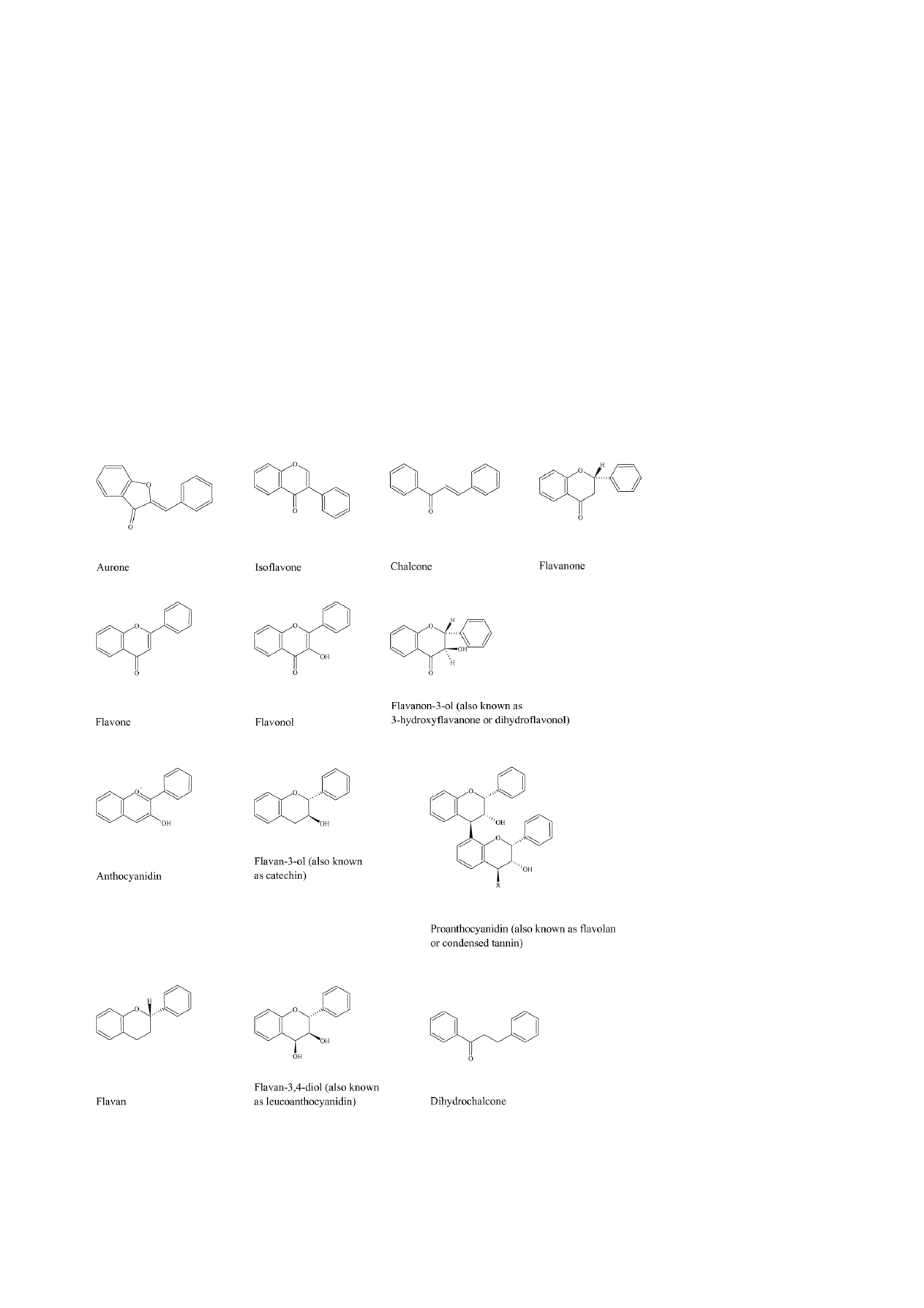
The basic structural feature of flavonoid compounds is the
2-phenyl-benzo[
␣]pyrane or flavane nucleus, which consists
of two benzene rings (A and B) linked through a heterocyclic
pyrane ring (C) (
. Flavonoids can be classified
according to biosynthetic origin. Some classes, for exam-
ple chalcones, flavanones, flavan-3-ols and flavan-3,4-diols,
are both intermediates in biosynthesis as well as end prod-
ucts that can accumulate in plant tissues. Other classes are
only known as end products of biosynthesis, for example
anthocyanidins, proanthocyanidins, flavones and flavonols.
Two additional classes of flavonoid are those in which the
2-phenyl side chain of flavanone isomerises to the 3 posi-
tion, giving rise to isoflavones and related isoflavonoids. The
Fig. 1. The skeleton structure of the flavones (a class of flavonoids), with
rings named and positions numbered
neoflavonoid is formed through further isomerisation to the 4
position
. Structures of the major classes of flavonoids are
given in
. The structures of specific compounds within
these classes that possess antimicrobial activity and that are
discussed in the present review are summarised in
Individual flavonoids may be assigned names in three dif-
ferent ways. Trivial names are employed extensively and
sometimes indicate flavonoid class or plant source. For exam-
ple, names ending in ‘inidin’ can denote an anthocyanidin,
names ending in ‘etin’ generally denote a flavonol, and
compounds tricin and hypolaetin have been extracted from
plants belonging to the genera Triticum and Hypolaena.
Flavonoids may also be named in a semi-systematic man-
ner based on trivial names such as flavone or chalcone as
the parent structure, e.g. 3,5,7,3
4
-pentahydroxyflavone or
3,3
,4
,5,7-pentahydroxyflavone. Lastly, flavonoids may be
given systematic chemical names, e.g. 3,4-dihydro-2-phenyl-
2H-1-benzopyran for flavan, but this method is cumbersome
and rarely used
. In the present review, trivial names will
be used wherever possible.
3. Medicinal properties of flavonoids
Increasingly, flavonoids are becoming the subject of medi-
cal research. They have been reported to possess many useful
properties, including anti-inflammatory activity, oestrogenic
activity, enzyme inhibition, antimicrobial activity
antiallergic activity, antioxidant activity
, vascular activ-
ity and cytotoxic antitumour activity
. For a group
of compounds of relatively homogeneous structure, the
flavonoids inhibit a perplexing number and variety of eukary-
otic enzymes and have a tremendously wide range of activi-
ties. In the case of enzyme inhibition, this has been postulated
to be due to the interaction of enzymes with different parts of
the flavonoid molecule, e.g. carbohydrate, phenyl ring, phe-
nol and benzopyrone ring
. Several reviews have been
written on the interaction between flavonoids and mammalian
cells, including comprehensive articles by Harborne and
Williams
and Middleton et al.
. An extensive review
on the biochemistry and medical significance of flavonoids
has also recently been produced by Havsteen
4. History of flavonoid use in antimicrobial treatment
For centuries, preparations that contain flavonoids as the
principal physiologically active constituents have been used
by physicians and lay healers in attempts to treat human
diseases
. For example, the plant Tagetes minuta (contain-
ing quercetagetin-7-arabinosyl-galactoside) has been used
extensively in Argentine folk medicine to treat infectious dis-
ease
. The healing properties of propolis (or ‘tzori’ in
Hebrew) are referred to throughout the Old Testament
and this balm was prescribed by Hippocrates (460–377 BC)
in Ancient Greece for the treatment of sores and ulcers
T.P.T. Cushnie, A.J. Lamb / International Journal of Antimicrobial Agents 26 (2005) 343–356
345
Fig. 2. The skeleton structures of the main classes of flavonoids: aurones
, isoflavones
, flavanones
, flavones
, flavonols
, flavanon-3-ols
, anthocyanidins
, flavan-3-ols
, proanthocyanidins (occur as dimers, trimers, tetramers and pentamers; R = 0, 1, 2 or 3
flavan-3-ol structures)
, flavans
, flavan-3,4-diols
and dihydrochalcones

346
T.P.T. Cushnie, A.J. Lamb / International Journal of Antimicrobial Agents 26 (2005) 343–356
Table 1
A summary of the structures of antimicrobial flavonoids discussed within the present review article (compiled from The Handbook of Natural Flavonoids
and individual research papers)
Compound
Substituents at carbon position:
2
3
4
5
6
7
8
2
3
4
5
6
Flavones and their glycosides
Acacetin
–
–
–
OH
–
OH
–
–
–
OCH
3
–
–
Apigenin
–
–
–
OH
–
OH
–
–
–
OH
–
–
Baicalin
–
–
–
OH
OH
OR1
–
–
–
–
–
–
Baicalein
–
–
–
OH
OH
OH
–
–
–
–
–
–
Chrysin
–
–
–
OH
–
OH
–
–
–
–
–
–
Gardenin A (demethylated)
–
–
–
OH
OH
OH
OH
–
OH
OH
OH
–
Genkwanin
–
–
–
OH
–
OCH
3
–
–
–
OH
–
–
Luteolin
–
–
–
OH
–
OH
–
–
OH
OH
–
–
Luteolin 7-glucoside
–
–
–
OH
–
OR2
–
–
OH
OH
–
–
7,8-Dihydroxyflavone
–
–
–
–
–
OH
OH
–
–
–
–
–
5,5
-Dihydroxy-8,2
,4
-trimethoxyflavone
–
–
–
OH
–
–
OCH
3
OCH
3
–
OCH
3
OH
–
5-Hydroxy-7,4
-dimethoxyflavone
–
–
–
OH
–
OCH
3
–
–
–
OCH
3
–
–
5,7,4
-Trihydroxy-3
,5
-dimethoxyflavone
–
–
–
OH
–
OH
–
–
CH
3
OH
CH
3
–
6,7,4
-Trihydroxy-3
,5
-dimethoxyflavone
–
–
–
–
OH
OH
–
–
CH
3
OH
CH
3
–
Isoflavones
6,8-Diprenylgenistein
–
–
–
OH
R3
OH
R3
–
–
OH
–
–
Sophoraisoflavone A
–
–
–
OH
–
OH
–
*
*
OH
–
–
Flavonols and their glycosides
Galangin
–
OH
–
OH
–
OH
–
–
–
–
–
–
Kaempherol
–
OH
–
OH
–
OH
–
–
–
OH
–
–
3-O-methylquercetin
–
OCH
3
–
OH
–
OH
–
–
OH
OH
–
–
Morin
–
OH
–
–
–
OH
–
OH
–
OH
OH
–
Myricetin
–
OH
–
OH
–
OH
–
–
OH
OH
OH
–
Quercetagetin
–
OH
–
OH
OH
OH
–
–
OH
OH
–
–
Quercetagetin-7-arabinosyl-galactoside
–
OH
–
OH
OH
OR4
–
–
OH
OH
–
–
Quercetin
–
OH
–
OH
–
OH
–
–
OH
OH
–
–
Quercetin-3-O-(2
-galloyl)-
␣-l-
arabinopyranoside
–
OR5
–
OH
–
OH
–
–
OH
OH
–
–
Quercetrin
–
OR6
–
OH
–
OH
–
–
OH
OH
–
–
Robinetin
–
OH
–
–
–
OH
–
–
OH
OH
OH
–
Rutin
–
OR7
–
OH
–
OH
–
–
OH
OH
–
–
3,2
-Dihydroxyflavone
–
OH
–
–
–
–
–
OH
–
–
–
–
3,6,7,3
,4
-Pentahydroxyflavone
–
OH
–
–
OH
OH
–
–
OH
OH
–
–
Flavan-3-ols
Catechin
–
OH
OH
–
–
OH
–
–
OH
–
OH
–
Epicatechin gallate
–
R8
–
OH
–
OH
–
–
OH
OH
–
–
Epigallocatechin
–
OH
–
OH
–
OH
–
–
OH
OH
OH
–
Epigallocatechin gallate
–
R8
–
OH
–
OH
–
–
OH
OH
OH
–
3-O-octanoyl-(+)-catechin
–
R9
–
OH
–
OH
–
–
OH
OH
–
–
3-O-octanoyl-(
−)-epicatechin
–
R9
–
OH
–
OH
–
–
OH
OH
–
–
Flavanon-3-ols
Dihydrofisetin
–
OH
–
–
–
OH
–
–
OH
OH
–
–
Dihydroquercetin
–
OH
–
OH
–
OH
–
–
OH
OH
–
–
Flavanones and their glycosides
Lonchocarpol A
–
–
–
OH
R3
OH
R3
–
–
OH
–
–
Naringenin
–
–
–
OH
–
OH
–
–
–
OH
–
–
Naringin
–
–
–
OH
–
OR7
–
–
–
OH
–
–
Pinocembrin
–
–
–
OH
–
OH
–
–
–
–
–
–
Ponciretin
–
–
–
OH
–
OH
–
–
–
OCH
3
–
–
Sophoraflavanone G
–
–
–
OH
–
OH
R10
OH
–
OH
–
–
3-Methyleneflavanone
–
CH
2
–
–
–
–
–
–
–
–
–
–
5,7,4
-Trihydroxy-8-methyl-6-(3-methyl-[2-
butenyl])-(2S)-flavanone
–
–
–
OH
R3
OH
CH
3
–
–
OH
–
–
Chalcones
Licochalcone A
–
R11
OH
–
OCH
3
–
–
–
–
OH
–
–
Licochalcone C
–
–
OH
R3
OCH
3
–
–
–
–
OH
–
–
2,4,2
-Trihydroxychalcone
OH
–
OH
–
–
–
–
OH
–
–
–
–
2,4,2
-Trihydroxy-5
-methylchalcone
OH
–
OH
CH
3
–
–
–
OH
–
–
–
–

T.P.T. Cushnie, A.J. Lamb / International Journal of Antimicrobial Agents 26 (2005) 343–356
347
Table 1 (Continued )
Compound
Substituents at carbon position:
2
3
4
5
6
7
8
2
3
4
5
6
Flavan-3,4-diols and anthocyanidins
Leucocyanidin
–
OH
OH
OH
–
OH
–
–
OH
OH
–
–
Pelargonidin chloride
–
Cl
–
OH
–
OH
–
–
–
OH
–
–
Flavans
6,4
-Dichloroflavan
–
–
–
–
Cl
–
–
–
–
Cl
–
–
7-Hydroxy-3
,4
-(methylenedioxy)flavan
–
–
–
–
–
OH
–
–
#
#
–
–
R1: Glucuronic acid; R2: glucose; R3: prenyl group; R4: arabinose–galactose; R5: (2
-galloyl)-
␣-l-arabinopyranoside; R6: rhamnose; R7: rhamnose–glucose;
R8: gallic acid; R9: octanoyl; R10: lavandulyl; R11: 3-methyl-1-butene.
–, no substitution; *, pyran ring between positions 2
and 3
; #, O-CH
2
-O between positions 3
and 4
.
Note: Hinokiflavone and robustaflavone are biflavonoids (also known as biflavonyls) consisting of two apigenin molecules joined through I-6-O-II-4
and
I-6-II-3
linkages, respectively.
The antimicrobial properties of propolis have been attributed
to its high flavonoid content and in particular the presence of
the flavonoids galangin and pinocembrin
chin (Scutellaria baicalensis) is yet another example. This
herbal medicine has been used systemically and topically for
thousands of years in China for the treatment of periodontal
abscesses and infected oral wounds. The flavone baicalein is
reported to be largely responsible for this plant’s antimicro-
bial effects
5. Toxicity of flavonoids
It has been suggested that because flavonoids are widely
distributed in edible plants and beverages and have previously
been used in traditional medicine, they are likely to have
minimal toxicity. However, this family of compounds has a
diverse range of activities in mammalian cells
and in
vivo confirmation of their side effects would be necessary for
a full evaluation of their practical usefulness in the field of
modern medicine
. Given that the selectivity of flavonoids
for eukaryotic enzymes appears to vary from compound to
compound
, such a study would need to assess the
toxicity of these phytochemicals on an individual basis.
6. Antifungal activity of flavonoids
Owing to the widespread ability of flavonoids to inhibit
spore germination of plant pathogens, they have been pro-
posed for use against fungal pathogens of man
. A new
prenylated flavanone recently isolated from the shrub Eysen-
hardtia texana has been identified as 5,7,4
-trihydroxy-8-
methyl-6-(3-methyl-[2-butenyl])-(2S)-flavanone and shown
to possess activity against the opportunistic pathogen
Candida albicans
. The flavonoid 7-hydroxy-3
,4
-
(methylenedioxy)flavan, isolated from Terminalia bellerica
fruit rind, has also been shown to possess activity against C.
albicans
. Two new flavones from Artemisia giraldi, iden-
tified as 6,7,4
-trihydroxy-3
,5
-dimethoxyflavone and 5,5
-
dihydroxy-8,2
,4
-trimethoxyflavone, together with 5,7,4
-
trihydroxy-3
,5
-dimethoxyflavone have been reported to
exhibit activity against Aspergillus flavus
, a species
of fungi that causes invasive disease in immunosuppressed
patients
. The activity of propolis against dermatophytes
and Candida spp. has been attributed at least partially to
its high flavonoid content
. Galangin, a flavonol com-
monly found in propolis samples
, has been shown to
have inhibitory activity against Aspergillus tamarii, A. flavus,
Cladosporium sphaerospermum, Penicillium digitatum and
Penicillium italicum
7. Antiviral activity of flavonoids
A recent area of research that is of particular interest is
the apparent inhibitory activity of some flavonoids against
human immunodeficiency virus (HIV). To date, most if not
all investigations have involved work with the pandemic HIV-
1 strain and its enzymes. In vitro studies have shown that
baicalin inhibits HIV-1 infection and replication. Inhibition
of HIV-1 entry into cells expressing CD4 and chemokine
co-receptors
, and antagonism of HIV-1 reverse transcrip-
tase
by the flavone O-glycoside have been demonstrated
by Li and colleagues. Baicalein
, robustaflavone and
hinokiflavone
have also been shown to inhibit HIV-1
reverse transcriptase, as have several catechins, but catechins
inhibit other DNA polymerases and their interaction with
the HIV-1 enzyme is therefore thought to be non-specific
in nature
. In addition, it has been demonstrated that
several flavonoids, including demethylated gardenin A and
3,2
-dihydroxyflavone, inhibit HIV-1 proteinase
. Robi-
netin, myricetin, baicalein, quercetagetin
and quercetin
3-O-(2
-galloyl)-
␣-l-arabinopyranoside
inhibit HIV-1
integrase, although there are concerns that HIV enzyme inhi-
bition by quercetagetin and myricetin is non-specific
. It
has also been reported that the flavonoids chrysin, acacetin
and apigenin prevent HIV-1 activation via a novel mech-
anism that probably involves inhibition of viral transcrip-
tion
. Interestingly, in a study by Hu and colleagues,
chrysin was reported to have the highest therapeutic index
of 21 natural and 13 synthetic flavonoids against HIV-1

348
T.P.T. Cushnie, A.J. Lamb / International Journal of Antimicrobial Agents 26 (2005) 343–356
Several research groups have investigated the relationship
between flavonoid structure and inhibitory activity against
HIV-1 and its enzymes
. Furthermore, at
least two groups have proposed mechanisms of action for
HIV-1 enzyme inhibition
Flavonoids also have inhibitory activity against a variety
of other viruses. For example, Selway reports that quercetin,
morin, rutin, dihydroquercetin, dihydrofisetin, leucocyani-
din, pelargonidin chloride and catechin possess activity
against up to seven types of virus, including herpes simplex
virus (HSV), respiratory syncytial virus, poliovirus and Sind-
bis virus
. Proposed antiviral mechanisms of action
include inhibition of viral polymerase and binding of viral
nucleic acid or viral capsid proteins
. In addition to the
flavonoids mentioned above, three proanthocyanidins from
Pavetta owariensis (with structural similarity to proantho-
cyanidin A2 and cinnamtannin B1 and B2) have been shown
to have activity against HSV and coxsackie B virus
. It
has also been demonstrated that two of the flavonoids found in
propolis, chrysin and kaempferol, inhibit viral replication of
HSV, human coronavirus and rotavirus
. More recently,
the flavonol galangin has been reported to have significant
antiviral activity against HSV and coxsackie B virus
Although naturally occurring flavonoids with antiviral
activity have been recognised since the 1940s, it is only
in the last 25 years that attempts have been made to syn-
thetically modify flavonoids for improved antiviral activity.
One such synthesised compound is 6,4
-dichloroflavan. How-
ever, despite showing strong in vitro activity, this compound
proved unsuccessful in clinical trials
Synergism has been demonstrated between various com-
binations of flavones and flavonols. For example, kaempferol
and luteolin show synergy against HSV. It has been suggested
that this is why propolis is more active than its individual com-
ponent compounds
. Synergism has also been reported
between flavonoids and other antiviral agents. Quercetin, for
example, potentiates the effects of 5-ethyl-2
-dioxyuridine
and acyclovir
against HSV and pseudorabies infec-
tion. Apigenin also enhances the antiviral activity of acyclovir
against these viruses
8. Antibacterial activity of flavonoids
8.1. Reports of flavonoids possessing antibacterial
activity
The antibacterial activity of flavonoids is being increas-
ingly documented. Crude extracts from plants with a history
of use in folk medicine have been screened in vitro for
antibacterial activity by many research groups. Flavonoid-
rich plant extracts from species of Hypericum
and Chromolaena
have been reported to possess
antibacterial activity. Many other phytochemical prepara-
tions with high flavonoid content have also been reported
to exhibit antibacterial activity
. Propolis has been
analysed on many occasions too, and samples containing high
concentrations of flavonoids are frequently reported to show
antibacterial activity
Many research groups have gone one step further and
either isolated and identified the structure of flavonoids
that possess antibacterial activity, or quantified the activity
of commercially available flavonoids. Examples of such
flavonoids are apigenin
, galangin
pinocembrin
, genkwanin
sophoraflavanone G and its derivatives
and naringenin
, epigallocatechin gallate
and its derivatives
, luteolin and luteolin 7-
glucoside
, quercetin, 3-O-methylquercetin and
various quercetin glycosides
and
kaempferol and its derivatives
Other
flavones
flavone
glyco-
sides
isoflavones
flavanones
, isoflavanones
, isofla-
vans
, flavonols
, flavonol glycosides
and chalcones
with antibac-
terial activity have also been identified.
Some researchers have reported synergy between nat-
urally occurring flavonoids and other antibacterial agents
against resistant strains of bacteria. Examples of these
include epicatechin gallate
and sophoraflavanone
G
. At least one group has demonstrated syn-
ergy between flavonoids with antibacterial activity
Others have synthetically modified natural flavones and
analysed them for antibacterial activity
. For
example, Wang and colleagues have complexed 5-hydroxy-
7,4
-dimethoxyflavone with a number of transition metals
and shown that this process increases antibacterial activ-
ity
. Another group reported increased antibacterial
activity of 3-methyleneflavanones when the B ring con-
tained bromine or chlorine substituents
. Two research
groups have described the use of flavonoids in vivo. In
a study by Vijaya and Ananthan, oral administration of
either 142.9 mg/kg quercetin or 214.3 mg/kg quercetrin pro-
tected guinea pigs against an induced Shigella infection that
killed untreated control animals
. More recently, Dasti-
dar and co-workers reported that intraperitoneal injection of
either 1.58 mg/kg sophoraisoflavone A or 3.16 mg/kg 6,8-
diprenylgenistein gave significant protection to mice chal-
lenged with
∼9.5 × 10
8
colony-forming units (CFUs) of
Salmonella typhimurium
8.2. Discrepancies between reports of flavonoid
antibacterial activity
When reports of the antibacterial activity of flavonoids are
compared, the results appear widely conflicting (
For example, it was published that apigenin had no activity
against S. aureus at concentrations up to 128
g/mL
a separate study in the same year reported that the flavone
inhibited the growth of 15 strains of MRSA and 5 sensi-
tive strains of S. aureus at concentrations between 3.9
g/mL

T.P.T. Cushnie, A.J. Lamb / International Journal of Antimicrobial Agents 26 (2005) 343–356
349
T
able
2
The
inhibitory
acti
vity
of
apigenin
against
numerous
species
of
Gram-positi
v
e
and
Gram-ne
gati
v
e
bacteria,
as
determined
by
v
arious
research
group
s
between
1980
and
2000
Staphylococcus
aur
eus
MRSA
Staphylococcus
alb
u
s
Staphylococcus
epidermidis
Enter
ococcus
faecalis
Bacillus
subtilis
Micr
ococcus
luteus
Esc
heric
h
ia
coli
Pseudomonas
aeruginosa
Pr
oteus
vulgaris
Pr
oteus
mir
abilis
Klebsiella
pneumoniae
Salmonella
typhimurium
Enter
obacter
aer
o
g
enes
Enter
obacter
cloacae
Stenotr
ophomonas
maltophilia
Khanna
et
al.
(DD)
++
−
Miski
et
al.
(DD)
−
−
−−−
−
P
alacios
et
al.
(DD)
−
++
Oksuz
et
al.
(DD)
−−
+
+++
+
Oksuz
et
al.
(BMAD)
+
+++
+
Ohemeng
et
al.
(BMID)
−
+
−−
−
Bashir
et
al.
(NS)
++
+
+
Aljancic
et
al.
(A
WD)
[70]
−
++
Basile
et
al.
(BMAD)
[71,72]
−
++
−
+
+++
+
Sato
et
al.
(AD)
[73]
++
DD,
disk
dif
fusion
assay;
BMAD,
broth
macrodilution
assay;
BMID,
broth
microdilution
assay;
NS,
assay
type
not
stated
in
report;
A
WD,
agar
well
dif
fu
sion
assay;
AD,
agar
dilution
assay;
+,
antibacterial
acti
vity
detected;
−
,
n
o
antibacterial
acti
vity
detected.
and 15.6
g/mL
. From
it can be seen that such
discrepancies could perhaps be attributed on occasion to dif-
ferent assays being used (e.g.
). Many
different assays are employed in flavonoid research, including
the agar dilution technique
, the paper disk diffusion assay
, the hole-plate diffusion method
, the cylinder diffu-
sion method
, the broth macrodilution technique
the broth microdilution technique
. In particular, assays
relying on diffusion of test flavonoids may not give a reliable
quantitative measure of antibacterial activity because a potent
antibacterial flavonoid may have a low rate of diffusion
However, it is clear from
that additional factors are
involved in causing these discrepancies because even groups
using the same assay are obtaining conflicting results (e.g.
). Such inconsistencies may be due to
variations within each assay. For example, different groups
using the agar dilution technique have used different sizes of
bacterial inoculum
. In a report by the National Com-
mittee for Clinical Laboratory Standards (NCCLS), inoculum
size was considered the single most important variable in sus-
ceptibility testing
. It should be noted that many groups
assaying flavonoid antibacterial activity have not quantified
the test bacterial suspension
and others have not
even standardised the size of their unenumerated inocula
. From the published work it is clear that,
in addition to inoculum size, there are many other variable
factors for each type of assay. These include volume of broth
or agar
, type of broth or agar
, size of wells
, size of paper disks
, strains of a particular bac-
terial species used
and incubation period
Recently, a set of guidelines was published for standard agar
dilution, broth macrodilution and broth microdilution meth-
ods
. This may help to reduce the number of conflicting
reports of flavonoid antibacterial activity in the future. How-
ever, it will remain necessary to consider carefully additional
variables such as the solvent used to dissolve test flavonoids
. It has previously been shown that precipitation
occurs when selected flavonoids are dissolved in organic
solvents and diluted with neutral polar solutions
itation of flavonoids in a minimum inhibitory concentration
(MIC) assay is likely to cause diminished contact between
bacterial cells and flavonoid molecules and may lead to false
negative reports of antibacterial activity. Also, in improp-
erly controlled experiments, flavonoid precipitation could be
misinterpreted as bacterial growth and further false negative
results may be recorded as a consequence. The structural
alteration of flavonoids such as galangin in alkaline solvents
is another matter for consideration
. If flavonoid salts
are formed and these have increased or decreased potency
compared with the parent structure, this may lead to false
positive/negative reports of antibacterial activity. Other vari-
ables worth noting include whether the test flavonoids are
obtained from a commercial or natural source
which companies
/natural products
the com-
pounds are from.

350
T.P.T. Cushnie, A.J. Lamb / International Journal of Antimicrobial Agents 26 (2005) 343–356
8.3. Structure–activity relationship for antibacterial
activity of flavonoids
The diverse range of cell functions affected by flavonoids
in eukaryotic systems is well documented
. Although
there have been comparatively few studies into the mecha-
nisms underlying flavonoid antibacterial activity, information
from published literature indicates that different compounds
within this class of phytochemicals may target different com-
ponents and functions of the bacterial cell
. If this is
the case, it is surprising that the small number of groups which
have investigated the relationship between flavonoid struc-
ture and antibacterial activity (summarised below) have been
able to identify common structural features among active
compounds. However, it may be that individual antibacte-
rial flavonoids have multiple cellular targets, rather than one
specific site of action. Alternatively, these common structural
features may simply be necessary for flavonoids to gain prox-
imity to or uptake into the bacterial cell.
Tsuchiya and colleagues sought to establish a structure–
activity relationship for flavanones by isolating a number
of differently substituted compounds and determining their
MICs against MRSA
. Their study indicated that 2
,4
- or
2
,6
-dihydroxylation of the B ring and 5,7-dihydroxylation
of the A ring in the flavanone structure was important for
anti-MRSA activity. Substitution at the 6 or 8 position with
a long chain aliphatic group such as lavandulyl (5-methyl-2-
isopropenyl-hex-4-enyl) or geranyl (trans-3,7-dimethyl-2,6-
octadienyl) also enhanced activity
. Interestingly, a recent
report by Stapleton and colleagues demonstrated that sub-
stitution with C
8
and C
10
chains also enhanced the anti-
staphylococcal activity of flavonoids belonging to the flavan-
3-ol class
Osawa et al. assessed the activity of a number of struc-
turally different flavonoids including flavones, flavanones,
isoflavones and isoflavanones based on the paper disk agar
diffusion assay
. It was shown that 5-hydroxyflavanones
and 5-hydroxyisoflavanones with one, two or three additional
hydroxyl groups at the 7, 2
and 4
positions inhibited the
growth of Streptococcus mutans and Streptococcus sobrinus.
These results correlate well with those of Tsuchiya and col-
leagues
. It was also reported by Osawa and colleagues
that 5-hydroxyflavones and 5-hydroxyisoflavones with addi-
tional hydroxyl groups at the 7 and 4
positions did not exhibit
this inhibitory activity
. However, when Sato et al. exam-
ined two isoflavones with hydroxyl groups at the 5, 2
and 4
positions using an agar dilution assay, intensive inhibitory
activity was detected against a wide range of streptococcal
species
. This may suggest that hydroxylation at position
2
is important for activity. Alternatively, the lack of activity
detected by Osawa et al. may simply be due to the poor diffu-
sion of flavones and isoflavones (compared with flavanones
and isoflavanones) through the medium.
A more recent paper
also reports the importance of
a hydroxyl group at position 5 of flavanones and flavones
for activity against MRSA, supporting the earlier findings of
Tsuchiya et al.
. It further states that chalcones are more
effective against MRSA than flavanones or flavones, and that
hydroxyl groups at the 2
position are important for the anti-
staphylococcal activity of these compounds. Methoxy groups
were reported to drastically decrease the antibacterial activ-
ity of flavonoids
. The importance of hydroxylation
at the 2
position for antibacterial activity of chalcones is
supported by earlier work from Sato and colleagues, who
found that 2,4,2
-trihydroxy-5
-methylchalcone and 2,4,2
-
trihydroxychalcone inhibited the growth of 15 strains of
cariogenic streptococci
As mentioned previously, Ward and colleagues syn-
thesised a number of halogenated derivatives of 3-
methyleneflavanone
. Substitution of the B ring was
found to enhance antibacterial activity, with 3
-chloro, 4
-
chloro and 4
-bromo analogues each being approximately
twice as effective as their parent compound against S. aureus,
and four times more active against Enterococcus faecalis.
Also, the 2
,4
-dichloro derivative exhibited a four- to eight-
fold improvement in activity against S. aureus and a two-
to four-fold improvement against E. faecalis. By contrast,
3-methylene-6-bromoflavanone was less potent than the par-
ent compound and the authors suggested that halogenation
of the A ring may diminish activity
. Clearly, however,
it would be necessary to prepare analogues with substitu-
tion at other A-ring positions before this could be said with
any certainty. In chalcones, neither fluorination nor chlo-
rination at position 4 of the B ring is reported to affect
antibacterial potency significantly
. Again, however,
other structural analogues of this class of flavonoids would
need to be synthesised and examined before the effect of
halogenation upon antibacterial activity could be properly
assessed.
8.4. Nature of flavonoid activity: bacteriostatic or
bactericidal?
Several research groups have attempted to determine
whether flavonoid activity is bacteriostatic or bactericidal
by conducting time–kill studies. In such experiments, epi-
gallocatechin gallate
, galangin
and 3-O-octanoyl-
(+)-catechin
have been shown to cause a reduction of
1000-fold or more in viable counts of MRSA-YK, S. aureus
NCTC 6571 and EMRSA-16, respectively. This would imme-
diately appear to suggest that flavonoids are capable of bac-
tericidal activity. However, it has recently been demonstrated
that 3-O-octanoyl-(
−)-epicatechin induces the formation of
pseudomulticellular aggregates both in antibiotic-sensitive
and antibiotic-resistant strains of S. aureus
. If this phe-
nomenon is induced by other compounds within the flavonoid
class (and liposomal studies suggest that this is the case for
epigallocatechin gallate
), questions are raised regarding
the interpretation of results from time–kill studies. It may
be that flavonoids are not killing bacterial cells but merely
inducing the formation of bacterial aggregates and thereby
reducing the number of CFUs in viable counts.

T.P.T. Cushnie, A.J. Lamb / International Journal of Antimicrobial Agents 26 (2005) 343–356
351
8.5. Antibacterial mechanisms of action of various
flavonoids
8.5.1. Inhibition of nucleic acid synthesis
In a study using radioactive precursors, Mori and col-
leagues showed that DNA synthesis was strongly inhibited
by flavonoids in Proteus vulgaris, whilst RNA synthesis was
most affected in S. aureus
. Flavonoids exhibiting this
activity were robinetin, myricetin and (
−)-epigallocatechin.
Protein and lipid synthesis were also affected but to a lesser
extent. The authors suggested that the B ring of the flavonoids
may play a role in intercalation or hydrogen bonding with the
stacking of nucleic acid bases and that this may explain the
inhibitory action on DNA and RNA synthesis
Ohemeng et al. screened 14 flavonoids of varying struc-
ture for inhibitory activity against Escherichia coli DNA
gyrase, and for antibacterial activity against Staphylococ-
cus epidermidis, S. aureus, E. coli, S. typhimurium and
Stenotrophomonas maltophilia
. It was found that E. coli
DNA gyrase was inhibited to different extents by seven of
the compounds, including quercetin, apigenin and 3,6,7,3
,4
-
pentahydroxyflavone. Interestingly, with the exception of
7,8-dihydroxyflavone, enzyme inhibition was limited to
those compounds with B-ring hydroxylation
. The
authors proposed that the observed antibacterial activity of
the seven flavonoids was due in part to their inhibition
of DNA gyrase. However, since the level of antibacterial
activity and enzyme inhibition did not always correlate,
they also suggested that other mechanisms were involved
More recently, Plaper and colleagues reported that
quercetin binds to the GyrB subunit of E. coli DNA gyrase and
inhibits the enzyme’s ATPase activity
. Enzyme binding
was demonstrated by isolating E. coli DNA gyrase and mea-
suring quercetin fluorescence in the presence and absence
of the gyrase subunits. The flavonoid-binding site was pos-
tulated to overlap with those of ATP and novobiocin, since
addition of these compounds interfered with quercetin fluo-
rescence. Inhibition of GyrB ATPase activity by quercetin
was also demonstrated in a coupled ATPase assay. This
research is in agreement with the earlier findings of Ohe-
meng et al.
and supports the suggestion that quercetin’s
antibacterial activity against E. coli may be at least partially
attributable to inhibition of DNA gyrase.
When screening natural products for type II topoisomerase
inhibitors, Bernard and co-workers found that the glycosy-
lated flavonol rutin was very effective
. This compound
exhibited antibacterial activity against a permeable E. coli
strain (a strain into which the envA1 allele had been incor-
porated
). Using enzyme assays and a technique
known as the SOS chromotest, it was shown that rutin selec-
tively promoted E. coli topoisomerase IV-dependent DNA
cleavage, inhibited topoisomerase IV-dependent decatena-
tion activity and induced the SOS response of the E. coli
strain. The group suggested that since topoisomerase IV is
essential for cell survival, the rutin-induced topoisomerase
IV-mediated DNA cleavage leads to an SOS response and
growth inhibition of E. coli cells
Within our own laboratory, a 4-quinolone-resistant S.
aureus strain was shown to have increased susceptibility
to the flavonol galangin compared with other 4-quinolone-
sensitive and -resistant strains
. Interestingly, this strain
possesses a distinct amino acid substitution (serine to pro-
line) at position 410 of the GrlB subunit. This may suggest
that topoisomerase IV and the relatively homologous gyrase
enzyme are involved in the antibacterial mechanism of action
of galangin. Clearly, however, further work with mutant
strains and purified enzymes would be necessary before this
could be verified.
8.5.2. Inhibition of cytoplasmic membrane function
A research team that had previously found sophorafla-
vanone G to have intensive antibacterial activity against
MRSA and streptococci
recently reported attempts
to elucidate the mechanism of action of this flavanone
. The effect of sophoraflavanone G on membrane flu-
idity was studied using liposomal model membranes and
compared with the less active flavanone naringenin, which
lacks 8-lavandulyl and 2
-hydroxyl groups. At concentra-
tions corresponding to the MIC values, sophoraflavanone
G was shown to increase fluorescence polarisation of the
liposomes significantly. These increases indicated an alter-
ation of membrane fluidity in hydrophilic and hydrophobic
regions, suggesting that sophoraflavanone G reduced the flu-
idity of outer and inner layers of membranes. Naringenin
also exhibited a membrane effect but at much higher con-
centrations. This correlation between antibacterial activity
and membrane interference was suggested to support the
theory that sophoraflavanone G demonstrates antibacterial
activity by reducing membrane fluidity of bacterial cells
Another group, Ikigai and colleagues, carried out research
on (
−)-epigallocatechin gallate, a strongly antibacterial cat-
echin found in green tea. Catechins are a group of flavonoids
that appear to have greater activity against Gram-positive
than Gram-negative bacteria
. In this study, liposomes
were again used as model bacterial membranes, and it was
shown that epigallocatechin gallate induced leakage of small
molecules from the intraliposomal space. Aggregation was
also noted in liposomes treated with the compound. The
group therefore concluded that catechins primarily act on
and damage bacterial membranes. It was not known how
this damage occurred but two theories were put forward.
First, catechins may perturb the lipid bilayers by directly
penetrating them and disrupting the barrier function. Alterna-
tively, catechins may cause membrane fusion, a process that
results in leakage of intramembranous materials and aggrega-
tion. Interestingly, the group also demonstrated that leakage
induced by epigallocatechin gallate was significantly lower
when liposome membranes were prepared containing nega-
tively charged lipids. It was therefore suggested that the low
catechin susceptibility of Gram-negative bacteria may be at

352
T.P.T. Cushnie, A.J. Lamb / International Journal of Antimicrobial Agents 26 (2005) 343–356
least partially attributable to the presence of lipopolysaccha-
ride acting as a barrier
As mentioned previously, Stapleton and colleagues found
that substitution with C
8
and C
10
chains increased the antibac-
terial activity of selected flavan-3-ols (catechins). The group
went on to show that cells of an MRSA clinical isolate treated
with (
−)-epicatechin gallate and 3-O-octanoyl-(+)-catechin,
respectively, exhibited moderately and highly increased lev-
els of labelling with the selectively permeable fluorescent
stain propidium iodide. In addition, when S. aureus cells were
grown in the presence of either (
−)-epicatechin gallate or 3-
O-octanoyl-(
−)-epicatechin and examined by transmission
electron microscopy, they were shown to form pseudomulti-
cellular aggregates
. This work constitutes a substantial
advance in the development of catechins as antibacterial
agents and lends support to Ikigai’s argument that catechins
act on and damage bacterial membranes.
It has also been demonstrated by Sato and colleagues that
the chalcone 2,4,2
-trihydroxy-5
-methylchalcone induces
leakage of 260 nm absorbing substances from S. mutans.
This observation generally indicates leakage of intracellular
material such as nucleotide, and the authors suggested that
2,4,2
-trihydroxy-5
-methylchalcone exerts its antibacterial
effect by changing the permeability of the cellular membrane
and damaging membrane function
In addition, the effect of galangin upon cytoplasmic
integrity in S. aureus has been investigated by measuring
loss of internal potassium
. When high cell densities
of S. aureus were incubated for 12 h in media containing
50
g/mL of the flavonol, a 60-fold decrease in the number
of CFUs was noted and cells lost ca. 20% more potassium
than untreated control bacteria. These data strongly suggest
that galangin induces cytoplasmic membrane damage and
potassium leakage. Whether galangin damages the mem-
brane directly, or indirectly as a result of autolysis or cell wall
damage and osmotic lysis, remains to be established however
In an investigation into the antimicrobial action of propo-
lis, Mirzoeva and colleagues showed that one of its con-
stituent flavonoids, quercetin, caused an increase in perme-
ability of the inner bacterial membrane and a dissipation of
the membrane potential
. The electrochemical gradient
of protons across the membrane is essential for bacteria to
maintain capacity for ATP synthesis, membrane transport and
motility. Mirzoeva et al. suggested that the effect of propo-
lis on membrane permeability and membrane potential may
contribute enormously to its overall antibacterial activity and
may decrease the resistance of cells to other antibacterial
agents. It was thought that this might explain the synergistic
effect that occurs between propolis and other antibiotics such
as tetracycline
and ampicillin
. The group also
demonstrated that the flavonoids quercetin and naringenin
significantly inhibited bacterial motility, providing further
evidence that the proton motive force is disrupted. Bacte-
rial motility and chemotaxis are thought to be important in
virulence as they guide bacteria to their sites of adherence
and invasion. Mirzoeva et al. suggested that the antimotil-
ity action of propolis components may have an important
role in inhibition of bacterial pathogenesis and the develop-
ment of infection
. The cytoplasmic membrane activ-
ity detected for quercetin by Mirzoeva and co-workers may
represent one of the additional mechanisms of antibacterial
action that was suspected to be present among the seven DNA
gyrase-inhibiting flavonoid compounds tested by Ohemeng
and colleagues
8.5.3. Inhibition of energy metabolism
Haraguchi and colleagues recently carried out an investi-
gation into the antibacterial mode of action of two retrochal-
cones (licochalcone A and C) from the roots of Glycyrrhiza
inflata
. These flavonoids demonstrated inhibitory activ-
ity against S. aureus and Micrococcus luteus but not against
E. coli, and in preliminary tests licochalcone A inhibited
incorporation of radioactive precursors into macromolecules
(DNA, RNA and protein). The group hypothesised that the
licochalcones may be interfering with energy metabolism
in a similar way to respiratory-inhibiting antibiotics, since
energy is required for active uptake of various metabolites
and for biosynthesis of macromolecules
the licochalcones were found to inhibit strongly oxygen con-
sumption in M. luteus and S. aureus but not in E. coli, which
correlated well with the observed spectrum of antibacterial
activity. The group further demonstrated that licochalcones
A and C effectively inhibited NADH-cytochrome c reduc-
tase, but not cytochrome c oxidase or NADH-CoQ reductase.
It was therefore suggested that the inhibition site of these
retrochalcones was between CoQ and cytochrome c in the
bacterial respiratory electron transport chain
Merck Research Laboratories recently reported that the
flavanone lonchocarpol A inhibits macromolecular synthesis
in Bacillus megaterium. Using radioactive precursors, it was
demonstrated that RNA, DNA, cell wall and protein synthesis
were all inhibited at concentrations similar to the MIC value
. This may represent another example of a flavonoid that
interferes with energy metabolism.
9. Concluding remarks
With regard to natural products, it is generally accepted
that phytochemicals are less potent anti-infectives than agents
of microbial origin, i.e. antibiotics
. However, new classes
of antimicrobial drug are urgently required and the flavonoids
represent a novel set of leads. Future optimisation of these
compounds through structural alteration may allow the devel-
opment of a pharmacologically acceptable antimicrobial
agent or group of agents. Existing structure–activity data
suggest that it might be possible, for example, to prepare
a potent antibacterial flavanone by synthesising a compound
with halogenation of the B ring as well as lavandulyl or ger-
anyl substitution of the A ring. Also, it is worth noting that
the rapid progress which is being made toward elucidation

T.P.T. Cushnie, A.J. Lamb / International Journal of Antimicrobial Agents 26 (2005) 343–356
353
of flavonoid biosynthetic pathways
may soon allow
the production of structural analogues of active flavonoids
through genetic manipulation. Screening of these analogues
might lead to the identification of compounds that are suffi-
ciently potent to be useful as antifungal, antiviral or antibac-
terial chemotherapeutics. In addition to the structural alter-
ation of weak and moderately active antimicrobial flavonoids,
investigation into the mechanisms of action of these com-
pounds is likely to be a productive area of research. Such
information may assist in the optimisation of a lead com-
pound’s activity, provide a focus for toxicological attention
and aid in the anticipation of resistance. Also, characterisa-
tion of the interaction between antimicrobial flavonoids and
their target sites could potentially allow the design of second-
generation inhibitors.
Acknowledgments
The authors are very grateful to Dr Paul Kong and
Dr Satyajit Sarker for critiquing preliminary drafts of the
manuscript and for advice on flavonoid classification and
structure. Thanks are extended to Dr Peter Taylor for insight-
ful comments regarding interpretation of data from time–kill
studies with flavonoids. Thanks also to Dr Derek Chapman,
Miss Vivienne Hamilton, Dr Bruce Thomson and Mrs Amina
Al-Mossawi for their kind support and encouragement.
References
[1] Infectious Diseases Society of America. Statement of the IDSA
concerning ‘Bioshield II: Responding to an ever-changing threat’.
Alexandria, VA: IDSA; 2004.
[2] Adcock H. Pharmageddon: is it too late to tackle growing resistance
to anti-infectives? Pharm J 2002;269:599–600.
[3] Jeu L, Piacenti FJ, Lyakhovetskiy AG, Fung HB. Voriconazole.
Clin Ther 2003;25:1321–81.
[4] De Clercq E. New developments in anti-HIV chemotherapy.
Farmaco 2001;56:3–12.
[5] Poole K. Overcoming antimicrobial resistance by targeting resis-
tance mechanisms. J Pharm Pharmacol 2001;53:283–94.
[6] Taylor PW, Stapleton PD, Paul Luzio J. New ways to treat bacterial
infections. Drug Discov Today 2002;7:1086–91.
[7] Anonymous. The global threat of antibiotic resistance (British Phar-
maceutical Conference 2000). Pharm J 2000;265:692–4.
[8] Kimberlin DW, Whitley RJ. Antiviral resistance: mechanisms, clin-
ical significance, and future implications. J Antimicrob Chemother
1996;37:403–21.
[9] Silver L, Bostian K. Screening of natural products for antimicrobial
agents. Eur J Clin Microbiol Infect Dis 1990;9:455–61.
[10] Havsteen B. Flavonoids, a class of natural products of high phar-
macological potency. Biochem Pharmacol 1983;32:1141–8.
[11] Middleton Jr E, Chithan K. The impact of plant flavonoids on mam-
malian biology: implications for immunity, inflammation and can-
cer. In: Harborne JB, editor. The flavonoids: advances in research
since 1986. London, UK: Chapman and Hall; 1993.
[12] Grange JM, Davey RW. Antibacterial properties of propolis (bee
glue). J R Soc Med 1990;83:159–60.
[13] Harborne JB, Baxter H. The handbook of natural flavonoids, Vols
1 and 2. Chichester, UK: John Wiley and Sons; 1999.
[14] Skibola CF, Smith MT. Potential health impacts of excessive
flavonoid intake. Free Radic Biol Med 2000;29:375–83.
[15] Harborne JB, Williams CA. Advances in flavonoid research since
1992. Phytochemistry 2000;55:481–504.
[16] Brown JP. A review of the genetic effects of naturally occur-
ring flavonoids, anthraquinones and related compounds. Mutat Res
1980;75:243–77.
[17] Muziol T, Cody V, Wojtczak A. Comparison of binding interac-
tions of dibromoflavonoids with transthyretin. Acta Biochim Pol
2001;48:885–92.
[18] Villemin D, Martin B, Bar N. Application of microwave in organic
synthesis; dry synthesis of 2-arylmethylene-3(2)-naphthofuranones.
Molecules 1998;3:88–93.
[19] Xu HX, Lee SF. Activity of plant flavonoids against antibiotic-
resistant bacteria. Phytother Res 2001;15:39–43.
[20] Middleton Jr E, Kandaswami C, Theoharides TC. The effects
of plant flavonoids on mammalian cells: implications for inflam-
mation, heart disease, and cancer. Pharmacol Rev 2000;52:673–
751.
[21] Havsteen BH. The biochemistry and medical significance of the
flavonoids. Pharmacol Ther 2002;96:67–202.
[22] Tereschuk ML, Riera MV, Castro GR, Abdala LR. Antimicrobial
activity of flavonoids from leaves of Tagetes minuta. J Ethnophar-
macol 1997;56:227–32.
[23] The Bible, Jeremiah 8, verse 22; Jeremiah 46, verse 11; Jeremiah
51, verse 8.
[24] Fearnley J. Bee propolis. London, UK: Souvenir Press Ltd.; 2001.
[25] Bosio K, Avanzini C, D’Avolio A, Ozino O, Savoia D. In vitro
activity of propolis against Streptococcus pyogenes. Lett Appl
Microbiol 2000;31:174–7.
[26] Hegazi AG, Abd El Hady FK, Abd Allah FA. Chemical composi-
tion and antimicrobial activity of European propolis. Z Naturforsch
[C] 2000;55:70–5.
[27] Pepeljnjak S, Jalsenjak I, Maysinger D. Growth inhibition of Bacil-
lus subtilis and composition of various propolis extracts. Pharmazie
1982;37:864–5.
[28] Tsao TF, Newman MG, Kwok YY, Horikoshi AK. Effect of Chi-
nese and western antimicrobial agents on selected oral bacteria. J
Dent Res 1982;61:1103–6.
[29] Tsuchiya H, Sato M, Miyazaki T, et al. Comparative study
on the antibacterial activity of phytochemical flavanones against
methicillin-resistant Staphylococcus aureus. J Ethnopharmacol
1996;50:27–34.
[30] Wachter GA, Hoffmann JJ, Furbacher T, Blake ME, Timmermann
BN. Antibacterial and antifungal flavanones from Eysenhardtia tex-
ana. Phytochemistry 1999;52:1469–71.
[31] Valsaraj R, Pushpangadan P, Smitt UW, et al. New anti-HIV-1,
antimalarial, and antifungal compounds from Terminalia bellerica.
J Nat Prod 1997;60:739–42.
[32] Zheng WF, Tan RX, Yang L, Liu ZL. Two flavones from Artemisia
giraldii and their antimicrobial activity. Planta Med 1996;62:160–2.
[33] Prescott LM, Harley JP, Klein DA. Microbiology. London, UK:
WCB/McGraw-Hill; 1999.
[34] Cafarchia C, De Laurentis N, Milillo MA, Losacco V, Puccini
V. Antifungal activity of Apulia region propolis. Parassitologia
1999;41:587–90.
[35] Afolayan AJ, Meyer JJ. The antimicrobial activity of 3,5,7-
trihydroxyflavone isolated from the shoots of Helichrysum aure-
onitens. J Ethnopharmacol 1997;57:177–81.
[36] Li BQ, Fu T, Dongyan Y, Mikovits JA, Ruscetti FW, Wang JM.
Flavonoid baicalin inhibits HIV-1 infection at the level of viral
entry. Biochem Biophys Res Commun 2000;276:534–8.
[37] Li BQ, Fu T, Yan YD, Baylor NW, Ruscetti FW, Kung HF. Inhibi-
tion of HIV infection by baicalin — a flavonoid compound purified
from Chinese herbal medicine. Cell Mol Biol Res 1993;39:119–24.
[38] Ono K, Nakane H, Fukushima M, Chermann JC, Barre-Sinoussi
F. Inhibition of reverse transcriptase activity by a flavonoid com-

354
T.P.T. Cushnie, A.J. Lamb / International Journal of Antimicrobial Agents 26 (2005) 343–356
pound, 5,6,7-trihydroxyflavone. Biochem Biophys Res Commun
1989;160:982–7.
[39] Lin YM, Anderson H, Flavin MT, et al. In vitro anti-HIV activ-
ity of biflavonoids isolated from Rhus succedanea and Garcinia
multiflora. J Nat Prod 1997;60:884–8.
[40] Moore PS, Pizza C. Observations on the inhibition of HIV-1 reverse
transcriptase by catechins. Biochem J 1992;288:717–9.
[41] Brinkworth
RI,
Stoermer
MJ,
Fairlie
DP.
Flavones
are
inhibitors of HIV-1 proteinase. Biochem Biophys Res Commun
1992;188:631–7.
[42] Fesen MR, Pommier Y, Leteurtre F, Hiroguchi S, Yung J, Kohn
KW. Inhibition of HIV-1 integrase by flavones, caffeic acid
phenethyl ester (CAPE) and related compounds. Biochem Phar-
macol 1994;48:595–608.
[43] Kim HJ, Woo ER, Shin CG, Park H. A new flavonol glycoside
gallate ester from Acer okamotoanum and its inhibitory activity
against human immunodeficiency virus-1 (HIV-1) integrase. J Nat
Prod 1998;61:145–8.
[44] Ono K, Nakane H, Fukushima M, Chermann JC, Barre-Sinoussi F.
Differential inhibitory effects of various flavonoids on the activities
of reverse transcriptase and cellular DNA and RNA polymerases.
Eur J Biochem 1990;190:469–76.
[45] Critchfield JW, Butera ST, Folks TM. Inhibition of HIV activation
in latently infected cells by flavonoid compounds. AIDS Res Hum
Retroviruses 1996;12:39–46.
[46] Hu CQ, Chen K, Shi Q, Kilkuskie RE, Cheng YC, Lee KH.
Anti-AIDS agents, 10. Acacetin-7-O-beta-D-galactopyranoside,
an anti-HIV principle from Chrysanthemum morifolium and a
structure–activity correlation with some related flavonoids. J Nat
Prod 1994;57:42–51.
[47] Selway JWT. Antiviral activity of flavones and flavans. In: Cody
V, Middleton E, Harborne JB, editors. Plant flavonoids in biology
and medicine: biochemical, pharmacological, and structure–activity
relationships. New York, NY: Alan R. Liss, Inc.; 1986.
[48] Yamada H. Natural products of commercial potential as medicines.
Curr Opin Biotechnol 1991;2:203–10.
[49] Balde AM, Van Hoof L, Pieters LA, Vanden Berghe DA, Vlietinck
AJ. Plant antiviral agents. VII. Antiviral and antibacterial proan-
thocyanidins from the bark of Pavetta owariensis. Phytother Res
1990;4:182–8.
[50] Cheng PC, Wong G. Honey bee propolis: prospects in medicine.
Bee World 1996;77:8–15.
[51] Meyer JJ, Afolayan AJ, Taylor MB, Erasmus D. Antiviral activity
of galangin isolated from the aerial parts of Helichrysum aure-
onitens. J Ethnopharmacol 1997;56:165–9.
[52] Amoros M, Simoes CM, Girre L, Sauvager F, Cormier M. Syner-
gistic effect of flavones and flavonols against herpes simplex virus
type 1 in cell culture. Comparison with the antiviral activity of
propolis. J Nat Prod 1992;55:1732–40.
[53] Mucsi I, Gyulai Z, Beladi I. Combined effects of flavonoids and
acyclovir against herpesviruses in cell cultures. Acta Microbiol
Hung 1992;39:137–47.
[54] Dall’Agnol R, Ferraz A, Bernardi AP, et al. Antimicrobial activity
of some Hypericum species. Phytomedicine 2003;10:511–6.
[55] El-Abyad MS, Morsi NM, Zaki DA, Shaaban MT. Preliminary
screening of some Egyptian weeds for antimicrobial activity.
Microbios 1990;62:47–57.
[56] Aladesanmi AJ, Sofowora A, Leary JD. Preliminary biological and
phytochemical investigation of two Nigerian medicinal plants. Int
J Crude Drug Res 1986;24:147–53.
[57] Al-Saleh FS, Gamal El-Din AY, Abbas JA, Saeed NA. Phyto-
chemical and biological studies of medicinal plants in Bahrain:
family Chenopodiaceae. Part 2. Int J Pharmacogn 1997;35:
38–42.
[58] Mahmoud MJ, Jawad AL, Hussain AM, Al-Omari M, Al-Naib A.
In vitro antimicrobial activity of Salsola rosmarinus and Adiantum
capillus-veneris. Int J Crude Drug Res 1989;27:14–6.
[59] Quarenghi MV, Tereschuk ML, Baigori MD, Abdala LR. Antimi-
crobial activity of flowers from Anthemis cotula. Fitoterapia
2000;71:710–2.
[60] Rauha JP, Remes S, Heinonen M, et al. Antimicrobial effects of
Finnish plant extracts containing flavonoids and other phenolic
compounds. Int J Food Microbiol 2000;56:3–12.
[61] Singh RK, Nath G. Antimicrobial activity of Elaeocarpus sphaer-
icus. Phytother Res 1999;13:448–50.
[62] Tarle D, Dvorzak I. Antimicrobial activity of the plant Cirsium
oleraceum (L.) Scop. Acta Pharm Jugosl 1990;40:569–71.
[63] Torrenegra RD, Ricardo AA, Pedrozo JP, Fuentes OC. Flavonoids
from
Gnaphalium
gracile
H.B.K.
Int
J
Crude
Drug
Res
1989;27:22–4.
[64] Park YK, Ikegaki M. Preparation of water and ethanolic extracts
of propolis and evaluation of the preparations. Biosci Biotechnol
Biochem 1998;62:2230–2.
[65] Khanna P, Sharma OP, Sehgal M, et al. Antimicrobial principles
from tissue culture of some plant species. Indian J Pharm Sci
1980;42:113–7.
[66] Palacios P, Gutkind G, Rondina RV, de Torres R, Coussio JD.
Genus Baccharis. II. Antimicrobial activity of B. crispa and B.
notosergila. Planta Med 1983;49:128.
[67] Oksuz S, Ayyildiz H, Johansson C. 6-Methoxylated and C-glycosyl
flavonoids from Centaurea species. J Nat Prod 1984;47:902–3.
[68] Ohemeng KA, Schwender CF, Fu KP, Barrett JF. DNA gyrase
inhibitory and antibacterial activity of some flavones (1). Bioorg
Med Chem Lett 1993;3:225–30.
[69] Bashir AK, Abdalla AA, Wasfi IA, Hassan ES, Amiri MH,
Crabb TA. Flavonoids of Limonium axillare. Int J Pharmacogn
1994;32:366–72.
[70] Aljancic I, Vajs V, Menkovic N, et al. Flavones and sesquiterpene
lactones from Achillea atrata subsp. multifida: antimicrobial activ-
ity. J Nat Prod 1999;62:909–11.
[71] Basile A, Giordano S, Lopez-Saez JA, Cobianchi RC. Antibacterial
activity of pure flavonoids isolated from mosses. Phytochemistry
1999;52:1479–82.
[72] Basile A, Sorbo S, Giordano S, et al. Antibacterial and allelo-
pathic activity of extract from Castanea sativa leaves. Fitoterapia
2000;71:S110–6.
[73] Sato Y, Suzaki S, Nishikawa T, Kihara M, Shibata H, Higuti
T. Phytochemical flavones isolated from Scutellaria barbata and
antibacterial activity against methicillin-resistant Staphylococcus
aureus. J Ethnopharmacol 2000;72:483–8.
[74] Nishino C, Enoki N, Tawata S, Mori A, Kobayashi K, Fukushima
M. Antibacterial activity of flavonoids against Staphylococcus epi-
dermidis, a skin bacterium. Agric Biol Chem 1987;51:139–43.
[75] Cushnie TPT, Hamilton VES, Lamb AJ. Assessment of the antibac-
terial activity of selected flavonoids and consideration of dis-
crepancies between previous reports. Microbiol Res 2003;158:
281–9.
[76] Pomilio AB, Buschi CA, Tomes CN, Viale AA. Antimicrobial
constituents of Gomphrena martiana and Gomphrena boliviana.
J Ethnopharmacol 1992;36:155–61.
[77] Pepeljnjak S, Kosalec I. Galangin expresses bactericidal activity
against multiple-resistant bacteria: MRSA, Enterococcus spp. and
Pseudomonas aeruginosa. FEMS Microbiol Lett 2004;240:111–6.
[78] Fukui H, Goto K, Tabata M. Two antimicrobial flavanones from
the leaves of Glycyrrhiza glabra. Chem Pharm Bull (Tokyo)
1988;36:4174–6.
[79] Hufford CD, Lasswell WL. Antimicrobial activities of constituents
of Uvaria chamae. Lloydia 1978;41:156–60.
[80] Bae EA, Han MJ, Kim DH. In vitro anti-Helicobacter pylori
activity of some flavonoids and their metabolites. Planta Med
1999;65:442–3.
[81] Kim DH, Bae EA, Han MJ. Anti-Helicobacter pylori activity of the
metabolites of poncirin from Poncirus trifoliata by human intestinal
bacteria. Biol Pharm Bull 1999;22:422–44.

T.P.T. Cushnie, A.J. Lamb / International Journal of Antimicrobial Agents 26 (2005) 343–356
355
[82] Cottiglia F, Loy G, Garau D, et al. Antimicrobial evaluation of
coumarins and flavonoids from the stems of Daphne gnidium L.
Phytomedicine 2001;8:302–5.
[83] Sakagami Y, Mimura M, Kajimura K, et al. Anti-MRSA activity of
sophoraflavanone G and synergism with other antibacterial agents.
Lett Appl Microbiol 1998;27:98–100.
[84] Sato M, Tsuchiya H, Takase I, Kureshiro H, Tanigaki S, Iinuma
M. Antibacterial activity of flavanone isolated from Sophora exigua
against methicillin-resistant Staphylococcus aureus and its combi-
nation with antibiotics. Phytother Res 1995;9:509–12.
[85] Tsuchiya H, Sato M, Iinuma M, et al. Inhibition of the growth
of cariogenic bacteria in vitro by plant flavanones. Experientia
1994;50:846–9.
[86] Ng TB, Ling JM, Wang ZT, Cai JN, Xu GJ. Examination of
coumarins, flavonoids and polysaccharopeptide for antibacterial
activity. Gen Pharmacol 1996;27:1237–40.
[87] Ramaswamy AS, Jayaraman S, Sirsi M, Rao KH. Antibacterial
action of some naturally occurring citrus bioflavonoids. Indian J
Exp Biol 1972;10:72–3.
[88] Ikigai H, Nakae T, Hara Y, Shimamura T. Bactericidal cate-
chins damage the lipid bilayer. Biochim Biophys Acta 1993;1147:
132–6.
[89] Kono K, Tatara I, Takeda S, Arakawa K, Hara Y. Antibacte-
rial activity of epigallocatechin gallate against methicillin-resistant
Staphylococcus aureus. Kansenshogaku Zasshi 1994;68:1518–22.
[90] Sakanaka S, Kim M, Taniguchi M, Yamamoto T. Antibacterial sub-
stances in Japanese green tea extract against Streptococcus mutans,
a cariogenic bacterium. Agric Biol Chem 1989;53:2307–11.
[91] Yam TS, Shah S, Hamilton-Miller JM. Microbiological activity
of whole and fractionated crude extracts of tea (Camellia sinen-
sis), and of tea components. FEMS Microbiol Lett 1997;152:
169–74.
[92] Yee YK, Koo MW. Anti-Helicobacter pylori activity of Chinese
tea: in vitro study. Aliment Pharmacol Ther 2000;14:635–8.
[93] Zhao WH, Hu ZQ, Okubo S, Hara Y, Shimamura T. Mechanism of
synergy between epigallocatechin gallate and beta-lactams against
methicillin-resistant Staphylococcus aureus. Antimicrob Agents
Chemother 2001;45:1737–42.
[94] Stapleton PD, Shah S, Hamilton-Miller JMT, et al. Anti-
Staphylococcus aureus activity and oxacillin resistance modu-
lating capacity of 3-O-acyl-catechins. Int J Antimicrob Agents
2004;24:374–80.
[95] Taguri T, Tanaka T, Kouno I. Antimicrobial activity of 10 different
plant polyphenols against bacteria causing food-borne disease. Biol
Pharm Bull 2004;27:1965–9.
[96] Miski M, Ulubelen A, Johansson C, Mabry TJ. Antibacterial
activity studies of flavonoids from Salvia palaestina. J Nat Prod
1983;46:874–5.
[97] Arima H, Danno G. Isolation of antimicrobial compounds from
guava (Psidium guajava L.) and their structural elucidation. Biosci
Biotechnol Biochem 2002;66:1727–30.
[98] el-Gammal AA, Mansour RM. Antimicrobial activities of some
flavonoid compounds. Zentralbl Mikrobiol 1986;141:561–5.
[99] Gutkind G, Norbedo C, Mollerach M, Ferraro G, Coussio JD,
de Torres R. Antibacterial activity of Achyrocline flaccida. J
Ethnopharmacol 1984;10:319–21.
[100] Jit S, Nag TN. Antimicrobial principles from in vitro tissue culture
of Tribulus alatus. Indian J Pharm Sci 1985;47:101–3.
[101] Van Puyvelde L, De Kimpe N, Costa J, et al. Isolation of flavonoids
and a chalcone from Helichrysum odoratissimum and synthesis of
helichrysetin. J Nat Prod 1989;52:629–33.
[102] Waage SK, Hedin PA. Quercetin 3-O-galactosyl-(1-6)-glucoside,
a compound from narrowleaf vetch with antibacterial activity.
Phytochemistry 1985;24:243–5.
[103] Sakar MK, Engelshowe R, Tamer AU. Isolation and antimicrobial
activity of flavonoids from Prunus spinosa L. flowers. Hacettepe
Universitesi Eczacilik Fakultesi Dergisi 1992;12:59–63.
[104] Alcaraz LE, Blanco SE, Puig ON, Tomas F, Ferretti FH. Antibac-
terial activity of flavonoids against methicillin-resistant Staphylo-
coccus aureus strains. J Theor Biol 2000;205:231–40.
[105] Encarnacion R, Ochoa N, Anthoni U, Christophersen C, Nielsen
PH. Two new flavones from Calliandra californica. J Nat Prod
1994;57:1307–9.
[106] Iniesta-Sanmartin E, Tomas-Barberan FA, Guirado A, Tomas-
Lorente F. Antibacterial flavonoids from Helichrysum picardii and
H. italicum. Planta Med 1990;56:648–9.
[107] Sato M, Fujiwara S, Tsuchiya H, et al. Flavones with antibac-
terial activity against cariogenic bacteria. J Ethnopharmacol
1996;54:171–6.
[108] el-Lakany AM, Abdel-Kader MS, Hammoda HM, Ghazy NM,
Mahmoud ZF. A new flavone glycoside with antimicrobial activity
from Carduus pycnocephalus L. Pharmazie 1997;52:78–9.
[109] Verma DK, Singh SK, Tripathi V. A rare antibacterial flavone glu-
coside from Lantana camara. Indian Drugs 1997;34:32–5.
[110] Dastidar SG, Manna A, Kumar KA, et al. Studies on the
antibacterial potentiality of isoflavones. Int J Antimicrob Agents
2004;23:99–102.
[111] Chacha M, Bojase-Moleta G, Majinda RR. Antimicrobial and rad-
ical scavenging flavonoids from the stem wood of Erythrina latis-
sima. Phytochemistry 2005;66:99–104.
[112] Biyiti L, Pesando D, Puiseux-Dao S. Antimicrobial activity of
two flavanones isolated from the Cameroonian plant Erythrina sig-
moidea. Planta Med 1988;54:126–8.
[113] Deng Y, Lee JP, Tianasoa-Ramamonjy M, et al. New antimi-
crobial flavanones from Physena madagascariensis. J Nat Prod
2000;63:1082–9.
[114] Kuroyanagi M, Arakawa T, Hirayama Y, Hayashi T. Antibacterial
and antiandrogen flavonoids from Sophora flavescens. J Nat Prod
1999;62:1595–9.
[115] Osawa K, Yasuda H, Maruyama T, Morita H, Takeya K, Itokawa
H. Isoflavanones from the heartwood of Swartzia polyphylla and
their antibacterial activity against cariogenic bacteria. Chem Pharm
Bull (Tokyo) 1992;40:2970–4.
[116] Li W, Asada Y, Yoshikawa T. Antimicrobial flavonoids from Gly-
cyrrhiza glabra hairy root cultures. Planta Med 1998;64:746–7.
[117] Simin K, Ali Z, Khaliq-Uz-Zaman SM, Ahmad VU. Structure and
biological activity of a new rotenoid from Pongamia pinnata. Nat
Prod Lett 2002;16:351–7.
[118] Faizi S, Ali M. Shamimin: a new flavonol C-glycoside from leaves
of Bombax ceiba. Planta Med 1999;65:383–5.
[119] Liu H, Orjala J, Sticher O, Rali T. Acylated flavonol glycosides
from leaves of Stenochlaena palustris. J Nat Prod 1999;62:70–5.
[120] Yadava RN, Reddy KI. A new bio-active flavonol glycoside
from the stems of Butea superba Roxb. J Asian Nat Prod Res
1998;1:139–45.
[121] Gafner S, Wolfender JL, Mavi S, Hostettmann K. Antifungal
and antibacterial chalcones from Myrica serrata. Planta Med
1996;62:67–9.
[122] Hamilton-Miller JMT, Shah S. Activity of the tea component epi-
catechin gallate and analogues against methicillin-resistant Staphy-
lococcus aureus. J Antimicrob Chemother 2000;46:852–3.
[123] Shiota S, Shimizu M, Mizushima T, et al. Marked reduction in
the minimum inhibitory concentration (MIC) of beta-lactams in
methicillin-resistant Staphylococcus aureus produced by epicate-
chin gallate, an ingredient of green tea (Camellia sinensis). Biol
Pharm Bull 1999;22:1388–90.
[124] Yam TS, Hamilton-Miller JMT, Shah S. The effect of a component
of tea (Camellia sinensis) on methicillin resistance, PBP2
syn-
thesis, and beta-lactamase production in Staphylococcus aureus. J
Antimicrob Chemother 1998;42:211–6.
[125] Stapleton PD, Shah S, Anderson JC, Hara Y, Hamilton-Miller JMT,
Taylor PW. Modulation of beta-lactam resistance in Staphylococ-
cus aureus by catechins and gallates. Int J Antimicrob Agents
2004;23:462–7.

356
T.P.T. Cushnie, A.J. Lamb / International Journal of Antimicrobial Agents 26 (2005) 343–356
[126] Arima H, Ashida H, Danno G. Rutin-enhanced antibacterial activi-
ties of flavonoids against Bacillus cereus and Salmonella enteritidis.
Biosci Biotechnol Biochem 2002;66:1009–14.
[127] Ayhan-Kilcigil G, Bozdag O, Tuncbilek M, Altanlar N, Ertan R.
Synthesis and antimicrobial activity of flavone-6-carboxaldehyde
oxime ether derivatives. Pharmazie 1999;54:228–9.
[128] Bozdag-Dundar O, Tuncbilek M, Altanlar N, Ertan R. Synthesis
and antimicrobial activity of flavone-3
-carboxaldehyde oxime ether
derivatives. Arzneimittelforschung 2003;53:522–5.
[129] Tuncbilek
M,
Bozdag
O,
Ayhan-Kilcigil
G,
Altanlar
N,
Buyukbingol E, Ertan R. Synthesis and antimicrobial activity of
some new flavonyl oxime ether derivatives. Arzneimittelforschung
1999;49:853–7.
[130] Wang SX, Zhang FJ, Feng QP, Li YL. Synthesis, characterization,
and antibacterial activity of transition metal complexes with 5-
hydroxy-7,4
-dimethoxyflavone. J Inorg Biochem 1992;46:251–7.
[131] Ward FE, Garling DL, Buckler RT, Lawler DM, Cummings
DP. Antimicrobial 3-methyleneflavanones. J Med Chem 1981;24:
1073–7.
[132] Vijaya K, Ananthan S. Therapeutic efficacy of medicinal plants
against experimentally induced shigellosis in guinea pigs. Indian J
Pharm Sci 1996;58:191–3.
[133] Mariee NK, Khalil AA, Nasser AA, al-Hiti MM, Ali WM. Isola-
tion of the antimicrobial alkaloid stemmadenine from Iraqi Rhazya
stricta. J Nat Prod 1988;51:186–7.
[134] Liu IX, Durham DG, Richards RME. Baicalin synergy with
beta-lactam antibiotics against methicillin-resistant Staphylococcus
aureus and other beta-lactam-resistant strains of S. aureus. J Pharm
Pharmacol 2000;52:361–6.
[135] National Committee for Clinical Laboratory Standards. Methods for
determining bactericidal activity of antimicrobial agents; Approved
guideline (M26-A), Vol. 19 (18). Wayne, PA: NCCLS; 2000.
[136] National Committee for Clinical Laboratory Standards. Methods
for dilution antimicrobial susceptibility tests for bacteria that grow
aerobically; Approved guideline (M7-A5), Vol. 20 (2). Wayne, PA:
NCCLS; 2000.
[137] Haraguchi H, Tanimoto K, Tamura Y, Mizutani K, Kinoshita T.
Mode of antibacterial action of retrochalcones from Glycyrrhiza
inflata. Phytochemistry 1998;48:125–9.
[138] Mori A, Nishino C, Enoki N, Tawata S. Antibacterial activity and
mode of action of plant flavonoids against Proteus vulgaris and
Staphylococcus aureus. Phytochemistry 1987;26:2231–4.
[139] Tsuchiya H, Iinuma M. Reduction of membrane fluidity by
antibacterial sophoraflavanone G isolated from Sophora exigua.
Phytomedicine 2000;7:161–5.
[140] Sato M, Tsuchiya H, Akagiri M, Takagi N, Iinuma M. Growth
inhibition of oral bacteria related to denture stomatitis by anti-
candidal chalcones. Aust Dent J 1997;42:343–6.
[141] Hilliard JJ, Krause HM, Bernstein JI, et al. A comparison of active
site binding of 4-quinolones and novel flavone gyrase inhibitors to
DNA gyrase. Adv Exp Med Biol 1995;390:59–69.
[142] Plaper A, Golob M, Hafner I, Oblak M, Solmajer T, Jerala R.
Characterization of quercetin binding site on DNA gyrase. Biochem
Biophys Res Commun 2003;306:530–6.
[143] Bernard FX, Sable S, Cameron B, et al. Glycosylated flavones
as selective inhibitors of topoisomerase IV. Antimicrob Agents
Chemother 1997;41:992–8.
[144] Normark S. Transduction and dominance studies of the envA gene
present in a chain-forming mutant of Escherichia coli K12. J Gen
Microbiol 1969;57.
[145] Normark S, Boman HG, Matsson E. Mutant of Escherichia coli
with anomalous cell division and ability to decrease episomally
and chromosomally mediated resistance to ampicillin and several
other antibiotics. J Bacteriol 1969;97:1334–42.
[146] Cushnie TPT, Lamb AJ. Assessment of the antibacterial activity
of galangin against 4-quinolone resistant strains of Staphylococcus
aureus. Phytomedicine, in press.
[147] Cushnie TPT, Lamb AJ. Detection of galangin-induced cytoplas-
mic membrane damage in Staphylococcus aureus by measuring
potassium loss. J Ethnopharmacol 2005;101:243–8.
[148] Mirzoeva OK, Grishanin RN, Calder PC. Antimicrobial action
of propolis and some of its components: the effects on growth,
membrane potential and motility of bacteria. Microbiol Res
1997;152:239–46.
[149] Stepanovic
S,
Antic
N,
Dakic
I,
Svabic-Vlahovic
M.
In
vitro antimicrobial activity of propolis and synergism between
propolis and antimicrobial drugs. Microbiol Res 2003;158:
353–7.
[150] Salvatore MJ, King AB, Graham AC, et al. Antibacterial activity
of lonchocarpol A. J Nat Prod 1998;61:640–2.
[151] Dixon RA, Howles PA, Lamb C, He XZ, Reddy JT. Prospects
for the metabolic engineering of bioactive flavonoids and related
phenylpropanoid compounds. Adv Exp Med Biol 1998;439:
55–66.
Document Outline
- Antimicrobial activity of flavonoids
- Introduction
- Flavonoids: occurrence, functions, structure and nomenclature
- Medicinal properties of flavonoids
- History of flavonoid use in antimicrobial treatment
- Toxicity of flavonoids
- Antifungal activity of flavonoids
- Antiviral activity of flavonoids
- Antibacterial activity of flavonoids
- Reports of flavonoids possessing antibacterial activity
- Discrepancies between reports of flavonoid antibacterial activity
- Structure-activity relationship for antibacterial activity of flavonoids
- Nature of flavonoid activity: bacteriostatic or bactericidal?
- Antibacterial mechanisms of action of various flavonoids
- Concluding remarks
- Acknowledgments
- References
Wyszukiwarka
Podobne podstrony:
Antioxidant and antimicrobial activity of extracts
A Review of the Antimicrobial Activity of Chitosan
[Open Life Sciences] Biological activity of new flavonoid from Hieracium pilosella L
Syntheses, structural and antimicrobial studies of a new N allylamide
antinoceptive activity of the novel fentanyl analogue iso carfentanil in rats jpn j pharmacol 84 188
Anticancer activity of Rubia cordifolia
Antibacterial Activity of Isothiocyanates, Active Principles in Armoracia Rusticana Roots
In Vitro Anticancer Activity of Ethanolic Extract
Cytotoxic Activities of Extracts of Medicinal Plants
89 1268 1281 Tool Life and Tool Quality Summary of the Activities of the ICFG Subgroup
Assessment of proliferative activity of thyroid Hürthle
Activity of urea
Metabolic Activities of the Gut Microora in Relation to Cancer
Antioxidant activity of tea polyphenols in vivo evidence from animal studies
aminoalkylindole analogs cannabimimetic activity of a class of compounds structurally distinct from
antinoceptive activity of the novel fentanyl analogue iso carfentanil in rats jpn j pharmacol 84 188
więcej podobnych podstron