
Not Bot Horti Agrobo, 2013, 41(2):414-426
Print ISSN 0255-965X; Electronic 1842-4309
Available online at www.notulaebotanicae.ro
Notulae Botanicae Horti Agrobotanici
Chemical Composition of Celandine (
Chelidonium majus L.) Extract
and its Effects on
Botrytis tulipae (Lib.) Lind Fungus and the Tulip
Marcel PARVU
1
*, Laurian VLASE
2
, Laszlo FODORPATAKI
3
, Ovidiu PARVU
4
, Oana ROSCA-
CASIAN
5
, Csaba BARTHA
3
, Lucian BARBU-TUDORAN
6
, Alina Elena PARVU
7
1
Babes-Bolyai University, Faculty of Biology and Geology, Department of Biology, 42 Republicii St,
400015 Cluj-Napoca, Romania;
marcel.parvu@ubbcluj.ro
(*corresponding author)
2
Iuliu Hatieganu University of Medicine and Pharmacy, Faculty of Pharmacy, Department of Pharmaceutical Technology
and Biopharmaceutics, 12 Ion Creanga St, 400010 Cluj-Napoca, Romania;
laurian.vlase@yahoo.com
3
Babes-Bolyai University, Hungarian Department of Biology and Ecology, 1 Mihail Kogalniceanu
St, 400084 Cluj-Napoca, Romania;
lfodorp@gmail.com
;
barthacsabi@gmail.com
4
Babes-Bolyai University, Faculty of Mathematics and Computer Science, 1 Mihail Kogalniceanu
St, 400084 Cluj-Napoca, Romania;
ovidiu.parvu@gmail.com
5
Babes-Bolyai University, A. Borza Botanical Garden, 42 Republicii St, 400015 Cluj-Napoca, Romania;
casioana@yahoo.com
6
Babes-Bolyai University, Electron Microscopy Center, 5-7 Clinicilor St, 400006 Cluj-Napoca, Romania;
lucianbarbu@yahoo.com
7
Iuliu Hatieganu University of Medicine and Pharmacy, Faculty of Medicine, Department of Pathophysiology,
3 Victor Babes St, 400012 Cluj-Napoca, Romania;
parvualinaelena@yahoo.com
Abstract
In this study, the content of chelidonine and berberine alkaloids, and sterols and phenols in the
Chelidonium majus plant extract were
analyzed. Subsequently, the effects of the extract on the germination and growth of
Botrytis tulipae fungus on nutritive medium were
compared to the effects of fluconazole. The plant extract was used at the minimum inhibitory concentration on
B. tulipae developed in
tulip leaves and the
in vivo effects were investigated. The influence of different concentrations of C. majus extract on the physiological
processes of the tulip (gas exchange parameters, photosynthetic light use efficiency, and induced chlorophyll fluorescence) were also
tested to assess the applicability of the extract for the protection of ornamental plants against fungal infection. Our results demonstrated
that 2% celandine extract does not significantly change the gas exchange parameters (transpiration rate, carbon dioxide uptake, and
stomatal conductivity) of leaves exposed for 2 h, and does not interfere with the photochemical processes in the leaves. However, in
higher concentrations, it increases the transpiration rate and net carbon dioxide influx. At concentrations of 15% and 20%, the extract
lowers the potential quantum yield efficiency of photosystem II and the vitality index of the photosynthetic apparatus. Therefore we
recommend the use of lower concentrations (≤6%) of celandine extract for the biological protection of tulips against gray mold.
Keywords: alkaloids, antifungal action, chlorophyll fluorescence, electron microscopy, leaf gas exchange, tulip fire
Introduction
The
Botrytis genus comprises over 20 species (Beever
and Weeds, 2007), and
Botrytis diseases are one of the
most common and widely distributed; they have been
identified on vegetables, ornamentals, fruits, and some
field crops worldwide (Agrios, 2005). The members of this
genus include
Botrytis cinerea Pers., Botrytis allii Munn,
Botrytis fabae Sardina, Botrytis paeoniae Oudem., and
Botrytis tulipae (Lib.) Lind (Elad et al., 2007).
Botrytis blight, which is also known as tulip fire, or tu-
lip mold, is the most common and destructive disease to
tulips, and is caused by the fungus
B. tulipae (Hong et al.,
2002; Staats
et al., 2007). The fungus attacks all parts of
the tulip and can rapidly kill its host’s tissue and continue
growing on the dead remains (Webster and Weber, 2007).
B. tulipae produces abundant gray mycelium and long,
branched conidiophores with one-celled, ovoid conidia.
The conidiophores and clusters of conidia form a grape-
like cluster (Agrios, 2005; Webster and Weber, 2007).
The ability of
B. tulipae to infect living host plants may
result from a combination of at least 4 factors: (1) posses-
sion of pathogenic factors (e.g., toxins and cell-wall de-
grading enzymes) that confer the ability to kill and invade
plant tissue; (2) the ability to avoid or counteract plant
resistance mechanisms; (3) the ability to survive outside
host-plant tissue under less favorable conditions (e.g., low
humidity and UV irradiation); and (4) the ability to re-
produce and disperse (Staats
et al., 2005).
Because tulips occupy an important position among
flowering plants cultivated worldwide and because gray

Parvu M. et al. / Not Bot Horti Agrobo, 2013, 41(2):414-426
415
stomatal conductance) and efficiency indicators of photo-
synthetic light use revealed by induced
in vivo chlorophyll
fluorescence. The main aim of this study was to introduce
the
C. majus extract to the practice of biological disease
management in tulip cultures.
Materials and methods
Plant material
Celandine (
C. majus L.) was collected from the A.
Borza Botanical Garden of Cluj-Napoca (46°45’36’’N and
23°35’13’’E) in April 2010 and was identified by Dr. M.
Parvu, Babes-Bolyai University of Cluj-Napoca. A vouch-
er specimen (CL 663 692) is deposited at the Herbarium
of Babes-Bolyai University, Cluj-Napoca, Romania.
Preparation of fungal colony
B. tulipae (Lib.) Lind was isolated from a tulip (CL
663 693) and was identified in the Mycology Labora-
tory, Babes-Bolyai University, Cluj-Napoca, Romania, by
Dr. M. Parvu. Colonies were obtained in Petri dishes on
Czapek-agar medium (BD Difco, Budapest, Hungary),
by inoculation in the central point with
B. tulipae spore
suspension (1 ∙ 10
5
conidia∙mL
-1
) and incubation at 22 °C
for 5 days.
Preparation of alcoholic plant extract
Fresh
C. majus herba (leaves, stems, and flowers frag-
ments of 0.5 – 1.0 cm) was extracted with 70% ethanol
(Merck, Bucuresti, Romania) in the Mycology Labora-
tory of Babes-Bolyai University, Cluj-Napoca, Romania
by cold repercolation method (Mishra and Verma, 2009;
Sundaram and Gurumoorthi, 2012), at room tempera-
ture, for 3 days (Sundaram and Gurumoorthi, 2012). The
C. majus extract, containing 1 g plant material in 1 mL of
35% ethanol (w/v), was obtained by filtration. From this
initial solution, dilutions were made with distilled water
to obtain final concentrations of 2%, 6%, 10%, 15%, and
20%.
Chemical composition of the C. majus extract
Determinations of chelidonine and berberine alkaloids
A high-performance liquid chromatography method
coupled with mass spectrometry (LC/MS) was accessed
to quantify the amounts of berberine and chelidonine in
the
C. majus extract (Wu et al., 2005).
The LC/MS system was an Agilent 1100 Series HPLC
system (Agilent Technology Co., Ltd., USA) that con-
sisted of a binary pump, degasser, autosampler, thermo-
stat operating at 48 °C, VL ion trap detector, and a UV
detector. Chromatographic separation was performed on
a Zorbax SB-C18 column (100mm ∙ 3.0mm i.d., 3.5µm;
Agilent) proceeded by a 0.5 µm online filter.
The mobile phase consisted of acetonitrile and 0.1%
(v/v) formic acid in water at 18:82 (v/v) and was delivered
mold is present each year in the crop, protection measures
against tulip fire are vital (Agrios, 2005). The application
of fungicides to control gray mold of plants is frequently
used; however, the control of
Botrytis in the field through
chemical sprays is only partially successful, especially in
cool, damp weather. Indeed,
Botrytis strains resistant to
several systemic fungicides, as well as some resistant to
broad-spectrum fungicides have been found in various
crops sprayed with these chemicals (Agrios, 2005; Web-
ster and Weber, 2007). Plant fungicides based on synthetic
chemicals cause severe and long-term environmental pol-
lution, are highly and acutely toxic, and are carcinogenic
to humans and animals (Strange and Scott, 2005). In ad-
dition, pathogens may become resistant to many of these
chemicals. Consequently, the aim of new antifungal strat-
egies is to develop drugs that combine low cost with sus-
tainability, high efficacy, restricted toxicity, and increased
safety for humans, animals, host plants and ecosystems.
Biological control has become popular worldwide because
fungicides of biological origin are biodegradable and have
been demonstrated to be specifically effective against tar-
get organisms (Barker and Rogers, 2006; Carrillo-Munoz
et al., 2006; Fatehi et al., 2005; Strange and Scott, 2005;
Ienaşcu
et al., 2008).
Therefore, identifying new methods to control gray
mold is an important requirement, in the protection of
cultivated plants. In particular, the biological control of
Botrytis species is very important and may be done via a
variety of methods, which include the use of microbial
antagonists (Elad and Stewart, 2004), and plant extracts
(Choi
et al., 2004; Pârvu and Pârvu, 2011; Wilson et al.,
1997). Plants are rich in a wide range of bioactive second-
ary metabolites such as tannins, terpenoids, alkaloids, and
flavonoids that are reported to have
in vitro antifungal
properties. In addition, a series of molecules that possess
antifungal activity against different strains of fungus have
been found in plants. These molecules may be directly
used or exploited as models to develop better molecules
(Arif
et al., 2011).
The
B. tulipae fungus is found every year on tulips from
Cluj-Napoca, Romania. We have studied the
in vitro and in
vivo effects of C. majus against gray mold on tulips because
previous studies have shown that the
C. majus extract has
an antifungal effect (Matos
et al., 1999; Pârvu et al., 2008)
against phytopathogenic fungi. In brief, we determined
the chemical composition of the
C. majus, specifically, the
content of chelidonine and berberine alkaloids, sterols,
and polyphenols. In addition, the antifungal activity of
C. majus on B. tulipae germination and growth was evalu-
ated and the
in vivo ultrastructural changes present in the
tulip leaves attacked by tulip fire and treated with the
C.
majus plant extract at the minimum inhibitory concentra-
tion (MIC) for 2 h. Finally, we investigated the effects of
different concentrations of
C. majus extracts on the physi-
ological processes of tulip plants, such as gas exchange pa-
rameters (transpiration rate, carbon dioxide uptake, and
Parvu M. et al. / Not Bot Horti Agrobo, 2013, 41(2):414-426
416
at a flow rate of 1 mL ∙ min
-1
. The autosampler injection
volume was set at 10 µL. The mass spectrometer operated
using an ESI source in the positive mode and was set for
isolation and fragmentation of the berberine molecular
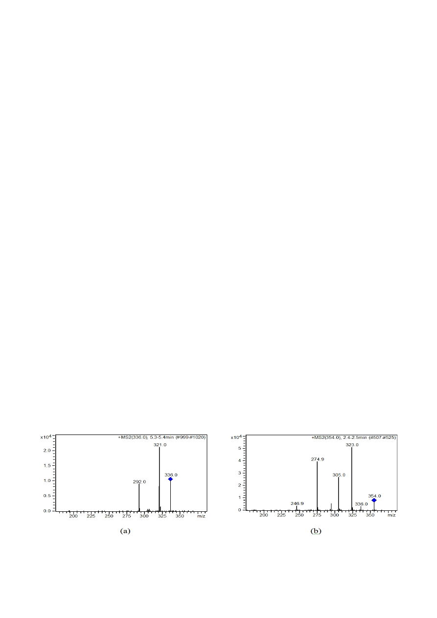
ion with m/z = 336 and the chelidonine ion with m/z =
354.
The quantification of berberine was based on the sum
of ions with m/z = 291.9 and 321.0 from the MS spec-
trum of the parent ion (Fig. 1a). Chelidonine was quanti-
fied based on the sum of the ions with m/z 275, 305, and
323 (Fig. 1b). The calibration curves were linear in the
range of 6.8–68 ng ∙ mL
-1
for berberine and 14–140 ng ∙
mL
-1
for chelidonine, with a correlation coefficient greater
than 0.997.
Identification and quantitative determinations of the
polyphenols
A high-performance liquid chromatography method
coupled with mass spectrometry (LC/MS) was used to
analyze the polyphenolic compounds in the
C. majus
plant extract. The method used was a previously published
HPLC method with minor changes (Nencu
et al., 2012;
Compaore
et al., 2012; Meda et al., 2011). The method is
suitable for qualitative (18 compounds) and quantitative
(14 compounds) analyses. In this study, 18 standards of
the polyphenolic compounds were used, namely, caftaric
acid, gentisic acid, caffeic acid, chlorogenic acid, paracou-
maric acid, ferulic acid, sinapic acid, hyperoside, isoquer-
citrin, rutoside, myricetol, fisetin, quercitrin, quercetol,
patuletine, luteolin, kaempferol, and apigenin.
Apparatus and chromatographic conditions
The experiments were performed using an Agilent 1100
HPLC Series system (Agilent) equipped with a degasser,
binary gradient pump, column thermostat, autosampler,
and UV detector. The HPLC system was coupled with an
Agilent 1100 mass spectrometer (LC/MSD ion trap VL).
For the separation, a reverse-phase analytical column was
employed (Zorbax SB-C18 100 x 3.0 mm i.d., 3.5 μm par-
ticle); the temperature was 48 °C. The compounds were
detected in both the UV and MS mode. The UV detec-
tor was set at 330 nm until 17.5 min, and then at 370 nm
for the remainder of the experiment. The MS system used
an electrospray ion source in the negative mode. The chro-
matographic data were processed using ChemStation and
DataAnalysis software from Agilent. The mobile phase
was a binary gradient prepared from methanol and a solu-
tion of 0.1% (v/v) acetic acid. The elution started with a
linear gradient, beginning with 5% methanol and ending
at 42% methanol, for 35 min; isocratic elution followed
for the next 3 min with 42% methanol. The flow rate was
1 mL ∙ min
-1
and the injection volume was 5 μL.
Polyphenols
The MS signal was used only for qualitative analysis
based on the specific mass spectra of each polyphenol. The
MS spectra obtained from a standard solution of poly-
phenols were integrated in a mass spectra library. Subse-
quently, the MS traces/spectra of the analyzed samples
were compared to spectra from the library, which allowed
the positive identification of compounds, based on spec-
tral matches. The UV trace was used for quantification of
the identified compounds following MS detection. Us-
ing the chromatographic conditions described above, the
polyphenols all eluted in less than 35 min (Tab. 1). Four
polyphenols could not be quantified under the chromato-
graphic conditions because of overlapping (caftaric acid
with gentisic acid and caffeic acid with chlorogenic acid).
However, all 4 compounds were selectively identified using
MS detection (qualitative analysis) based on differences in
their molecular mass and MS spectra. The detection limits
were calculated as the minimal concentration required to
produce a reproducible peak with a signal-to-noise ratio of
>3. The quantitative determinations were performed us-
ing an external standard method. Calibration curves in the
0.5–50 μg ∙ L
-1
range with good linearity (R2 > 0.999) for
a 5-point plot were used to determine the concentration of
the polyphenols in the plant samples.
Fig. 1. ESI/MS/MS spectra of berberine (a) and chelidonine (b)

Parvu M. et al. / Not Bot Horti Agrobo, 2013, 41(2):414-426
417
(147.3, 149.3, 161.3, and 175.3) for campesterol, ≤395
(163.3, 173.2, 187.3, 199.3, and 227.2) for stigmasterol
and ≤397 (160.9, 174.9, 188.9, 202.9, and 214.9) for si-
tosterol. The quantitative experiments were performed us-
ing an external standard method. Calibration curves in the
60–3000 ng ∙ mL
-1
range with good linearity (R2 > 0.99)
for a 7-point plot were used to determine the concentra-
tion of the sterols in the plant samples.
Determination of antifungal activity
The antifungal activity of the
C. majus extract, expressed
as the MIC, was determined by the agar-dilution assay,
and was compared to the antimycotic drug fluconazole
(2 mg ∙ mL
-1
, Krka, Novo Mesto, Slovenia) and a control
(nutritive medium and 35% ethanol). The percentage of
mycelial growth inhibition (P) at each concentration was
calculated using the formula P = (C-T) × 100/C, where C
is the diameter of the control colony and T is the diameter
of the treated colony (Nidiry and Babu, 2005).
Statistical analysis
Statistical analyses were performed using the program
R environment, version 2.14.1. The results for each group
were expressed as mean ± standard deviation. Data were
evaluated by analysis of variance (ANOVA). A P value of
≤ 0.05 was considered statistically significant. The correla-
tion analysis was performed by the Pearson test. Measure-
ments of physiological processes in tulip leaves treated
for 2 h with different concentrations of
C. majus extract
were performed in triplicate, and the post-ANOVA Tukey
HSD test was used to analyze the significance of differ-
ences between treatments and control.
In vivo effect of C. majus extract against B. tulipae
Fresh tulip leaves from the field that were infected by
B. tulipae were sprayed with the plant extract of C. ma-
jus at the MIC (6 %) and compared to the control tulips
leaves after 2 h.
Identification and quantitative determinations of the
sterols
The LC/MS technique was also used to analyze the ste-
rols from the
C. majus plant extract. The method used was
a previously published HPLC method with minor changes
(Sanchez-Machado
et al., 2004; Khalaf et al., 2011). Three
standards were used for the quantitative analysis, namely,
beta-sitosterol, stigmasterol, and cholesterol.
Apparatus and chromatographic conditions
The analyses were performed using an Agilent 1100
HPLC Series system equipped with a G1322A degasser,
G1311A binary pump, and G1313A autosampler. For
the separation, we used a reverse-phased Zorbax SB-C18
analytical column (100 mm ∙ 3.0 mm i.d., 5 µm particles)
fitted with a precolumn Zorbax SB-C18, both operated
at 40 °C. The mobile phase was prepared from methanol
and acetonitrile 10:90 (v/v) isocratic elution. The flow
rate was 1 mL ∙ min
-1
and the injection volume was 4 μL.
All solvents were filtered through 0.5-mL Sartorius filters
and degassed using ultrasound. MS/MS detection using
multiple reaction monitoring (MRM) of specific daugh-
ter ions was used for each sterol. The HPLC was coupled
with an Agilent ion trap 1100 VL mass detector, equipped
with an atmospheric pressure chemical ionization (APCI)
interface working in the positive ion mode. The operat-
ing conditions were: nitrogen gas, flow rate of 7 L ∙ min
-1
,
heater at 400 °C, ion source temperature of 250 °C, nitro-
gen nebuliser at 50 psi, and capillary voltage of 4000 V. All
chromatographic data were processed using ChemStation
software and Data Analysis from Agilent.
Sterols
Under our chromatographic conditions, the reten-
tion times of the 5 analyzed sterols were 3.2 min for er-
gosterol, 3.9 min for brassicasterol, 4.9 min for both stig-
masterol and campesterol (co-elution), and 5.7 min for
beta-sitosterol. The ions monitored by the MRM method
were ≤379 (253.3, 295.3, and 309.3) for ergosterol, ≤381
(201.3, 203.3, 215.2, and 217.3) for brassicasterol, ≤383
Tab. 1. Retention times (tR) for the investigated polyphenols
Peak no.
Phenolic
compound
t
R
+ SD (min)
Peak no.
Phenolic
compound
t
R
+ SD (min)
1
Caftaric acid
*
2.10 + 0.06
11
Myricetin
20.70 + 0.06
2
Gentisic acid
*
2.15 + 0.07
12
Fisetin
22.60 + 0.15
3
Caffeic acid
*
5.60 + 0.04
13
Quercitrin
23.00 + 0.13
4
Chlorogenic acid
*
5.62 + 0.05
14
Quercetol
26.80 + 0.15
5
p-coumaric acid
8.7 + 0.08
15
Patuletin
28.70 + 0.12
6
Ferulic acid
12.2 + 0.10
16
Luteolin
29.10 + 0.19
7
Sinapic acid
14.3 + 0.10
17
Kaempferol
31.60 + 0.17
8
Hyperoside
18.60 + 0.12
18
Apigenin
33.10 + 0.15
9
Isoquercitrin
19.60 + 0.10
10
Rutoside
20.20 + 0.15
*overlapping UV peaks, qualitative analysis performed using MS detection
Parvu M. et al. / Not Bot Horti Agrobo, 2013, 41(2):414-426
418
ciently weak (0.04 µM ∙ m
-2
∙ s
-1
) so as not to produce any
significant variable fluorescence. A single saturating flash
(2,000 µmol ∙ m
-2
∙ s
-1
for 0.5 s) was applied to reach the
maximal fluorescence Fm. After the decline of the signal,
the actinic light was turned on (100 µmol ∙ m
-2
∙ s
-1
) to in-
duce the kinetics. The determined parameters were the
initial fluorescence (F
0
), the maximal fluorescence (Fm),
the F
v
/F
m
ratio (F
v
or variable fluorescence, which is the
difference between the maximal and initial fluorescence),
the modulated maximal fluorescence (F
m
’), the steady state
fluorescence (F
s
), the effective quantum use efficiency (Φ)
representing the ratio (F
m
’ – F
s
)/F
m
’, as well as the vitality
index (relative fluorescence decrease, Rfd) expressed as the
ratio (F
m
– F
s
)/F
s
(Baker, 2008; Bartha and Fodorpataki,
2007; Horvath
et al., 1996). The experimental conditions
were identical to those for the leaf gas exchange measure-
ments, and the same leaves were used for the determina-
tion of the gas exchange parameters.
Results
The chelidonine and berberine alkaloids, polyphenols,
and sterols present in the
C. majus plant extract were de-
termined.
The chromatograms of chelidonine (Fig. 2) and ber-
berine (Fig. 3) from the
C. majus extract revealed the fol-
lowing levels: 26.09 µg ∙ mL
-1
as berberine base or 31.65 µg
∙ mL
-1
as berberine ∙ HCl ∙ 2H
2
O, and 304.62 µg ∙ mL
-1
as
chelidonine base. The retention time for chelidonine was
2.4 min and 5.3 min for berberine.
The non-hydrolyzed sample (Fig. 4) contains rutoside
(31.8 µg ∙ mL
-1
), whereas the hydrolyzed sample was deter-
mined to contain: p-coumaric acid (4.05 µg ∙ mL
-1
), ferulic
The samples of the treated leaves and control leaves were
examined by transmission electron microscopy (TEM)
with a JEOL JEM 1010 electron microscope (Japan Elec-
tron Optics Laboratory Co., Tokyo, Japan). Conidia con-
trols of
B. tulipae isolated from the tulip leaf surface were
examined by scanning electron microscopy (SEM) with a
JEOL JSM 5510 LV electron microscope (Hayat, 2000).
Measurement of leaf gas exchange
Specific gas exchange parameters were measured us-
ing a Ciras-2 leaf gas-exchange system (PP Systems) and
a PLC6 automatic leaf cuvette. The photon flux density
was set to 500 μM ∙ m
-2
∙ s
-1
, the air temperature in the leaf
cuvette was 26 °C, the reference carbon dioxide concentra-
tion was 340 ppm, and the reference relative air humidity
was 75%. Measurements of transpiration rate, net carbon
dioxide uptake, and stomatal conductivity were performed
at midday, on the fully expanded leaves of the tulip (
Tu-
lipa gesneriana cv. ‘Rococo’). Three leaves from each plant
were examined at 2 h after different concentrations of cel-
andine extracts were sprayed in a thin continuous layer on
the leaves. The leaves were maintained for the 2 h under
constant environmental conditions created in a vegetation
chamber (Pinheiro
et al., 2008).
Measurement of induced chlorophyll fluorescence
parameters
The parameters of the induced chlorophyll
a fluores-
cence were measured using a pulse amplitude modulated
chlorophyll fluorometer (PAM-FMS2, Hansatech), on 3
leaves of each plant. The leaves were left in the dark for 10
min prior to the measurements to terminate all previous
photochemical reactions. The modulated light was suffi-
Fig. 2. Chromatogram of chelidonine from the
Chelidonium
majus extract. The chelidonine peak is marked “1”
Fig. 3. Chromatogram of berberine from the
Chelidonium majus
extract. The berberine peak is marked “2”
Fig. 4. Chromatogram of polyphenol rutoside from non-hydro-
lyzed sample of
Chelidonium majus extract. The rutoside peak
is marked “1”
Fig. 5. Chromatogram of polyphenols from hydrolyzed sample
of
Chelidonium majus extract. The peaks are marked: “1” p-cou-
maric acid; “2” ferulic acid; “3” quercetol; “4” kaempherol
Parvu M. et al. / Not Bot Horti Agrobo, 2013, 41(2):414-426
419
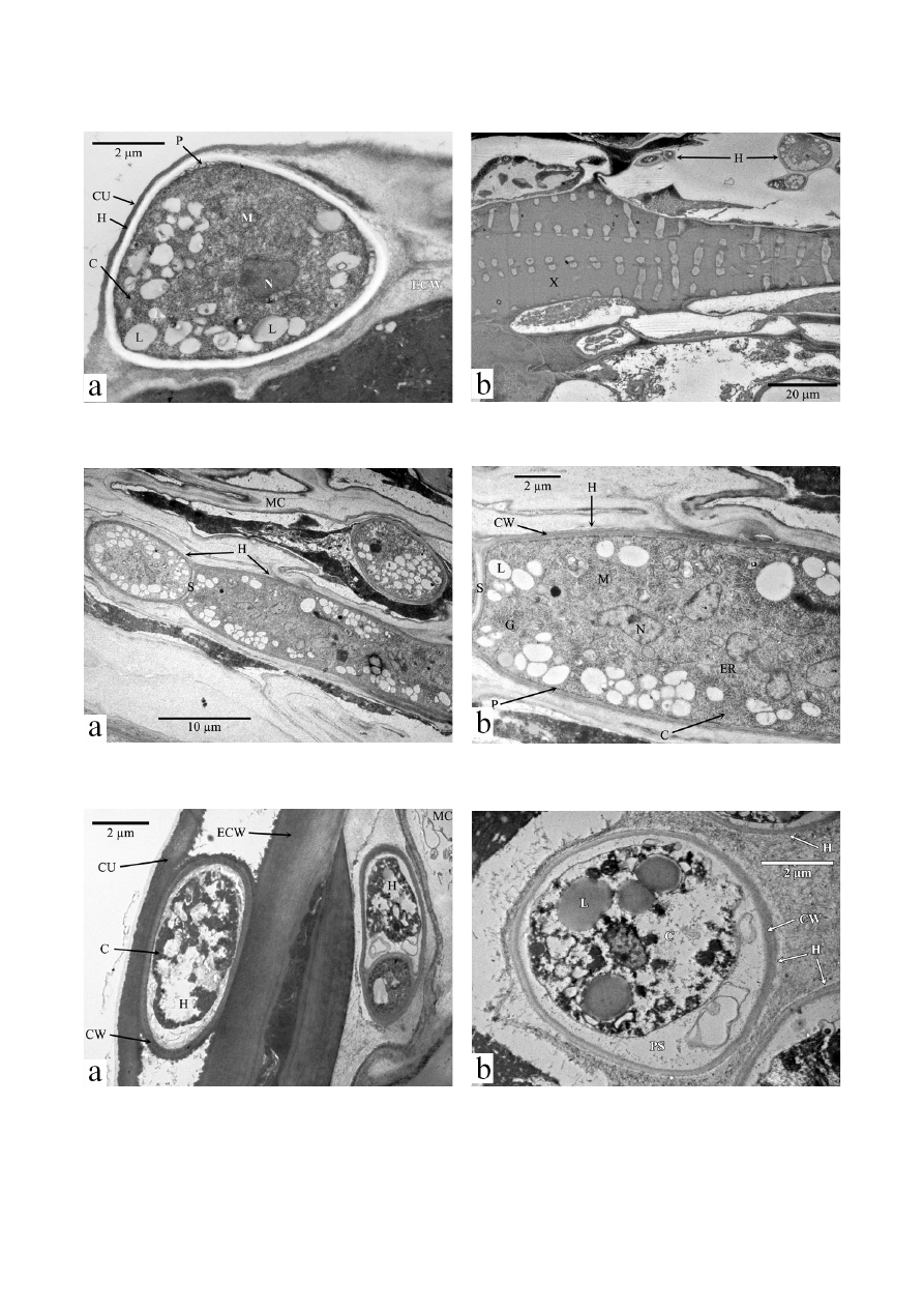
The
B. tulipae control hyphae were observed in the
attacked tulip leaf below the cuticle (Fig. 7a), in the leaf
mesophyll, and near the xylem vessel (Fig. 7b). At the ul-
trastructural level,
B. tulipae appears to have septate hy-
phae with regular cell walls and plasma membranes, as
well as cytoplasmic matrices with nuclei, mitochondria,
endoplasmic reticulum, lipid bodies, and glycogen (Figs.
8a and b). The parasitic activity of
B. tulipae destroyed the
attacked leaf tissues (Figs. 9a and b).
When treated with
C. majus plant extract at the MIC
for 2 h,
B. tulipae hyphae appeared damaged at the cel-
lular level. Specifically, the organelles were partly and/or
entirely destroyed, the cytoplasm was degenerated, and
electron dense material appeared in the hyphal cells. In ad-
dition, the outside of the cell wall had an irregular shape
and the plasma membrane was mostly destroyed and did
not adhere to the cell wall. Furthermore, precipitation of
the entire cytoplasm and destroyed organelles and nuclei
were seen. Because of these effects, the morpho-functional
relationship between the cell wall and cytoplasm was dam-
aged and a less electron dense band was formed between
the altered cytoplasm and cell wall (Figs. 9a and b).
acid (0.81 ∙ µgmL
-1
), quercetol (7.88 µg ∙ mL
-1
), and kaem-
pherol (1.21 µg ∙ mL
-1
) (Fig. 5).
The analyzed sample of
C. majus extract contains stig-
masterol (0.225 µg ∙ mL
-1
) and beta-sitosterol (0.191 µg ∙
mL
-1
).
The
C. majus plant extract had a significant inhibitory
effect on the mycelial growth of
B. tulipae on culture me-
dium. The
C. majus MIC is 6% and the Fluconazole MIC
is 12% (Tab. 2).
The
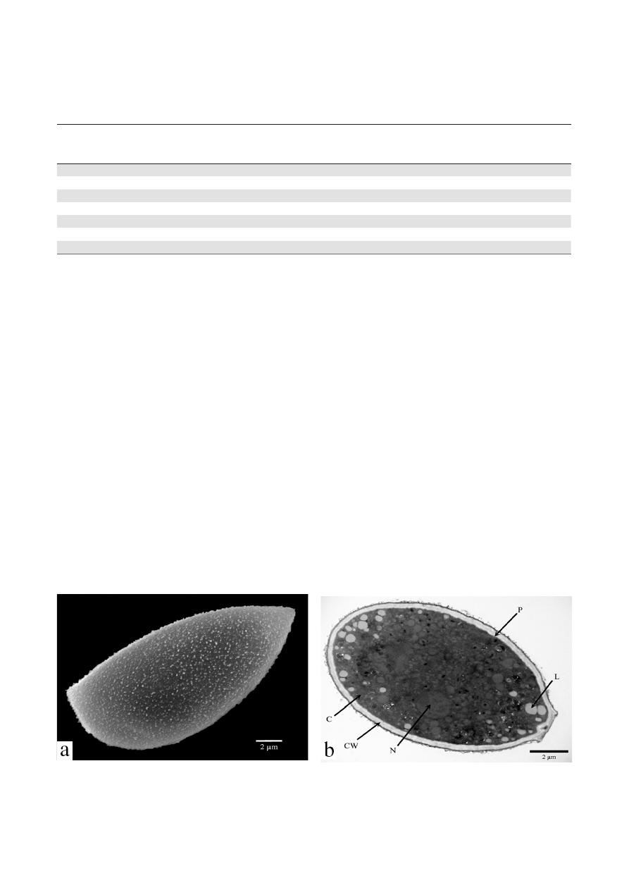
B. tulipae control conidia were observed by elec-
tron microscopy. The SEM micrographs of the
B. tulipae
control revealed unicellular conidia with numerous ran-
domly positioned protuberances (Fig. 6a). At the ultra-
structural level, the
B. tulipae control conidia contained
a regular cell wall with a 2-layer structure, plasma mem-
brane, cytoplasmic matrix with nucleus, and various cel-
lular organelles, and lipids. The external cell wall layer was
thin and electron dense, whereas the inner wall was thick,
uniform, and less electron dense. The plasma membrane
was tightly adhered to the cell wall. The cytoplasmic ma-
trix (cytosol) was uniformly distributed, and the nucleus
was ≤2 µm in diameter and ovoid or spherical (Fig. 6b)
Tab. 2.
In vitro effects of the Chelidonium majus extract on mycelial growth of Botrytis tulipae compared with the effects of the
synthetic fungicide fluconazole
Chelidonium majus
extract conc.
(%)
Botrytis tulipae
a
Colony diameter
(mm)
P
b
(%)
Fluconazole
conc.
(%)
Botrytis tulipae
c
Colony diameter
(mm)
P
d
(%)
C
62
0
C
62
0
1
60
3.22 ± 0.23
2
44
29.03 ± 0.23
2
46
25.80 ± 0.15
4
32
48.38 ± 0.15
3
30
51.61 ± 0.15
6
21
66.12 ±0.10
4
15
75.80 ± 0.15
8
12
80.64 ± 0.15
5
3
95.16 ± 0.16
10
4
93.54 ± 0.15
6
0
100 ± 0.21
12
0
100 ± 0.17
a
Mycelial growth of
B. tulipae at 5 days after inoculation in the presence of C. majus;
b
Inhibition % of radial growth in the presence of
C. majus;
c
Mycelial growth of
B.
tulipae at 5 days after inoculation in the presence of fluconazole;
d
Inhibition % of radial growth in the presence of fluconazole;
C, 35% aq. EtOH;
Colony diameter is expressed as mean ± SE of 6 replicates
Fig. 6. Visualization of
Botrytis tulipae conidium
a. Scanning electron micrograph showing protuberances on surface of cell wall. b. Transmission electron micrograph of an oblique section showing cell ultrastructure. CW
cell wall; C cytoplasm; L lipids; N nucleus; P plasma membrane
Parvu M. et al. / Not Bot Horti Agrobo, 2013, 41(2):414-426
420
Fig. 7. Transmission electron micrograph of a tulip leaf cross section showing the
Botrytis tulipae fungus
a. Hyphae (H) between the epidermal cell wall (ECW) and cuticle (CU) of the epidermis. b. Hyphae (H) in the leaf mesophyll, near the xylem (X). C cytoplasm; L lipids;
M mitochondrion; N nucleus; P plasma membrane
Fig. 8. Transmission electron micrograph of an oblique section of
Botrytis tulipae control hyphae from the tulip leaf
a. Septate hyphae (H) in the mesophyll cells (MC). b. Detailed micrograph of septate hyphae (H) in the mesophyll. C cytoplasm; CW cell wall; ER endoplasmic reticulum;
G glycogen; L lipids; M mitochondrion; N nucleus; P plasma membrane; S septum
Fig. 9. Transmission electron micrograph of a tulip leaf cross section showing irreversible ultrastructural changes
in Botrytis tulipae
hyphae treated with
Chelidonium majus plant extract at the MIC
a. Hyphae between the epidermal cell wall (ECW) and cuticle (CU) of the epidermis and hyphae (H) in the mesophyll cells (MC). b. hyphae (H) in the mesophyll cells
(MC). C cytoplasm; CW cell wall; L lipids; PS. periplasmic space
Parvu M. et al. / Not Bot Horti Agrobo, 2013, 41(2):414-426
421
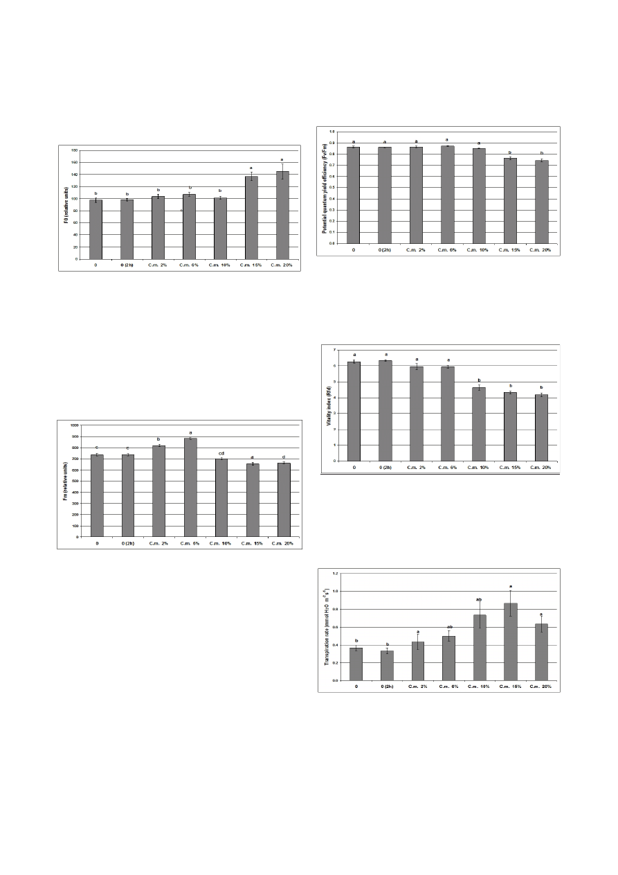
fects were noted on the investigated gas exchange param-
eters (Figs. 14-16).
The ground chlorophyll fluorescence was not signifi-
cantly affected when the tulip leaves were covered for 2 h
with solutions of 2%, 6%, and 10% extracts of celandine
(Fig. 10).
The maximal chlorophyll fluorescence is more sensitive
than the ground fluorescence, and its values were increased
in the presence of 2% and 6% extracts, and decreased by
10%, 15%, and 20% celandine extracts (Fig. 11).
The potential quantum yield efficiency of photosystem
II was not affected by the extract when it was sprayed on
the tulip leaves at concentrations ≤ 15% (Fig. 12).
The vitality index of the photosynthetic apparatus
starts to decrease significantly when the extract concentra-
tion is ≥ 10% (Fig. 13).
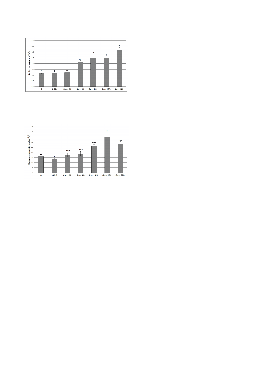
Progressive and statistically significant increases in all
3 gas exchange parameters (transpiration rate, net carbon
dioxide uptake, and stomatal conductivity) were regis-
tered following treatment of the tulip leaves with concen-
trations of celandine extracts of ≥ 6% for 2 h. When the
extract was used at concentrations of 2% no significant ef-
Fig. 10. Ground chlorophyll fluorescence (F
0
) in the dark-adapt-
ed tulip leaves treated for 2 h with different concentrations of
ce-landine (C.m.) extract
0, control leaves; 0 (2 h), control leaves after 2 h. Bars represent the standard
error obtained from 3 independent experiments. The letters indicate significant
differences at
p ≤ 0.05 according to the Tukey HSD test
Fig. 11. Maximal chlorophyll fluorescence (F
m
) in the dark-
adapted tulip leaves treated for 2 h with different concentra-
tions of celandine (C.m.) extract
0, control leaves; 0 (2 h), control leaves after 2 h. Bars represent the standard
error obtained from 3 independent experiments. The letters indicate significant
differences at
p ≤ 0.05 according to the Tukey HSD test
Fig. 12. Potential quantum yield efficiency of photosystem II
Values are based on the ratio between the variable and maximal chlorophyll
fluorescence (F
v
/F
m
) in the dark-adapted tulip leaves treated for 2 h with different
concentrations of celandine (C.m.) extracts. 0, control leaves; 0 (2 h), control
leaves after 2 h. Bars represent the standard error obtained from 3 independent
experiments. The letters indicate significant differences at
p ≤ 0.05 according to the
Tukey HSD test
Fig. 13. Vitality index of the photosynthetic apparatus
Values are based on the relative chlorophyll fluorescence decrease (R
fd
) in the tulip
leaves treated for 2 h with different concentrations of celandine (C.m.) extracts.
0, control leaves; 0 (2 h), control leaves after 2 h. Bars represent the standard
error obtained from 3 independent experiments. The letters indicate significant
differences at
p ≤ 0.05 according to the Tukey HSD test
Fig. 14. Transpiration rate of the tulip leaves treated for 2 h with
different concentrations of celandine (C.m.) extract
0, control leaves; 0 (2 h), control leaves after 2 h. Bars represent the standard
error obtained from 3 independent experiments. The letters indicate significant
differences at
p ≤ 0.05 according to the Tukey HSD test
Parvu M. et al. / Not Bot Horti Agrobo, 2013, 41(2):414-426
422
ophyll cells (Figs. 7 and 8). The ultrastructure of xylem
cells (Fig. 7b) is not affected by the fungus and the fungus
hyphae appear only in the leaf mesophyll tissues.
The infection of host plants by
B. tulipae is mediated
by numerous extracellular enzymes and metabolites. Each
of these compounds plays a role in different stages of the
infection process. Cutinases, lipases, and some cell wall-
degrading enzymes facilitate penetration of the host sur-
face, whereas toxins, oxalate, and reactive oxygen species
enable host cell death. Several cell wall-degrading enzymes
contribute to the conversion of host tissue into fungal bio-
mass, and other enzymes such as laccases and proteases are
involved in pathogenesis (Kars and van Kan, 2007).
Fungicide-resistant
Botrytis strains have been identi-
fied in various crops (Agrios, 2005). In addition, plant
fungicides based on synthetic chemicals are both pollut-
ants and toxic (Barker and Rogers, 2006; Carrillo-Munoz
et al., 2006; Fatehi et al., 2005; Strange and Scott, 2005;
Ienaşcu
et al., 2008). Therefore, the biological control of
Botrytis fungi with plant extracts is one of the important
measures for enhancing farming techniques.
An important objective of our study was to test the
in
vitro action of C. majus extract on mycelium growth of B.
tulipae and determine the MIC of the plant extract (Tab.
2). Previous studies suggested that
C. majus possesses an-
tifungal properties, and therefore, this extract is a prom-
ising source of active compounds against fungi such as
Fusarium spp. (Matos et al., 1999), B. cinerea (Pârvu et al.,
2008), and
Candida species (Meng et al., 2009).
Our study examined leaves attacked by gray mold and
treated with
C. majus extract at the MIC to demonstrate
the
in vivo inhibitory properties of the extract against B.
tulipae (Fig. 9). The C. majus plant extract caused irrevers-
ible ultrastructural changes that abolished the cell wall’s
barrier function and its ability to activate cell wall-bound
enzymes. The morpho-functional integrity of fungal cell
components is required for viability and germination ca-
pacity (Isaac, 1992). The
B. tulipae hyphae treated with C.
majus extract revealed precipitation of the cytoplasm and
destruction of organelles and nuclei that caused loss of vi-
ability and germination capacity.
In addition to the ultrastructural changes observed in
B. tulipae hyphae treated with the plant extract (Fig. 9),
the antimicrobial compounds from the
C. majus extract
induced important changes at the molecular level. The
C. majus extract contains a large number of alkaloids and
polyphenols, and is therefore, known for its antimicrobial
activity (Meng
et al., 2009; Nawrot et al., 2007; Zuo et al.,
2011). The main alkaloids identified in
C. majus extracts
are chelidonine, chelerythrine, sanguinarine, coptisine,
and berberine (Sárközi
et al., 2006a; Zuo et al., 2011).
Formaldehyde formation due to demethylation is re-
sponsible for the antimicrobial activity of these alkaloids
(Sárközi
et al., 2006b).
The alkaloids berberine, chelidonine, chelerythrine,
sanguinarine, and coptisine (Wink, 1998) from the
C.
Discussion
B. tulipae is the only Botrytis species able to infect tu-
lip (Staats
et al., 2005) and to abundantly produce sporu-
lating gray mycelium on infected tissue (Yohalem
et al.,
2003). The mitotically produced spores, macroconidia,
can be transported long distances by wind (Agrios, 2005;
Webster and Weber, 2007). This parasitic fungus overwin-
ters in the soil as mycelium in decaying plant debris and as
sclerotia, which are melanized mycelial survival structures
(Agrios, 2005; Yohalem
et al., 2003).
The
B. tulipae conidia are ellipsoidal or obovoid, uni-
cellular (Hong
et al., 2002) and have numerous randomly
positioned protuberances (Fig. 6a); however, these protu-
berances are fewer than those present in
B. cinerea conidia
(Pârvu
et al., 2008). Hydration and redrying causes these
protuberances to disappear (Doss
et al., 1997). The cell
wall of the conidia has 2 layers and appears dark (Fig. 6b)
because of melanin, which protects the spores from en-
zyme action and probably UV radiation (Epton and Rich-
mond, 1980).
B. tulipae fungus penetrates tulip leaves and produces
irreversible ultrastructural changes in epidermal and mes-
Fig. 15. Net carbon dioxide uptake by tulip leaves treated for 2 h
with different concentrations of celandine (C.m.) extract
0, control leaves; 0 (2 h), control leaves after 2 h. Bars represent the standard
error obtained from 3 independent experiments. The letters indicate significant
differences at
p ≤ 0.05 according to the Tukey HSD test
Fig. 16. Stomatal conductivity of the tulip leaves treated for 2 h
with different concentrations of celandine (C.m.) extract
0, control leaves; 0 (2 h), control leaves after 2 h. Bars represent the standard
error obtained from 3 independent experiments. The letters indicate significant
differences at
p ≤ 0.05 according to the Tukey HSD test

Parvu M. et al. / Not Bot Horti Agrobo, 2013, 41(2):414-426
423
must be effective against parasites and minimally affect
the vital processes of the host plant.
In vivo induced chlo-
rophyll fluorescence is a sensitive, non-destructive tool for
the study of environmental impacts on the primary ener-
gy-conversion processes of photosynthesis. The potential
quantum use efficiency of photosynthesis is reflected by
the ratio between the variable and the temporary maximal
fluorescence yield (F
v
/F
m
) in dark-adapted leaves. The F
v
/
F
m
value is one of the most relevant functional markers of
photosynthetic energy conversion, and therefore is used
for detection of various stress factors that interfere with
photochemical reactions in chloroplasts. Its drop below
the value of 0.8 is directly related to the disturbed photo-
chemical reactions that occur in photosystem II of thyla-
koid membranes in chloroplast, which leads to a less effi-
cient photosynthetic use of the absorbed light energy. The
initial fluorescence of the dark-adapted leaves (F
0
), which
is induced by a very weak red light flash, is related to the
organization and energy transfer capacity of the light-har-
vesting antennae. The maximal fluorescence (F
m
), which is
generated by a flash of saturating red light, is related to the
activity of the electron acceptors of photosystem II. One of
the most sensitive parameters of induced chlorophyll fluo-
rescence is the relative fluorescence decrease (R
fd
), which
is also known as the vitality index. This value is dependent
on the difference between the temporary maximal fluores-
cence yield in dark-adapted samples and the steady state
fluorescence level in constantly illuminated samples. The
pulse amplitude modulation of chlorophyll fluorescence is
obtained by regular saturating flashes on a background of
a constant actinic light (Baker, 2008).
Because the ground chlorophyll fluorescence of the
dark-adapted leaves was not significantly affected, one can
deduce that the organization and energy transfer func-
tion of the light-harvesting pigment antennae of the leaves
were not impaired by extract concentrations ≤15%. The
registered values of maximal chlorophyll fluorescence in-
dicate that small amounts of the celandine extract slightly
stimulate photochemical reactions on the acceptor side
of photosystem II (e.g., reduction of quinine acceptors),
whereas higher extract concentrations moderately inhibit
them, without causing a dramatic decline in the process.
The fact that potential quantum yield efficiency of photo-
system II was not affected by the extract implies that the
overall conversion of light energy into storable chemical
energy is not altered when plants are treated with dilute
extracts of celandine. This is also valid for the vitality index
of the photosynthetic apparatus, which is more sensitive
than quantum efficiency, and decreases only upon treat-
ment with extract concentrations reaching or exceeding
10% (Figs. 10-13). Based on the parameters of induced
chlorophyll fluorescence, one can state that the photosyn-
thetic light conversion capacity of tulip leaves is not re-
duced by the application of celandine extracts at concen-
trations of 2% or 6%.
majus plant extract induce changes at the molecular level
in
B. tulipae hyphae. Specifically, alterations involve dis-
turbed DNA/RNA and related enzymes, as well as altera-
tions to the cytoskeleton, ribosomal protein biosynthesis,
and membrane permeability (Wink, 1998; Wink, 2008;
Rosenkranz and Wink, 2008). The alkaloid berberine is
present in
C. majus extracts and Berberis extracts (Sárközi
et al., 2006a; Zuo et al., 2011). Berberine inhibits esteras-
es, DNA and RNA polymerases, cellular respiration, and
acts in DNA intercalation (Aniszewski, 2007).
The alkaloids from
C. majus are poisonous to B. tulipae
fungus because they inhibit processes like DNA replica-
tion and RNA transcription that are vital for the microor-
ganism (Wink, 1998).
Other antifungal compounds identified in the
C.
majus extract were the phenolic compounds rutoside, p-
coumaric acid, ferulic acid, quercetol, and kaempherol
(Tab.1). The mechanisms of action thought to be respon-
sible for phenolic toxicity involve enzyme inhibition by
the oxidized compounds, possibly through reaction with
sulfhydryl groups or nonspecific interactions with the pro-
teins (Arif
et al., 2009). In addition, antifungal phenolics
from plants with action against phytopathogenic fungi
B.
cinerea, Cercospora beticola, Colletotrichum circinans, Cla-
dosporium herbarum, Fusarium oxysporum, Phytophthora
infestans, Venturia inaequalis, Verticillium albo-atrum have
been identified (Lattanzio
et al., 2006).
The other antimicrobial compounds identified in the
C. majus extract were the sterols stigmasterol (0.225 µg
∙ mL
-1
) and beta-sitosterol (0.191 µg ∙ mL
-1
). The most
abundant plant sterols are sitosterol, campesterol, and
stigmasterol (Moreau
et al., 2002), and the antifungal
activity of plant sterols (Sharma and Kumar, 2009) and
sterols from
Ganoderma annulare mushroom have been
described (Smania
et al., 2003). Moreover, free flavonoids
and sterols of the
Tridax procumbens plant extract com-
pletely inhibited spore germination of the
F. oxysporum
phytopathogenic fungus (Sharma and Kumar, 2009).
Our results reveal that the
C. majus extract contains
important antifungal compounds like alkaloids, phenols,
and sterols. Importantly, these results clarify the antifungal
activity of
C. majus against phytopathogenic fungi (Matos
et al., 1999; Pârvu et al., 2008) such as B. tulipae. The C.
majus extract at MIC (6%) caused irreversible changes in
B. tulipae hyphae, and therefore, in vivo studies examining
the effect of the extract on tulips that have not been at-
tacked by gray mold is required.
Whenever environmental stress factors directly or in-
directly influence the energetic processes that occur during
photosynthesis, they cause specific changes in the various
parameters associated with induced chlorophyll fluores-
cence (Baker and Oxborough, 2004). This enables a quick
in situ evaluation of alterations to photosynthesis in tulip
leaves treated with antifungal celandine extracts. This is
important because any agent used in pest management

Parvu M. et al. / Not Bot Horti Agrobo, 2013, 41(2):414-426
424
II, increased upon treatment with lower concentrations
(2% and 6%) of extract, but declined when the leaves were
sprayed with higher concentrations (10-20%). The main
efficiency parameters of photosynthesis, such as potential
quantum yield efficiency and overall vitality index, were
not affected by the extract when its concentration was
≤10%.
In conclusion, we recommend the use of the celandine
extract in concentration of 6% for the efficient protection
of tulips against the attack of gray mold.
Acknowledgment
These studies were financially supported by the Roma-
nian Ministry of Education and Research from the CNC-
SIS grants 46/220/2006, 43/220/2007, and PNII–IDEI
2272/2009-2011.
References
Agrios GN (2005). Plant Pathology 5
th
ed. Elsevier, Amsterdam,
510 p.
Allen MT, Pearcy RW (2000). Stomatal behavior and photosyn-
thetic performance under dynamic light regimes in a season-
ally dry tropical rain forest. Oecologia 122:470-478.
Aniszewski T (2007). Alkaloids-secrets of Life Alkaloid Chem-
istry, Biological Significance, Applications and Ecological
Role. Elsevier, Amsterdam, 181 p.
Arif T, Bhosale JD, Kumar N, Mandal TK, Bendre RS, Lavekar
GS, Dabur R (2009). Natural products – antifungal agents
derived from plants. J Asian Nat Prod Res 11:621-638.
Arif T, Mandal TK, Dabur R (2011). Natural products: Anti-
fungal agents derived from plants, p. 283-311. In: Tiwari
VK (Ed.). Opportunity, Challenge and Scope of Natural
Products in Medicinal Chemistry. Research Signpost, Ker-
ala (India).
Baker NR (2008). Chlorophyll fluorescence: a probe of photo-
synthesis
in vivo. Annu Rev Plant Biol 59:89-113.
Baker NR, Oxborough K (2004). Chlorophyll fluorescence as
a probe of photosynthetic productivity, p. 65-82. In: Pa-
pageorgiou GC, Govindjee (Eds.). Chlorophyll a Fluores-
cence: A Signature of Photosynthesis. Springer, Dordrecht.
Barker KS, Rogers PD (2006). Recent insights into the mecha-
nisms of antifungal resistance. Curr Infect Dis Rep 8:449-
456.
Bartha L, Fodorpataki L (2007). Physiological reactions of the
succulent CAM plant
Bryophyllum daigremontianum to in-
creased salinity. Contrib Bot 42:47-56.
Beever RE, Weeds PL (2007). Taxonomy and genetic variation
of
Botrytis and Botryotinia, p. 29–52. In: Elad Y, Williamson
B, Tudzynski P, Delen N (Eds.).
Botrytis: Biology, Pathology
and Control. Springer, Dordrecht.
Carrillo-Munoz AJ, Giusiano G, Ezkurra PA, Quindos G
(2006). Antifungal agents: mode of action in yeast cells. Rev
Esp Quimioter 19:130-139.
Gas exchange processes through the leaf surfaces may
be directly influenced by any substance that is sprayed
onto the leaves, and may thus, penetrate the cuticle or en-
ter the leaf through open stomata. This can lead to distur-
bances in carbon dioxide supply for photosynthetic car-
bon assimilation, or to an impaired regulation of stomatal
movements that can threaten the water equilibrium and
inorganic nutrient uptake of the whole plant. Therefore,
it is important to investigate the effect of depositing cel-
andine extracts on leaves on gas exchange processes. In the
water economy of plants, the most important gas exchange
parameter is transpiration rate, whereas for the photosyn-
thetic carbon assimilation, net carbon dioxide uptake is a
crucial prerequisite. The overall dynamics of gas exchange
on the leaf surface is indicated by stomatal conductivity.
For example, the intensity of the gas exchange processes
per unit leaf area strongly decreases during drought and
salt stress, as well as under the influence of different air
pollutants (Allen and Pearcy, 2000; Medrano
et al., 2002;
Hetherington and Woodward, 2003).
The results of the gas exchange measurements suggest
that if the celandine extract is sprayed on tulip leaves at
concentrations higher than 2%, it may cause increased
transpirational water loss, which may also be beneficial if
excess water is present in the soil and the higher suction
force enhances the uptake of inorganic nutrients from the
soil, and may ensure a better carbon dioxide supply to the
leaves through the more widely open stomata. No inhibi-
tion of gas exchange processes were detected even when the
celandine extract was applied at higher concentrations.
Conclusions
The
C. majus plant extract exhibited strong in vitro
and
in vivo fungicidal activity against B. tulipae. The C.
majus plant extract at the MIC caused severe ultrastruc-
tural changes in the tulip leaf hyphae that lead to loss of
viability. The
Botrytis strains have a high resistance to con-
ventional fungicides, and therefore, we propose that
C.
majus is a good in vivo biological treatment against fungal
infections like gray mold (Pârvu
et al., 2008), tulip mold,
and other species (Matos
et al., 1999). After 2 h of surface
treatment under growth chamber conditions, low concen-
trations (2%) of celandine extract did not affect the main
physiological parameters associated with leaf gas exchange
and photosynthetic light-use efficiency. The transpira-
tion rate, net carbon dioxide influx, and overall stomatal
conductivity increased at concentrations greater than 6%,
thereby indicating stimulated stomatal opening, which fa-
vors carbon assimilation but may impair water economy.
The ground fluorescence of chlorophyll
a indicated that
light harvesting by photosynthetic antenna pigments was
affected only by higher concentrations (15% and 20%) of
celandine extract. The maximal chlorophyll fluorescence
yield of dark-adapted leaves, which is related to the pho-
tochemical processes on the acceptor side of photosystem

Parvu M. et al. / Not Bot Horti Agrobo, 2013, 41(2):414-426
425
Studia UBB Chemia 56:97-102.
Lattanzio V, Lattanzio VMT, Cardinali A (2006). Role of phe-
nolics in the resistance mechanisms of plants against fungal
pathogens and insects, p. 23-67. In: Imperato F (Ed.). Phy-
tochemistry: Advances in Research. Trivandrum, Kerala
(India).
Matos OC, Baeta J, Silva MJ, Pinto Ricardo CP (1999). Sensi-
tivity of
Fusarium strains to Chelidonium majus L. extracts. J
Ethnopharmacol 66:151-168.
Meda RNT, Vlase L, Lamien-Meda A, Lamien CE, Muntean D,
Tiperciuc B, Oniga I, Nacoulma OG (2011). Identification
and quantification of phenolic compounds from
Balanites
aegyptiaca (L) Del (Balanitaceae) galls and leaves by HPLC-
MS. Nat Prod Res 25:93-99.
Medrano H, Escalona MH, Bota JM, Gulias J, Flexas J (2002).
Regulation of photosynthesis of C
3
plants in response to
progressive drought: stomatal conductance as a reference
parameter. Ann Bot 89:895-905.
Meng FY, Zuo GY, Hao XY, Wang GC, Xiao HT, Zhang JQ, Xu
GL (2009). Antifungal activity of the benzo[c]phenanthri-
dine alkaloids from
Chelidonium majus Linn against resis-
tant clinical yeast isolates. J Ethnopharmacol 125:494-496.
Mishra R, Verma DL, (2009). Antifungal activity and flavonoid
composition of
Wiesnerella denudata. Steph Acad Arena
1:42-45.
Moreau R, Whitaker B, Hicks K (2002). Phytosterols, phy-
tostanols, and their conjugates in foods: structural diversity,
quantitative analysis, and health-promoting uses. Prog Lipid
Res 41:457-500.
Nawrot R, Lesniewicz K, Pienkowska J, Gozdzicka-Jozefiak A
(2007). A novel extracellular peroxidase and nucleases from
a milky sap of
Chelidonium majus. Fitoterapia 78:496-501.
Nencu I, Vlase L, Istudor V, Dutu LE, Gird CE (2012). Prelimi-
nary research regarding the therapeutic uses of
Urtica dioica
L. Note I. The polyphenols evaluation. Farmacia 60:493-
500.
Nidiry ESJ, Babu CSB (2005). Antifungal activity of tuberose
absolute and some of its constituents. Phytother Res 19:447-
449.
Pârvu M, Pârvu AE (2011). Antifungal plant extracts, p.1055-
1062. In: Méndez-Vilas A (Ed.). Science Against Microbial
Pathogens: Communicating Current Research and Tech-
nological Advances. Formatex Research Center, Badajoz
(Spain).
Pârvu M, Pârvu AE, Crăciun C, Barbu-Tudoran L, Tămaş M
(2008). Antifungal activities of
Chelidonium majus extract
on
Botrytis cinerea in vitro and ultrastructural changes in its
conidia. J Phytopathol 156:550-552.
Pinheiro HA, Silva JV, Endres L, Ferreira VM, Camara CA,
Cabral FF, Oliveira JF, Carvalho LWT, Santos JM, Filho
BGS (2008). Leaf gas exchange, chloroplastic pigments and
dry matter accumulation in castor bean (
Ricinus communis
L) seedlings subjected to salt stress conditions. Ind Crop
Choi GJ, Jang KS, Kim JS, Lee SW, Cho JY, Cho KY, Kim JC
(2004).
In vivo antifungal activities of 57 plant extracts
against six plant pathogenic fungi. Plant Pathol J 20:184–
191.
Compaore M, Lamien CE, Lamien-Meda A, Vlase L, Kien-
drebeogo M, Ionescu C, Nacoulma OG (2012). Antioxi-
dant, xanthine oxidase and lipoxygenase inhibitory activities
and phenolics of
Bauhinia rufescens Lam. (Caesalpiniaceae).
Nat Prod Res 26:1069-1074.
Doss RP, Christian JK, Potter SW, Soeldner AH, Chastagner
GA (1997). The conidial surface of
Botrytis cinerea and sev-
eral other
Botrytis species. Can J Bot 75:612-617.
Elad Y, Stewart A (2004). Microbial control of
Botrytis spp., p.
223–241. In: Elad Y, Williamson B, Tudzynski P, Delen N
(Eds.).
Botrytis: Biology, Pathology and Control. Springer,
Dordrecht.
Elad Y, Williamson B, Tudzynski P, Delen N (2007).
Botrytis
spp. and diseases they cause in agricultural systems – an in-
troduction, p. 1–8. In: Elad Y, Williamson B, Tudzynski P,
Delen N (Eds.).
Botrytis: Biology, Pathology and Control.
Springer, Dordrecht.
Epton HAS, Richmond DV (1980). Formation, structure and
germination of conidia, p. 41-83. In: Coley-Smith JR, Ver-
hoeff K, Jarvis WR (Eds.). The Biology of
Botrytis. Academ-
ic Press, London.
Fatehi M, Saleh TM, Fatehi-Hassanabad Z, Farrokhfal K, Davo-
di SJ (2005). A pharmacological study on
Berberis vulgaris
fruit extract. J Ethnopharmacol 102:46-52.
Hayat MA (2000). Principles and Techniques of Electron Mi-
croscopy: Biological Applications. University Press, Cam-
bridge, 1 p.
Hetherington AM, Woodward FI (2003). The role of stomata in
sensing and driving environmental change. Nature 424:901-
908.
Hong SK, Kim WG, Cho WD, Kim HG (2002). Occurrence
of tulip fire caused by
Botrytis tulipae in Korea. Plant Pathol
J 18:106-108.
Horvath G, Droppa M, Fodorpataki L, Istokovics A, Garab G,
Oettmeier W (1996). Acridones: a chemically new group of
protonophores. Proc Nat Acad Sci USA 93:3876-3880.
Ienaşcu IMC, Lupea AX, Hădăruga D, Hădăruga N, Pope-
scu IM (2008). The antimicrobial activity and quantita-
tive structure – biological activity relationships evaluation
of some novel 2-hydroxybenzamide derivatives. Revista de
Chimie 59:247-250.
Isaac S (1992). Fungal-Plant Interactions. Chapman & Hall,
London, 10 p.
Kars I, van Kan JAL (2007). Extracellular enzymes and metabo-
lites involved in pathogenesis of
Botrytis, p. 99-118. In: Elad
Y, Williamson B, Tudzynski P, Delen N (Eds.).
Botrytis: Bi-
ology, Pathology and Control. Springer, Dordrecht.
Khalaf I, Corciova A, Vlase L, Ivănescu B, Lazăr D (2011). LC/
MS analysis of sterolic compounds from
Glycyrrhiza glabra.

Parvu M. et al. / Not Bot Horti Agrobo, 2013, 41(2):414-426
426
method for quantitative estimation of L-Dopa from
Mu-
cuna pruriens. Int Res J Pharm 3:300-304.
Webster JW, Weber RWS (2007). Introduction to Fungi. Uni-
versity Press, Cambridge, 434 p.
Wilson CL, Solar JM, El Ghaouth A, Wisniewski ME (1997).
Rapid evaluation of plant extracts and essential oils for an-
tifungal activity against
Botrytis cinerea. Plant Dis 81:204-
210.
Wink M (1998). Modes of action of alkaloids, p. 301-326. In:
Roberts MF, Wink M (Eds.). Alkaloids: Biochemistry, Ecol-
ogy, and Medicinal Applications. Plenum Press, New York.
Wink M (2008). Ecological roles of alkaloids, p. 3-23. In: Fat-
torusso E, Taglialatela-Scafati O (Eds.). Modern Alkaloids
Structure, Isolation, Synthesis and Biology. WILEY-VCH
Verlag GmbH & Co. KGaA, Weinheim.
Wu W, Song F, Yan C, Liu Z, Liu S (2005). Structural analyses
of protoberberine alkaloids in medicine herbs by using ESI-
FT-ICR-MS and HPLC-ESI-MS(n). J Pharm Biomed Anal
37:437-46.
Yohalem DS, Nielsen K, Nicolaisen M (2003). Taxonomic and
nomenclatural clarification of the onion neck rotting
Botry-
tis species. Mycotaxon 85:175-182.
Zuo G-Y, Meng F-Y, Han J, Hao X-Y, Wang G-C, Zhang Y-L,
Zhang Q (2011).
In vitro activity of plant extracts and al-
kaloids against clinical isolates of extended-spectrum
β-lactamase (ESBL)-producing strains. Molecules 16:5453-
5459.
Prod 27:385-392.
Rosenkranz V, Wink M (2008). Alkaloids induce programmed
cell death in bloodstream forms of trypanosomes (
Trypano-
soma b. brucei). Molecules 13:2462-2473.
Sanchez-Machado DI, Lopez-Hernandez J, Paseiro-Losada P,
Lopez-Cervantes J (2004). An HPLC method for the quan-
tification of sterols in edible seaweeds. Biomed Chromatogr
18:183-190.
Sárközi Á, Janicsák G, Kursinszki L, Kéry Á (2006a). Alkaloid
composition of
Chelidonium majus L. studied by different
chromatographic techniques. Chromatographia 63:81-86.
Sárközi Á, Móricz ÁM, Ott PG, Tyihák E, Kéry Á (2006b).
Investigation of
Chelidonium alkaloids by use of a complex
bioautographic system. J Planar Chromatogr 19:267-272.
Sharma B, Kumar P (2009). In vitro antifungal potency of some
plant extracts against
Fusarium oxysporum. Int J Green
Pharm 3:63-65.
Smania EF, Delle Monache F, Smania A, Yunes RA, Cuneo RS
(2003). Antifungal activity of sterols and triterpenes isolated
from
Ganoderma annulare. Fitoterapia 74:375-377.
Staats M, van Baarlen P, van Kan JA (2005). Molecular phylog-
eny of the plant pathogenic genus
Botrytis and the evolution
of host specificity. Mol Biol Evol 22:333-346.
Staats M, van Baarlen P, van Kan JAL (2007). AFLP analysis
of genetic diversity in populations of
Botrytis elliptica and
Botrytis tulipae from the Netherlands. Eur J Plant Pathol
117:219-235.
Strange RN, Scott PR (2005). Plant disease: a threat to global
food security. Annu Rev Phytopathol 43:83-116.
Sundaram U, Gurumoorthi P (2012). Validation of HPTLC
Wyszukiwarka
Podobne podstrony:
Metallurgy Chemical Compositions of Alloys
THE CHEMICAL COMPOSITION AND SENSORY QUALITY OF PORK
Comparision of vp;atile composition of cooperage oak wood
A chemical analog of curcumin as an improved inhibitor of amyloid abetaoligomerization
Chemical Composition and in Vitro Antifungal Activity Screening
Composition of surface oxide film of titanium with culturing
chemical behaviour of red phosphorus in water
the effect of sowing date and growth stage on the essential oil composition of three types of parsle
Influence of different microwave seed roasting processes on the changes in quality and fatty acid co
Effects of topography and composition of titanium surface ox
the effect of water deficit stress on the growth yield and composition of essential oils of parsley
Natural variations detected in the isotopic composition of copper possible applications to archeolo
Thermal and chemical modification of titanium
Elemental composition of willow short rotation crops biomass depending on variety and harvest cycle
Elemental composition of willow short rotation crops biomass depending on variety and harvest cycle
więcej podobnych podstron