
COMMISSIONED PAPER
Rabbit dentistry
A. Meredith
(1)
Dental disease is one of the most common reasons for presentation of a rabbit to the veterinary surgeon, although
this fact may not be immediately apparent. Anorexia, weight loss, facial swelling, ocular discharge, lack of grooming,
accumulation of caecotrophs and fly strike should all alert the practitioner to the possibility of dental disease, and a
full dental examination should be carried out. Even in rabbits with no apparent clinical signs, assessment of the teeth
should always be an essential part of the clinical examination, as early detection and treatment of disease is more
likely to have a good outcome. Unfortunately, many rabbits are presented with later stages of disease, where cure is
not possible and palliative treatment is all that is achievable. The majority of cases of dental disease are preventable
by the feeding of a natural high fibre diet, and thus owner education is vital.
SUMMARY
55
EX0TICS AND CHILDREN’S PETS
Dental Anatomy and Physiology
The dental formula of the rabbit is: 2 x ( I 2 / 1 C 0 / 0 P 3 / 2 M
3 / 3). Rabbits do have a deciduous dentition, but this is of no
clinical signifi cance as it is shed within the fi rst few days after
birth.
Rabbits have six unpigmented incisor teeth. There are four
maxillary incisors, two labially, which have a single vertical
groove in the midline, and two rudimentary “peg teeth” located
palatally. There is a large diastema between the incisor and
premolar teeth. The premolar teeth are similar in form to the
molar teeth, and are usually described together as the ‘cheek
teeth’. They are closely apposed and form a single functional
occlusal grinding surface. The premolars and molars have a
groove on the buccal surface formed by infolding of enamel.
Slower wear of the enamel at the circumference of the teeth
and the infolding compared to the softer dentine creates ridges,
which are matched by depressions in the opposite tooth, and
increase grinding effi ciency. It should be noted that normal
rabbits frequently have a small vertical ridge along the lingual
surface of the cheek teeth – this should not be confused for
abnormal “spikes” which are always lateral (see below).
All teeth erupt continuously and do not have a true anatomical
roots (aradicular (= without a root) hypsodont (=high crowned)).
Roots are more correctly described as “reserve crowns”, thus
much of the crown is subgingival. Some refer to the visible oral
portion as the clinical crown. Because of the continued eruption
of rabbit teeth, the periodontal ligament has fi ner collagen fi brils
and is relatively weak.
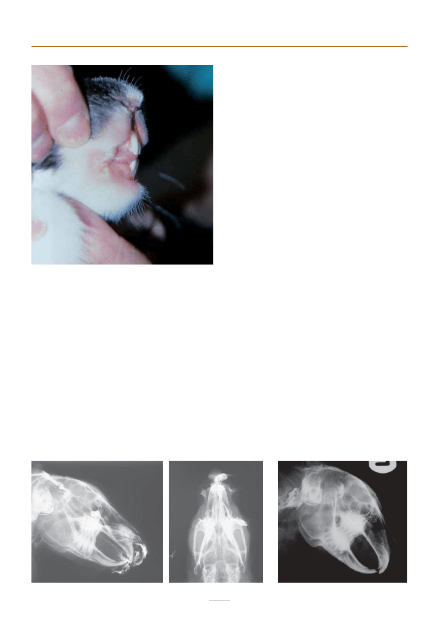
The fi rst incisor teeth have a chisel-like occlusal surface (Fig 1). The
thicker layer of labial enamel means that the lingual side wears
more quickly, forming the chisel shape of the cutting surface.
At rest the tips of the mandibular incisors fi t between the fi rst
and second maxillary incisors. Functionally the incisor teeth are
used with a largely vertical scissor-like slicing action to cut food.
During incisor use the cheek teeth are out of occlusion. Incisor
wear, growth and eruption are balanced in a normal rabbit at a
rate of about 3mm per week.
Cut food is prehended by the lips and passed to the back of the
mouth for grinding. Food is ground by the cheek teeth with a
wide lateral chewing action, concentrating on one side at a time.
The mandible is narrower than the maxilla, and the cheek teeth
are brought into occlusion by lateral mandibular movement. The
mandible is moved caudally to allow chewing, and the incisors
are separated during this phase.
The natural rabbit diet of grasses and other leafy plants is highly
abrasive as it has a high content of silicate phytoliths, so there is
normally rapid wear of the cheek teeth, around 3mm per month
in a wild rabbit, balanced by equally rapid tooth growth and
eruption. Mandibular incisors and cheek teeth grow and erupt
faster than maxillary teeth.
Maxillary and mandibular bone growth, development and
maintenance is also dependent on the mechanical stresses to
(1)Anna Meredith MA VetMB CertLAS DZooMed MRCVS RCVS Recognised Specialist in Zoo and Wildlife Medicine Head of Exotic Animal Service
University of Edinburgh Royal (Dick) School of Veterinary Studies Easter Bush Veterinary Centre Midloathian GB- EH25 9RG
E-mail:anna.meredith@ed.ac.uk
This paper was commissioned by
FECAVA for publication in EJCAP.
56
Rabbit dentistry - A. Meredith
which it is subjected. Rabbits which do not spend prolonged
periods chewing typically show poor jaw bone development,
or atrophy, at muscle insertions. This is most prominent in the
area of insertion of the pterygoid (medial) and masseter (lateral)
muscles into the ramus; the bone in this area may be so thin that
it is transparent or there may even be a perforation where the
bone has atrophied completely.
The nasolacrimal duct of the rabbit passes close to the apex of
the maxillary incisors and the fi rst maxillary premolar. (Fig 2)
Clinical signs of dental disease
Dental disease is one of the most common reasons for presentation
of a rabbit to the veterinary surgeon, although this fact may not
be immediately apparent. The commonest signs are:
– Anorexia
– Weight loss
– Facial swellings/asymmetry
– Ocular discharge
– Lack of grooming
– Accumulation of caecotrophs
– Fly strike (myiasis)
Any of these should all alert the practitioner to the possibility of
dental disease, and a full dental examination should be carried
out. Even in rabbits with no apparent clinical signs, assessment
of the teeth should always be an essential part of the clinical
examination, with as detailed an examination as is possible in a
conscious animal being performed.
Clinical examination
A dental examination should be preceded by a full history,
including a detailed dietary history. Clinical examination should
include:
– Facial palpation – for any bony or soft tissue swellings,
especially palpation of the ventral border of the mandible
where elongated apices may be present.
– Assessment of degree of lateral movement of the mandible
– Examination of length, quality and occlusion of the incisors
– Examination of the cheek teeth
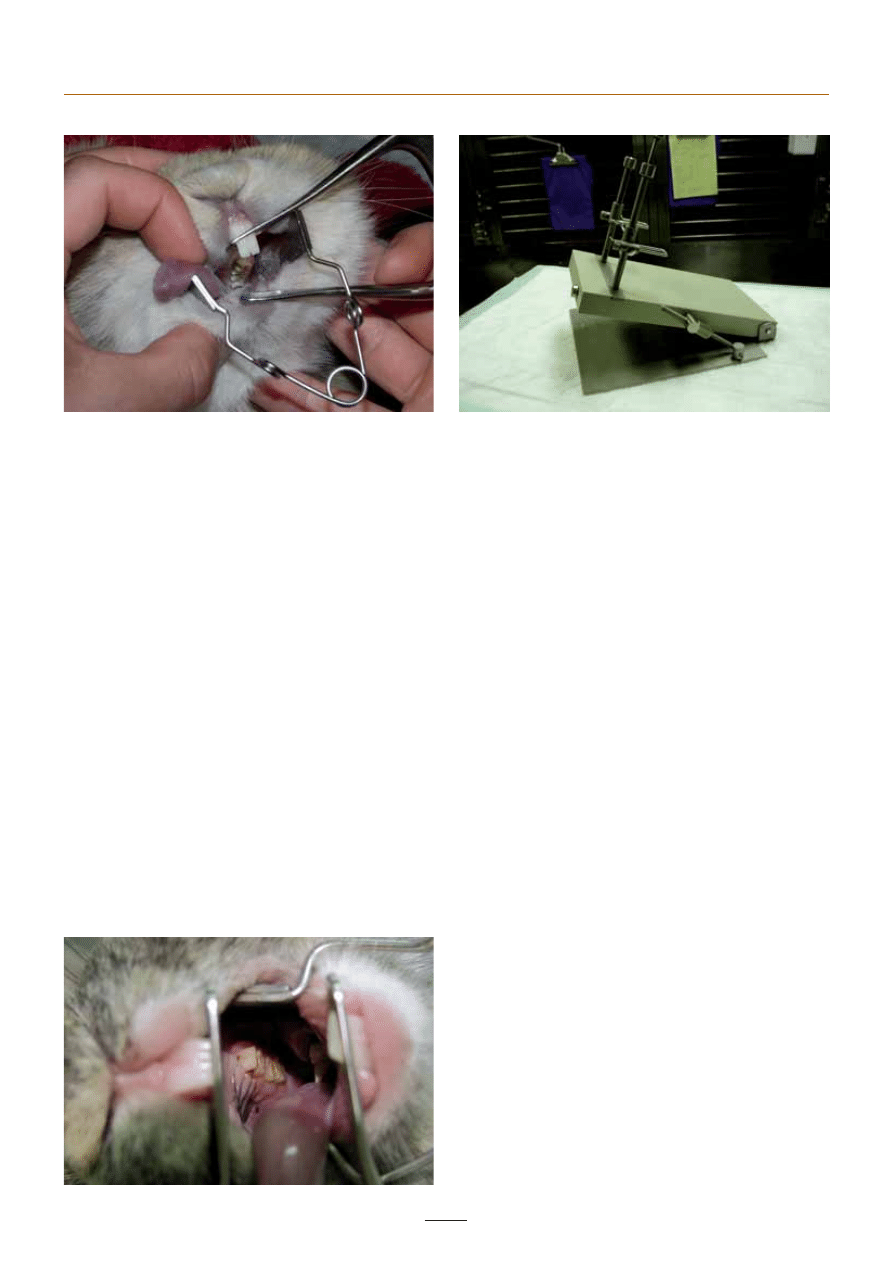
An initial examination of the cheek teeth can be carried out
in the conscious animal, with use of an otoscope, although it
must be recognised that visibility and detection of abnormalities
will be limited. It is estimated that conscious examination will
reveal only 50% of abnormalities, however. If dental disease is
suspected or lesions are detected in the conscious examination,
examination under deep sedation or anaesthesia must be
performed. This requires the use of specialist gags and cheek
retractors to enable good visualisation (Fig 3). Even then, it is
estimated that only 75% of lesions will be detected, with the
remainder only being picked up on post-mortem examination
(D A Crossley personal communication).
1. Normal incisors, demonstrating the chisel-shaped occlusal
surface
2. Contrast radiography of the nasolacrimal duct, lateral and DV views
4. Normal lateral skull radiograph
57
EJCAP - Vol. 17 - Issue 1 April 2007
Radiography
Abnormalities of the reserve crown and apex can only be
assessed radiographically, and radiography is an essential part of
a complete dental examination, enabling a full diagnosis, staging
and a judgement of prognosis [17]. Computed tomography (CT)
is also a very useful diagnostic tool, especially for assessment of
dental-associated abscesses, and is being used more widely.
Standard views are dorsoventral and lateral, plus a rostrocaudal
view is also useful. After assessment of these, oblique views may
be necessary to separate superimposed areas of interest.
When interpreting radiographs, possession of radiographs of
a normal animal ( Fig 4), and a normal prepared skull, can be
very useful. However, it should be recognised that there is a
great variety in shape and structure of rabbit skulls depending
on breed. The main points to assess are:
– Clinical (supragingival) crown length
– Position of the apices (elongation/intrusion)
– Degree of rostral convergence of the palatine bone and the
ventral border of the mandible. In a normal animal there is
generally some convergence, while elongation of the cheek
teeth leads to this being lost and the palatine bone and ventral
border of the mandible becoming parallel or even slightly
divergent. There is some breed variation, however.
– Shape of occlusal surfaces – incisors should be chisel-shaped,
cheek teeth should show an even zigzag pattern, even when
superimposed on the lateral view. Waves or steps may be
detected.
– Alveolar bone quality. There should be a fi ne lucent line
between the alveolar bone and the subgingival crown. If this
is blurred it can indicate ankylosis. Areas of increased bone
lucency may indicate infection or abscessation
Dental disease
Tooth elongation – eruption rate exceeding wear rate
This is the probably the commonest cause of dental disease in pet
rabbits and presents as a progressive pattern of abnormalities.
Rabbits on a low fi bre and high carbohydrate diet have reduced
tooth wear or attrition, resulting in elongation of the crown.
It is noticeable that rabbits consuming a low fi bre mixed grain
or pelleted diet tend to crush these items with an “up and
down” motion rather than the lateral grinding motion employed
when eating a highly fi brous diet. Defi ciency of calcium and
vitamin D as a result of selective feeding and lack of exposure
to sunlight respectively, have also been proposed as causative
or exacerbating factors, [9,12] although opinions vary on the
signifi cance of these.
Elongation causes occlusion of the cheek teeth at rest, resulting
in increased intrusive pressure. As elongation continues, the
mandible and maxilla are forced apart (seen radiographically as
the palatine shadow and ventral border of the mandible becoming
more parallel [13] and the masseter muscles stretched, which
also results in increased intrusive pressure. The teeth start to
intrude (apices become palpable as bony mandibular swellings)
and the crowns tip and/or rotate. Clinically, slight elongation of
the supragingival crown is diffi cult to appreciate, but it is more
obvious radiographically. As elongation and disrupted eruption
continue the altered forces and reduction in lateral movement
during chewing lead to the formation of ‘spurs’ on the lingual
occlusal surface of the mandibular cheek teeth and the buccal
3. a) Visualisation of the cheek teeth requires anaesthesia and the use of incisor gags and cheek pouch retractors. (Picture courtesy D.A
Crossley) b) A table top gag is also commercially available for this purpose, and allows single-handed oral inspection
5. A large lingual spur is visible on the left mandibular premolar
in this rabbit (Picture courtesy D.A Crossley)
58
Rabbit dentistry - A. Meredith
surface of the maxillary cheek teeth (Fig 5). Spurs or spikes, even
as small as 0.1mm, are always signifi cant and indicate a relatively
advanced stage of disease, and can cause great discomfort and
pain.
Elongation of the cheek teeth prevents the mouth from closing
fully (Fig 6). This separates the incisor teeth reducing their wear
until they have elongated suffi ciently to compensate. Beyond
a certain level of elongation the incisors no longer function
adequately and occlusal wear abnormalities become apparent,
i.e. a secondary incisor malocclusion and elongation occurs (Fig
7). Thus any rabbit presenting as an adult (>3-4 months) with
incisor problems should always be checked for cheek tooth
disease.
Elongation of the maxillary cheek teeth can impinge on the
nasolacrimal duct and cause bony distortion and blockage,
resulting in ocular discharge, with or without associated
infection. Elongation of the maxillary incisors can have the same
effect on the duct more rostrally. Contrast radiography of the
nasolacrimal duct is a useful technique (See Fig 2).
The exact pattern of disease progression varies amongst
individuals and depends on the degree of elongation and
dysplasia. In many rabbits severe dysplasia may eventually result
in complete cessation of growth due to ankylosis and resorption
of the teeth (see below), which, perhaps paradoxically, can
improve or even resolve the associated clinical signs.
Jaw length abnormalities
Primary incisor malocclusion and overgrowth is seen with
mandibular prognathism/maxillary brachygnathism in some
dwarf and lop breeds (Fig 8). In these cases the problem can be
detected at a very early age. It is common for the mandibular
incisors to become straighter preventing any correction of the
problem in mild cases. The maxillary incisors are not worn, but
contact with the mandible maintains occlusal pressure so the
tight spiral curvature of growth continues, the teeth eventually
penetrating the palate or cheek if left untreated. Regular crown
reduction or, preferably incisor extraction, is indicated for
affected animals.
Traumatic injury
Separation of the mandibular symphysis is the most common
accidental injury. Pulp exposure may occur associated with
6. a) Wild rabbit mandible, showing short cheek teeth. b) A domestic rabbit mandible, demonstrating elongation of the cheek teeth. (Picture
courtesy D.A Crossley)
7. Lateral skull radiograph showing marked cheek tooth elongation
and a secondary (acquired) incisor malocclusion (Picture courtesy
D.A Crossley)
8. Primary incisor malocclusion
59
EJCAP - Vol. 17 - Issue 1 April 2007
both dental fractures and trimming by a veterinary surgeon.
If the exposure is small and the blood supply to the pulp is
undamaged it may heal unaided, but many cases require partial
vital pulpectomy and vital pulp therapy, a specialist procedure.
In untreated cases pulpitis and pulp necrosis are common, with
the formation of abscesses around the premolar tooth roots
days to months later (Fig 9).
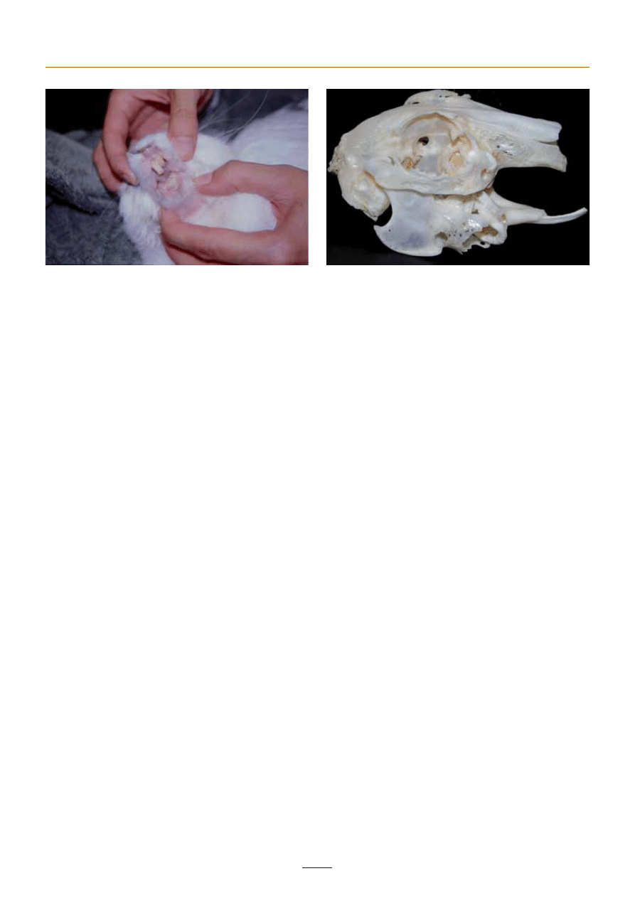
Periodontal disease and facial abscesses
Periodontal disease is common in rabbits, especially as the weak
structure of periodontal ligament renders it more likely to injury
and food impaction Elongation is a signifi cant factor, especially
with the cheek teeth, as this causes disruption of the tightly
packed occlusal surface and the opening up of gaps (diastemas)
between the teeth. Periodontal infection, often with anaerobic
oral bacteria such as Fusobacterium species, or Staphylococcus.
or Streptococus spp. [16] may spread to the tooth apex, leading
to endodontic lesions as the infection affects the pulp. Abscesses
frequently result from periodontal infection, or mucosal damage
caused by dental ‘spikes’. Unfortunately most dental abscesses
result in gross changes in the surrounding tissues including the
alveolar bone, so that there are residual problems even if the
abscess is successfully treated. If not treated early, abscesses
tend to behave as expansile masses, and they can displace teeth
(Fig 10).
Dental caries and resorption
High carbohydrate diets, reduced attrition and arrested eruption
predispose to caries (demineralisation), which can totally destroy
the exposed crown and progress subgingivally stimulating
resorption. Resorptive lesions are also seen associated with
periodontal disease and abscesses. If affected animals survive
long enough, replacement resorption may eventually result in
the disappearance of most of the cheek teeth. Affected rabbits
often do well on a suitably processed diet, though there are
continuing problems with progressive eruption remaining non-
occluding teeth.
Prevention and treatment of dental
disease
If rabbits are fed on fresh and dried grasses and other herbage,
dental disease is generally rare. Unfortunately the incidence in
some, particularly extreme dwarf and lop breeds, approaches
100% whatever their diet.
Coronal reduction
When detected in its very earliest stages, uncomplicated tooth
elongation can be corrected simply by dietary change. Established
tooth overgrowth may be helped by repeating burring at 4 to
6 week intervals. Radiographic assessment of tooth roots is
essential in all cases before undertaking treatment.
Incisors
In the unlikely event that problems are restricted to the incisor
teeth then these can easily be trimmed back to a normal length
and shape, or if repeated treatment is needed they can be
extracted. Incisor trimming can be performed without diffi culty
in conscious animals using either high or low speed dental
equipment. A high speed handpiece rotating at 2-400,000
times a second will cut the teeth with minimal effort, but care
should be taken to avoid overheating. Low speed burrs can also
be used but they are less effi cient, and should only be applied
for a maximum of 5 seconds before removal to allow cooling.
Diamond discs are hazardous and not recommended. Taper
fi ssure burrs are most effi cient with either high or low speed
handpieces, and soft tissues should be protected, e. g by placing
a wooden tongue depressor behind the incisors. The aim is to
restore normal crown height and the chisel shape. Care should
be taken not to expose the pulp. In the normal incisor pulp is
unlikely to extend more than 3mm above the gingival, but this
may be much more ( up to 17mm maxillary, 27mm mandibular)
in the overgrown incisor [6]. If exposed, vital pulp therapy using
calcium hydroxide cement is required, generally a specialist
procedure. Clippers should never be used as they leave sharp
edges and longitudinal cracks in the teeth and will often expose
the pulp. Clipping also releases a considerable amount of energy
9. Pus present at the mandibular incisors, which have stopped
growing, as a result of pulpitis and abscessation subsequent to
repeated trimming with nail clippers
10. Prepared skull showing extensive bony distortion associated
with mandibular and maxillary tooth root abscessation (Picture
courtesy D.A Crossley)
60
Rabbit dentistry - A. Meredith
into the tooth, concussing the pulp, and damaging the highly
innervated periodontal and periapical tissues, causing pain.
Cheek teeth
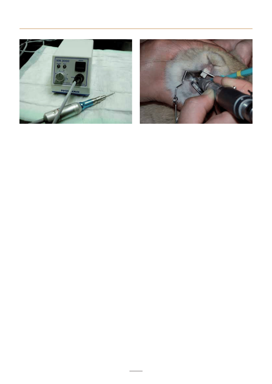
Coronal reduction of cheek teeth requires general anaesthesia,
and specialist mouth gags and cheek dilators. A straight slow
speed dental handpiece (Fig 11) with a long-shanked taper
fi ssure burr is recommended. A burr protector may be used (Fig
12). Avoidance of soft tissue trauma is vital, but can be diffi cult
due to the limited space and visualisation. Moistening the teeth
with a damp cotton bud can help prevent the burr “walking
off” the tooth. Hand held molar clippers may be used initially
to remove large spikes. There is little point in simply removing
sharp edges or ‘spikes’ as the main problem, tooth elongation,
is not then addressed. Hand held rasps are often too coarse and
not favoured by the author, as the forces applied can lead to
tearing the periodontal ligament and loosening teeth. However,
if powered equipment is not available, molar clippers (Fig 13)
and fi ne diamond rasps may be used.
The aim of coronal reduction is to shorten the crown and
attempt to restore the normal occlusal pattern. The stage of
disease will infl uence the treatment – in the early stages where
apical changes are minimal, restoration of normal anatomy and
function may be possible, but unfortunately this is seldom the
case as rabbits are not presented until the disease has reached a
later stage. In later stages, where changes in tooth morphology
are extensive, burring is palliative only, to remove painful spikes
and spurs and reduce crown height. Where changes are very
severe and eruption has ceased due to ankylosis or major damage
to the periapical tissues, coronal reduction is not indicated as
the teeth cannot grow again to restore occlusion and chewing
ability will be removed. In summary, coronal reduction is
advocated until eruption has ceased. Coronal reduction takes
teeth out of occlusion, removing intrusive pressure, so allowing
teeth to erupt as normally as possible. Radical reduction may
expose sensitive dentine and cause discomfort. Burring removes
the transverse occlusal ridging so chewing effi ciency is greatly
reduced until occlusion is resumed and ridging re-forms through
differential wear. It also may take some time for the jaw muscles
to recover their ability to contract fully after radical coronal
reduction. Repeated treatments, initially at 4-6 week intervals,
are generally necessary, but these intervals will generally increase
as the pattern of cheek tooth eruption becomes apparent
Early caries may be eliminated by burring away the affected
tissue. However, they often re-form unless the diet is corrected
and the coronal reduction may result in abnormal wear of
opposing teeth. Periodontal pockets deeper than 3mm are
diffi cult to clean in rabbits. Standard subgingival curettes may
be used but small dental excavators are often more effective.
Deeper pocketing is usually associated with abscessation in
which case the tooth will need extracting. This will also result in
abnormal wear of opposing teeth.
Extraction of teeth
Principles of extraction in rabbits are the same as for removal of
brachydont teeth in cats and dogs, i.e:
– Assessment
– Treatment planning
– Anaesthesia
– Cleansing of the operative fi eld
– Incision of the gingival attachment
– Severance of the periodontal ligament
– Enlargement of the alveolus
– Removal of supporting alveolar bone if necessary
– Gentle lifting of the detached tooth from its socket
– Encouragement of formation of a stable alveolar blood clot
Analgesia must be provided in the post-operative period. The
rabbit should be bright, alert and eating within 2-4 hours
postoperatively following appropriate anaesthesia and analgesia.
If substantial soft tissue or bone trauma was present (or created
iatrogenically) then a nasogastric tube may be used for nutritional
supplementation until the rabbit is able to eat normally. The
animal should be weighed daily in the post-operative period to
ensure weight loss does not occur. Food items must be prepared
in bite sized particles; vegetables may be chopped or grated. If
11. An example of a low speed dental machine and handpiece
12. Coronal reduction of cheek teeth using a low speed handpiece with
taper fissure burr and protector (Picture courtesy D.A Crossley)
61
EJCAP - Vol. 17 - Issue 1 April 2007
the animal does not eat voluntarily within 4 hours, nutritional
and fl uid support must be instigated. The normal rabbit uses the
incisors for grooming, so if these have been removed the rabbit
should be groomed regularly to prevent matting of the coat.
Incisor removal
Radiography is required before incisor removal to establish any
associated pathology and molar involvement [2]. The gingival
attachment around the incisor is cut using a hypodermic needle
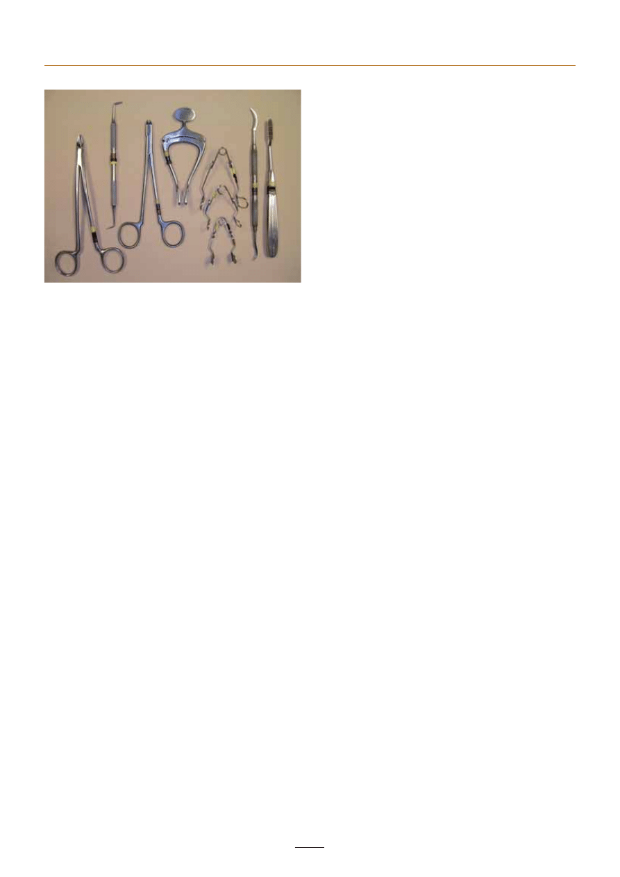
or a no 11 scalpel blade. An incisor elevator/luxator (See Fig 13)
(or blunted hypodermic needle) is then inserted along the mesial
aspect of the tooth to break down the periodontal ligament. The
elevator should follow the line of the tooth taking into account
its natural curvature. Gentle but sustained pressure is exerted on
the mesial and distal aspect of the tooth until it is loosened – it
is generally not necessary to luxate the ligament on the buccal
or lingual/palatal surfaces as it is so weak here. Once loosened,
the tooth should be gently rotated and pressed back into the
socket to destroy apical germinal tissue – failure to do this will
result in tooth regrowth, and even when this is done incisors will
occasionally regrow [14]. Alternatively, the apical tissue may be
debrided with a small curette after extraction of the tooth. The
tooth is then extracted using gentle traction. Excessive traction
may result in fracture of the teeth especially if they are of poor
quality. All 6 incisors should be removed; the small incisors (peg
teeth) require minimal luxation. The alveolus may be packed
with an anticoagulant sponge to limit haemorrhage in the post
operative period. The gingiva may be left to heal by granulation,
or closed with fi ne (5/0) absorbable suture material. If a tooth
breaks, the rabbit can be re-presented a few weeks later when
the crowns have re-erupted for completion of the extraction. If
the periapical tissues have been damaged, regrowth may not
occur and surgery may be required to retrieve the stump before
it serves as a nidus for infection or progresses to tooth root
abscessation.
Cheek tooth extraction
Cheek tooth extraction can be very diffi cult unless the tooth
is already loosened by periodontal disease. The most common
cause for extraction is in association with facial abscess
treatment (see below). Some abnormal cheek teeth may be
extracted per os by simple traction if the periodontal ligament
is weak or root pathology is such that the tooth is loose. The
curvature of the tooth should be taken into account when
attempting to extract the tooth. If the periodontal ligament is
still intact, it may be broken down using a modifi ed elevator
and the tooth extracted orally (see Fig 13 for molar elevator/
luxator and extraction forceps). The small size of the oral cavity
relative to the instrument makes intra-oral manipulation of the
tooth diffi cult. Once loosened the tooth should be intruded
into its alveolus and manipulated to help destroy any remaining
germinal tissue prior to removal. The pulp should remain in the
extracted tooth. If not, the germinal tissues are probably intact
and should be actively curetted using a sterile instrument. If the
germinal tissues are left intact the tooth will regrow, possibly as
a normal tooth, but more likely with gross deformity, in some
cases forming a pseudo-odontoma within the jaw bone.
Ankylosis of the tooth makes extraction very diffi cult and
an open technique is required. The removal of a molar via a
buccotomy incision, removal of alveolar bone and replacement
of a gingival fl ap requires careful technique and intensive post-
operative care to ensure a successful recovery.
It should be remembered that each molar opposes with two
others. These teeth may need corrective trimming following
extraction of one opposing tooth and so the rabbit should be
checked regularly.
Treatment of dental abscesses
The three main components of successful dental abscess
treatment are:
– Surgical removal/debridement of the abscess and any
associated teeth and infected bone
– Local antibiosis
– Systemic antibiosis
Surgical removal should be extracapsular where possible and
all associated teeth and infected bone must be removed. A
common reason for recurrence of abscesses, in the author’s
opinion, is failure to perform suffi ciently aggressive surgery.
Radiography is an essential part of the pre-surgical assessment,
in order to identify which tooth/teeth are involved and the
extent of involvement of the surrounding tissues.
Local antibiosis may be achieved in several ways. Installation
of antibiotic-impregnated polymethylmethacrylate (AIPMMA)
beads into the defect created by surgical removal is a common
technique that allows locally high antibiotic levels with little
systemic absorption [1,15]. Systemic antibiotics are given for
2-3 weeks post-operatively. The choice of antibiotic should
preferably be based on culture and sensitivity results. PMMA
with gentamicin already incorporated may be purchased
directly (e.g Refobacin® Bone Cement R
(a)
). Pre-made beads
are available (e.g Septopal®
(b)
) but these are often too large
for use in rabbits. AIPMMA beads are rapidly encapsulated by
13. Dental equipment available for rabbits. From left to right: molar
cutters, Crossley molar elevator/luxator, molar extraction forceps,
incisor gag, cheek dilators, Crossley incisor elevator/luxator, rasp
(d)

fi brous tissue, after which only tissues up to 3mm away receive
the high concentrations of antibiotic. Thus placing them within
the abscess capsule will be ineffective. The author and others
(David Crossley personal communication) have had good success
fi lling the surgically-created defect with doxycycline gel (e.g
Atridox®
(c)
). This is also useful for packing defects secondary to
periapical infection. Both these techniques involve closure of the
wound, enclosing the implant. AIPMMA beads do not generally
need to be removed, as they are biologically inert. Packing the
cavity with calcium hydroxide is favoured by some but has been
reported to cause serious tissue damage and necrosis [1].
An alternative technique of achieving local antibiosis is to
marsupialise the surgical cavity and allow it to heal by granulation,
while fl ushing with or instilling antibacterial/antibiotic solutions.
This technique has the advantage of allowing more control
over continued treatment of the site and easier monitoring and
detection of recurrence.
Systemic antibiosis is generally not necessary for more than 2-3
weeks post-operatively in case surgery causes a bacteraemia.
However, in cases where complete excision is not possible,
long term systemic antibiosis may be necessary. Long term use
of antibiotics that have good effi cacy against the anaerobic
organisms involved with dental abscesses, such as penicillin
G (by subcutaneous injection, never orally) are anecdotally
reported to have good success in preventing progression of
abscesses, or helping to achieve a cure when combined with
surgical debridement.
(a) Biomet Cementing Technologies AB, Forskaregatan 1, SE-275
37Sjöbo,Sweden www.bonecement.com
(b) BioMet Europe, Dordrecht, Netherlands
(c) CollaGenex Pharmaceuticals Inc. 41 University Drive, Suite 200
Newtown, PA 18940
(d)
Veterinary Instrumentation Limited, Broadfi eld Road, Sheffi eld, S8
OXL United Kingdom. www.vetinst.com
References and further reading
Note: The following references are not referred to in the text and are
intended as suggested futher reading. 3, 4, 5, 7, 8, 10, 11, 18
[1] BENNETT (R.A.) - Managing abscesses of the head. BSAVA
Congress Scientifi c Proceedings, 2001, 15-16
[2] BROWN (S.A.) - Surgical removal of incisors in the rabbit. Journal
of Small Animal Exotic Medicine, 1992, 1(4):150-153
[3
CROSSLEY (D.A.) - Clinical aspects of lagomorph dental anatomy:
the rabbit (Orytolagus cuniculus). J Vet Dent, 1995, 12(4):137-
140.
[4] CROSSLEY (D.A.) - Prevention and treatment of dental problems
in pet rabbits and rodents. Proceedings of DVG, Hanover, August
1997.
[5] CROSSLEY (D.A.) Dental disease in lagomorphs and rodents.
In: Kirk’s Current Veterinary Therapy XIII, ed. Bonagura JD. WB
Saunders, Philadelphia, 2000, 1133-1137.
[6] CROSSLEY (D.A.) - Risk of pulp exposure when trimming rabbit
incisor teeth. Proceedings of the 10th European Veterinary Dental
Society Annual Congress, Berlin, 2001, 175-196.
[7] GORREL (C.) - Dental diseases in lagomorphs and rodents. In:
Veterinary Dentistry for the General Practitioner, Saunders,
London, 2004, 175-196.
[8] HARCOURT-BROWN (F.M.) - A review of clinical conditions in
pet rabbits associated with their teeth. Veterinary Record, 1995,
137:341-346.
[9] HARCOURT-BROWN (F.M.) - Calcium defi ciency, diet and dental
disease in pet rabbits. Veterinary Record 1996, 139: 567-571.
[10] HARCOURT-BROWN (F.M.) - Diagnosis, treatment and prognosis
of dental disease in pet rabbits. In Practice, 1997, 19:407-421.
[11] HARCOURT-BROWN (F.M.) - Dental diseases. In :Textbook of
Rabbit Medicine, Butterworth Heinemann, 2002, 165-205.
[12] HARCOURT-BROWN (F.M.), BAKER (S.J.) - Parathyroid hormone,
haematological and biochemical parameters in relation to dental
disease and husbandry in rabbits. JSAP, 2001, 42(3):130-136
[13] HOBSON (P.) - Dentistry. In : Manual of Rabbit Medicine and
Surgery, BSAVA Publications, 2006, 184-196.
[14] STEENKAMP (G.), CROSSLEY (D.A.) - Incisor tooth regrowth in
a rabbit following complete extraction. Veterinary Record, 1999,
145: 585-586.
[15] TOBIAS (K.M.), SCHNEIDER (R.K.), BESSER (T.E.) - Use of
antimicrobial-impregnated polymethylmethacrylate. JAVMA,
1996, 208: 841-844
[16] TYRRELL (K.L.), CITRON (D.M.), JRENKINS (J.R.), GOLDSTEIN
(E.J.) - Periodontal bacteria in rabbit mandibular and maxillary
abscesses. J Clin Micro, 2002, 40:1044-1047.
[17] VERSTRAETE (F.J.M.), CROSSLEY (D.A.), HORNOF (W.J.) -
Diagnostic imaging of dental disease in rabbits. Proceedings of
18th Annual Veterinary Dental Forum, Fort worth, Texas, 2004.
[18] WIGGS (R.), LOBPRISE (H.) - Dental and oral disease in rodents
and lagomorphs. In : Veterinary Dentistry – Principles and Practice,
Lippincott-Raven, Philadelphia, 1997, 518-537.
Rabbit dentistry - A. Meredith
Wyszukiwarka
Podobne podstrony:
Rabbit dentistry1 (2)
BSAVA Manual of Rabbit Surgery Dentistry and Imaging
BSAVA Manual of Rabbit Surgery Dentistry and Imaging
2005 4 JUL Veterinary Dentistry
2013 3 MAY Clinical Veterinary Dentistry
Dentistry?
Dentist Theme
Magiczne przygody kubusia puchatka 33 RABBIT'S HARD FUCK
checklist dentists
Magiczne przygody kubusia puchatka 16 FOLLOW THE WHITE RABBIT
dentista, dottore słownictwo
2005 4 JUL Veterinary Dentistry
THE TALE OF PETER RABBIT
dentist
ANIMALS (4 16 14) RABBIT
White Rabbit WEBSTER, K
happy easter heineken easter playboy bunny rabbit eggs demotivational poster 1270399591
więcej podobnych podstron