
J
OURNAL OF
C
LINICAL
M
ICROBIOLOGY
, Sept. 1995, p. 2485–2488
Vol. 33, No. 9
0095-1137/95/$04.00
10
Copyright
q 1995, American Society for Microbiology
Detection of Measles Virus RNA in Urine Specimens
from Vaccine Recipients
PAUL A. ROTA,* ALI S. KHAN, EDISON DURIGON,† THOMAS YURAN,
YVONNE S. VILLAMARZO,
AND
WILLIAM J. BELLINI
Division of Viral and Rickettsial Diseases, National Center for Infectious Diseases, Centers for
Disease Control and Prevention, Atlanta, Georgia 30333
Received 7 March 1995/Returned for modification 18 April 1995/Accepted 30 May 1995
Analysis of urine specimens by using reverse transcriptase-PCR was evaluated as a rapid assay to identify
individuals infected with measles virus. For the study, daily urine samples were obtained from either 15-
month-old children or young adults following measles immunization. Overall, measles virus RNA was detected
in 10 of 12 children during the 2-week sampling period. In some cases, measles virus RNA was detected as early
as 1 day or as late as 14 days after vaccination. Measles virus RNA was also detected in the urine samples from
all four of the young adults between 1 and 13 days after vaccination. This assay will enable continued studies
of the shedding and transmission of measles virus and, it is hoped, will provide a rapid means to identify
measles infection, especially in mild or asymptomatic cases.
Despite the existence of an effective vaccine, measles virus
continues to cause sporadic outbreaks and epidemics of dis-
ease in the United States and throughout the world. Most
recent outbreaks have involved either children who were too
young to be vaccinated or older children and teenagers (5 to 19
years), most of whom had been previously vaccinated (3, 8).
Because of the sporadic nature of outbreaks in populations
with high rates of vaccination, the altered presentation of clin-
ical signs that occurs in ‘‘mild measles’’ infections (1, 11, 20),
and the presence of other exanthem-causing infections, effec-
tive public health measures to control measles outbreaks are
more dependent on laboratory confirmation of infection than
on diagnosis based on clinical presentation. Currently available
diagnostic techniques, which include virus isolation, viral anti-
gen detection, and serologic antibody studies, are very sensitive
and specific. However, these techniques are labor intensive,
require specimen collection by medically trained personnel,
and would be inappropriate for screening large numbers of
individuals.
The detection of measles virus RNA in urine by using re-
verse transcriptase-PCR (RT-PCR) would be a potentially
rapid means of detecting measles infections with a clinical
specimen which is more readily and conveniently accessible
than serum or nasopharyngeal aspirates. Collection of urine
specimens could be done in the absence of medical profession-
als, and on-site specimen-processing requirements are mini-
mal. Measles virus can be isolated from urine specimens from
infected individuals for as long as 10 days after the onset of the
rash (16, 28), and measles antigen has been detected by im-
munofluorescence in urine samples from asymptomatic case
contacts (5).
(This work was presented at the Annual Meeting of the
American Society for Virology, Madison, Wis., July 1994.)
Since urine specimens from naturally infected individuals
were unavailable at the time this study was conducted, the
RT-PCR assay was evaluated by using specimens obtained
from recently vaccinated individuals. In all cases, RNA was
extracted from urinary sediment by the guanidinium acid-phe-
nol method (9) and resuspended in 25
ml of RNase-free water.
For the measles virus-specific RT-PCR, a nested set of prim-
ers that hybridized to the nucleoprotein (N) gene was used
(MV41, CAT TAC ATC AGG ATC CGG; and MV42, GTA
TTG GTC CGC CTC ATC). The internal primers (MV43,
digoxigenin [DIG] -GA GCC ATC AGA GGA ATC A; and
MV44, DIG-CA TGT TGG TAC CTC TTG A) were 5
9 la-
beled with DIG. The target sequences for these primers are
located between bases 57 and 389 of the coding region of the
N gene, and these sequences are conserved among the N genes
of all wild-type measles viruses examined thus far (24). DIG-
5
9-labeled primers that amplified beta-actin mRNA (BA4 and
BA1) were used as controls for RNA extraction.
Before the RT reaction, the RNA was heated to 95
8C for 90
s and then placed on ice. The RT reaction mixture contained
50 mM Tris-HCl (pH 8.3), 8 mM MgCl
2
, 30 mM KCl, 5 mM
dithiothreitol, 1 mM each deoxynucleoside triphosphate
(dNTP), 10
mM each forward and reverse primer (MV41 and
MV42 or BA1 and BA4), 24 U of avian myeloblastosis virus
RT, and 40 U of human placental RNase inhibitor. The reac-
tion mixture was incubated at 42
8C for 45 min and then at 958C
for 5 min.
For PCR, 5
ml of the cDNA sample was added to a 45-ml
PCR mixture containing 10 mM Tris-HCl (pH 8.3), 50 mM
KCl, 1.5 mM MgCl
2
, 0.01% gelatin, 200
mM each dNTP, 5 mM
each primer (MV43 and MV44 or BA1 and BA4) and 5 U of
Taq DNA polymerase. PCR conditions were as follows: 94
8C
for 1 min, 50
8C for 1 min, and 728C for 1 min. After 39 cycles,
20
ml of each sample was analyzed by electrophoresis on a
1.5% agarose gel. DNA was visualized by ethidium bromide
staining and UV illumination. Immunochemiluminescence de-
tection of PCR products that were not visible after ethidium
bromide staining was performed as described previously (10).
In all RT-PCR assays, samples containing water were used
as contamination controls. Positive control RNA was extracted
from Vero cells that had been infected with measles virus, and
negative control RNA was extracted from uninfected Vero
cells or from urine specimens donated by laboratory personnel
who had not recently been vaccinated. For measurement of the
* Corresponding author. Mailing address: Measles Section, MS
G-17, REVB, Centers for Disease Control and Prevention, 1600
Clifton Rd., Atlanta, GA 30333. Phone: (404) 639-3308. Fax: (404)
639-1307. Electronic mail address: rota@beryl.biotech.cdc.gov.
† Present address: Department of Microbiology, University of Sao
Paulo, Sao Paulo, Brazil.
2485
sensitivity of the RT-PCR assay, measles virus N gene RNA
was synthesized in vitro from a plasmid template by using T7
RNA polymerase (25).
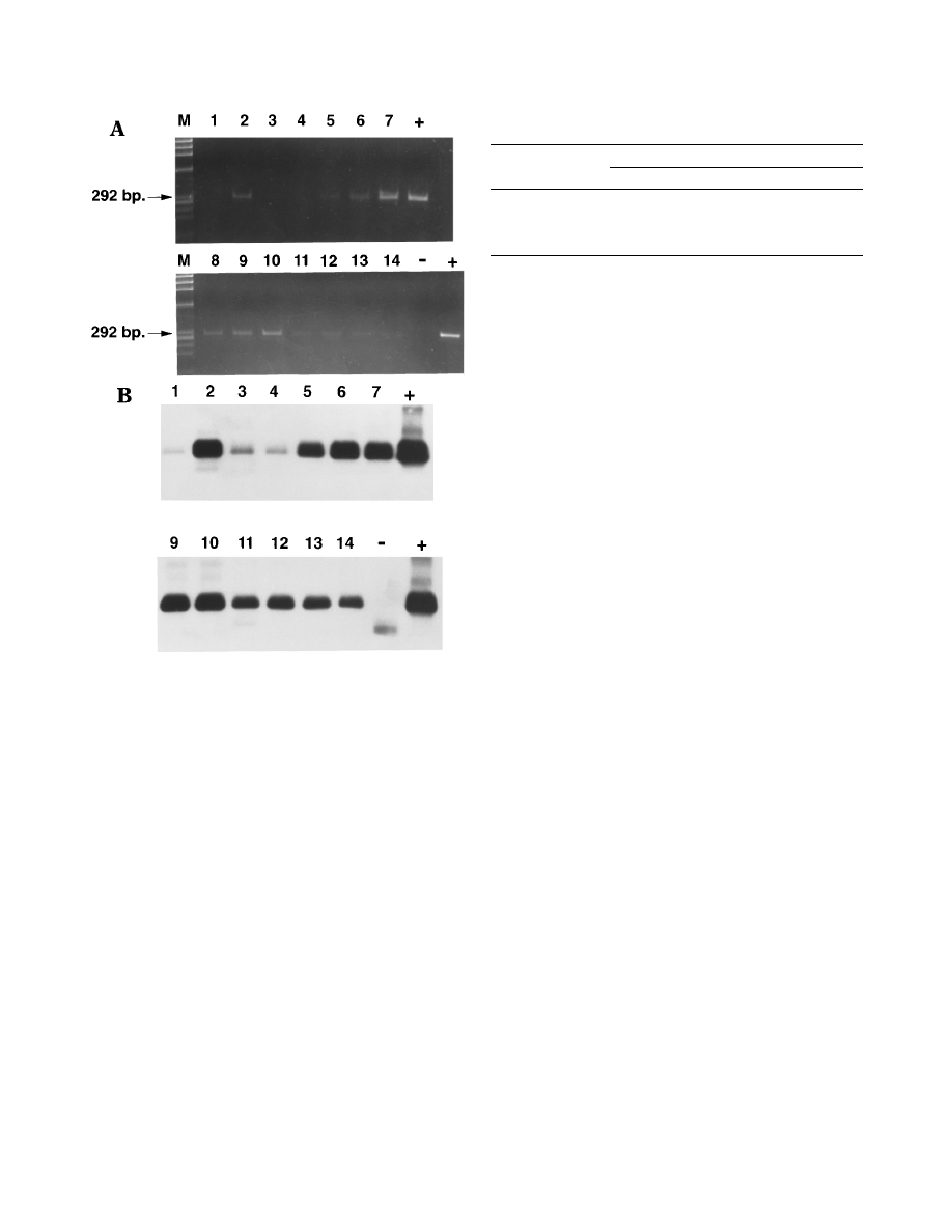
The product generated by the RT-PCR amplification of
measles virus RNA was a 292-bp DNA fragment that was end
labeled with DIG during the reaction (Fig. 1). The addition of
DIG to the second set of PCR primers increased the sensitivity
of detection of the PCR product by approximately 100-fold. As
little as 1.5 fg of in vitro-synthesized measles virus N gene
RNA could be detected, an amount that represents approxi-
mately 10
4
RNA molecules. The number of N gene mRNA
molecules in a single infected cell has been estimated to be
1,000 to 10,000 (7). Therefore, the RT-PCR assay was able to
detect as few as 1 to 10 infected cells.
First-voided morning urine samples were collected daily
from 12 children (age 15 months) over a 14-day period follow-
ing routine initial measles-mumps-rubella vaccination. The
time between collection and sample processing varied from 24
to 72 h. Many of the samples were highly contaminated with
bacteria upon arrival in the laboratory, and the volume of urine
obtained varied between 5 and 50 ml per specimen. A total of
144 specimens were received from the 12 children over the
14-day study period (there were 24 missing specimens).
The quality of the RNA extracted from all samples was
assessed by using exon-specific primers to amplify a 300-bp
region of beta-actin mRNA. The RNA extracted from the
urine samples appeared to be free of DNA contamination,
since the 300-bp actin PCR product was obtained after DNase,
but not RNase, treatment of the sample and there was no
evidence for a higher-molecular-weight PCR product that
would have been produced by amplification of the beta-actin
gene. No actin mRNA was amplified from 57 (52%) of 144
samples, indicating that extensive RNA breakdown had oc-
curred during storage, shipment, or RNA extraction (Table 1).
Measles virus RNA was detected in 48 (70%) of 69 actin-
positive samples and in 8 (11%) of 75 actin-negative samples
by using either the ethidium bromide or chemiluminescence
detection method (Table 1). The detection of measles virus
RNA in samples that were negative for actin mRNA could be
attributed to increased sensitivity of the measles virus PCR or,
more likely, to increased stability of measles virus RNA, which
would be associated with nucleocapsid structures. Overall,
measles virus RNA was detected in 56 (39%) of 144 samples.
Sequence analysis of several of the PCR products confirmed
that the appropriate region of the N gene of the measles
vaccine strain, Moraten (Attenuvax; Merck, Sharp and
Dohme, West Point, Pa.), was being amplified (23). Urine
samples donated by laboratory staff were processed in parallel
to the samples obtained from the vaccinated children. No mea-
sles virus RNA was detected in any of these control samples
(data not shown).
In some cases, measles virus RNA was detectable as early as
1 day after vaccination. In four samples, RNA was detected as
late as 14 days after vaccination. In Fig. 1, which shows the
results from one individual, the PCR product is visible by
ethidium bromide staining for 10 of the samples, but all of the
samples are positive with the chemiluminescence detection
method.
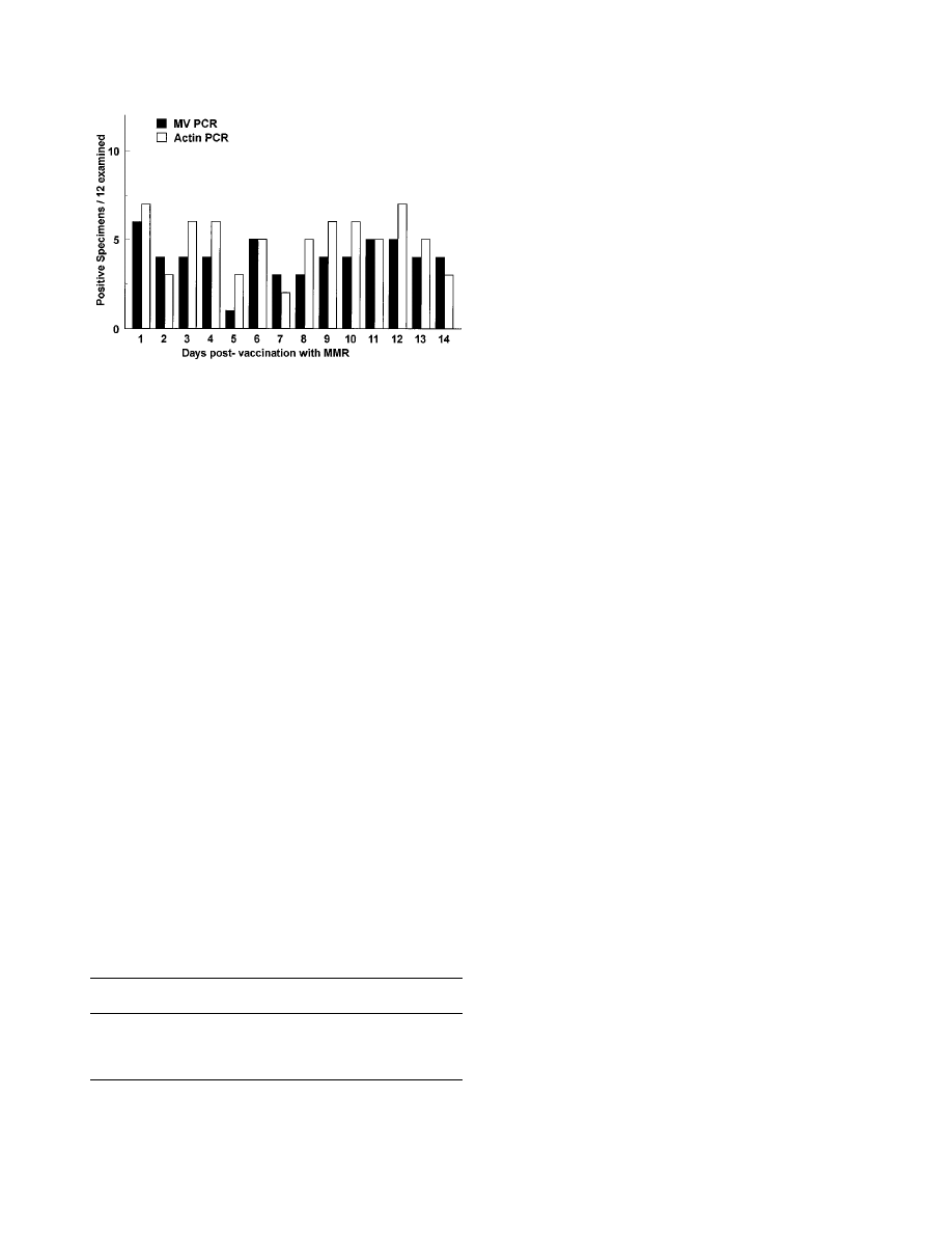
The number of measles virus-positive specimens remained
relatively constant during the 14-day sampling interval, with
between 1 and 6 of the 12 specimens positive for measles virus
RNA on any day (Fig. 2). Overall, measles virus RNA was
detected in at least one specimen from 10 (83%) of 12 of the
children. Of the 27 samples from the two children in whom
measles virus RNA was not detected, only 1 sample was pos-
itive for actin mRNA. This extensive RNA degradation was
probably due to poor specimen handling at the collection site.
For this study, the average numbers of actin-positive samples
and measles virus-positive samples were 5.1 and 4.6 per child,
respectively.
Urine specimens were also obtained from four healthy
young adults (ages 21 to 32 years) for 14 days after they re-
ceived a booster dose of measles-mumps-rubella vaccine.
These samples were of better quality than those obtained from
the young children, since larger volumes were obtained and the
times between collection, refrigeration, and RNA extraction
were shorter. In these cases, RNA was extracted from a max-
FIG. 1. RT-PCR analysis of urine samples from a single vaccinated child. (A)
Agarose gel electrophoretic analysis of PCR products after ethidium bromide
staining. Lane numbers indicate the day postvaccination for each sample. Posi-
tive (
1) and negative (2) controls and molecular size markers (M) are also
shown. (B) Chemiluminescence detection of PCR products from the RT-PCRs
shown in panel A (the sample from day 8 is missing).
TABLE 1. Detection of measles virus RNA in urine samples from
recently vaccinated children (n
5 12) by RT-PCR
Actin
mRNA
No. of samples with measles virus RNA:
Present
Absent
Total
Present
48
21
69
Absent
8
67
75
Total
56
88
144
2486
NOTES
J. C
LIN
. M
ICROBIOL
.
imum volume of 100 ml of urine; 81% of these samples were
positive for actin mRNA. While measles virus RNA was de-
tected in all four individuals (Table 2), it was detected in fewer
of the samples and in samples from only two of the individuals
after day 2. This suggests that preexisting immunity may have
reduced the extent of replication or shedding of the vaccine
virus.
During acute infection, measles virus is routinely isolated
from the urine for as many as 10 days after the onset of the
rash (16, 28). Viral antigen has been detected in multinucleate,
giant cells found in urinary sediment by using immunofluores-
cence (19) before or after cell culture amplification (22). De-
tailed microscopic and immunofluorescence studies have
shown that these antigen-bearing cells are exfoliative cells from
proximal renal tubules, collecting tubules, epithelial cells of
Bowman’s capsule, and the transitional epithelium of the renal
pelvis, ureter, and urinary bladder (6, 18, 27), suggesting that
the urinary tract is infected during measles infection. Other
morbilliviruses, such as canine distemper virus and phocine
distemper virus, also infect epithelial cells in the urinary tract
(2, 4, 14).
Measles virus antigen has been detected in the urinary sed-
iments of vaccinated individuals by immunofluorescence (19)
or, more recently, by RT-PCR (26a). In the study by Llanes-
Rodas and Liu (19) in 1966, the urine samples were obtained
during a measles vaccine trial. In that case, the test vaccine was
an earlier passage of the Edmonston virus (12) that had greater
reactogenicity than the more attenuated vaccine, Moraten
(17). In our study, individuals received the Moraten strain of
measles vaccine as measles-mumps-rubella vaccine.
Measles virus RNA was detected by RT-PCR in the urine
specimens from several of the vaccinated children as late as 14
days after vaccination. Because our research protocol was lim-
ited to only 14 days of specimen collection, we were unable to
determine the upper limit for the duration of viral RNA in
urine. In the previous study by Llanes-Rodas and Liu (19),
measles virus antigen was detected in urine as late as 16 days
after vaccination.
The finding that several of the urine samples were positive
for measles virus RNA as early as 1 day after vaccination was
surprising. In the previous study (19), none of the urine spec-
imens were positive by immunofluorescence before day 4.
Since a single cycle of viral replication would be expected to
take 17 to 24 h, it is unlikely that the RT-PCR detected the
progeny of virus replicating in the urinary tract. Rather, this
observation suggests that shortly after vaccination the input
virus or viral antigen, in the form of nucleocapsids, is deposited
directly into the bladder via interstitial fluid. This finding also
demonstrates the increased sensitivity of RT-PCR compared
with the immunofluorescence techniques that were used in
earlier studies (18). Unfortunately, most of the specimens were
of such poor quality that cytological studies or reisolation of
vaccine virus was not attempted. In the previous study (19),
attempts to isolate vaccine virus on cell culture were unsuc-
cessful.
The changing epidemiology of measles, in the form of mild
measles cases in previously vaccinated individuals (1, 11, 20),
suggests that more asymptomatic or subclinical cases might be
occurring. The frequency of such infections, which would not
meet the standard case definition of the Centers for Disease
Control and Prevention, is not known. Also, it is not known
whether individuals who do not display the full range of clinical
signs characteristic of measles infection are capable of trans-
mitting the virus to other susceptible individuals. In one pre-
vious study, urine samples from 5 of 12 measles case contacts
were positive for measles virus antigen even though only 1 of
these 5 contacts developed clinical signs (5).
In general, RT-PCR has proven to be a rapid and sensitive
method to detect measles virus RNA in a variety of clinical
specimens (13, 15, 21, 24, 26, 28). Successful RT-PCR ampli-
fication of measles virus RNA from urine samples now allows
the detection of measles virus RNA from a specimen that can
be obtained from a large number of individuals by noninvasive
means. We plan to use this assay to define further the extent of
asymptomatic or mild infection in case contacts during an
outbreak, to determine the role that these cases play in the
transmission of measles, and to measure the shedding patterns
of vaccine recipients. In future surveys, more care will need to
be taken to obtain and process the specimen in a manner that
minimizes RNA degradation.
We thank Joseph A. Wilber and J. David Smith of the Georgia
Department of Health and Human Resources, Ricks Eclemaus of the
Fulton County Health Department, Edward Lifshitz of Rutgers Uni-
versity, and Lyn Finelli of the New Jersey Department of Health for
supporting this study; Gloria Abley and Susan Estep for assistance in
recruiting study subjects; and Thomas Guyrick for specimen handling
and transport.
Financial support for this work was provided by the National Vac-
cine Program and the World Health Organization.
REFERENCES
1. Adcock, L. M., J. D. Bissey, and R. D. Feigin. 1992. A new look at measles.
Infect. Dis. Clin. N. Am. 6:133–148.
2. Appel, M. J. 1987. Canine distemper virus, p. 13–159. In M. J. Appel (ed.),
Virus infections of carnivores. Elsevier Science Publishers, New York.
FIG. 2. Time course of detection of measles virus RNA in urine specimens
from vaccinated children. Bar heights indicate numbers (total
5 12) of measles
virus (MV)-positive and actin-positive samples on each day of sampling. MMR,
measles-mumps-rubella vaccine.
TABLE 2. Detection of measles virus RNA in urine samples from
recently vaccinated young adults
a
Patient
no.
Age (yr)
No. of days
b
Days measles virus
positive
c
1
21
15
1, 2
2
24
13
1, 2
3
26
16
1, 2, 3, 4, 5, 6, 10, 13
4
32
14
9, 11
a
All subjects were positive for measles virus immunoglobulin G as determined
by enzyme immunoassay.
b
Number of days after vaccination that specimens were obtained.
c
Days on which measles virus RNA was detected by RT-PCR.
V
OL
. 33, 1995
NOTES
2487

3. Atkinson, W. L., and W. A. Orenstein. 1992. The resurgence of measles in the
United States, 1989–1990. Annu. Rev. Med. 43:451–463.
4. Blixenkrone-Moller, M. 1993. Biological properties of phocine distemper
virus and canine distemper virus. APMIS 34(Suppl.):5–51.
5. Boyd, J. F. 1983. A fourteen-year study to identify measles antigen in urine
specimens by fluorescent-antibody methods. J. Infect. 6:163–170.
6. Boyd, J. F., and N. Nedelkoska. 1967. Further observations on inclusion-
bearing cells in urinary sediment in infectious diseases. J. Clin. Pathol.
20:
835–840.
7. Cattaneo, R. G., A. Rebmann, A. Schmid, K. Baczko, V. ter Meulen, and
M. A. Billeter.
1987. Altered transcription of a defective measles virus ge-
nome derived from a diseased brain. EMBO J. 6:681–688.
8. Centers for Disease Control. 1992. Measles surveillance—United States.
Morbid. Mortal. Weekly Rep. 41(Suppl. 6):1–12.
9. Chomczynski, P., and N. Sacchi. 1987. Single-step method of RNA isolation
by acid guanidinium thiocyanate-phenol-chloroform extraction. Anal. Bio-
chem. 162:156–159.
10. Durigon, E. L., D. D. Erdman, B. C. Anderson, B. P. Holloway, and L. J.
Anderson.
1994. Immunochemiluminescent Southern blot assay for poly-
merase chain reaction detection of human parvovirus B19 DNA. Mol. Cell.
Probes 8:199–204.
11. Edmonson, M., D. Addiss, J. McPherson, J. Berg, S. Circo, and J. Davis.
1990. Mild measles and secondary vaccine failure during a sustained out-
break in a highly vaccinated population. JAMA 163:2467–2471.
12. Enders, J. F., and T. C. Peebles. 1965. Propagation in tissue cultures of
cytopathogenic agents from patients with measles. Proc. Soc. Exp. Biol. Med.
86:
277–286.
13. Esolen, L. M., B. J. Ward, T. R. Monench, and D. E. Griffin. 1993. Infection
of monocytes during measles. J. Infect. Dis. 168:47–52.
14. Fairchild, G., M. Wyman, and E. F. Donovan. 1967. Fluorescent antibody
test for canine distemper infection: detection of viral antigen in epithelial
tissues of experimentally infected dogs. Am. J. Vet. Res. 28:761–768.
15. Godec, M. S., D. M. Asher, P. T. Swoveland, Z. A. Eldadah, S. M. Feinstone,
L. G. Goldfarb, C. J. Gibbs, and D. C. Gajdusek.
1990. Detection of measles
virus genomic sequences in SSPE brain tissue by the polymerase chain
reaction. J. Med. Virol. 30:237–244.
16. Gresser, I., and S. L. Katz. 1960. Isolation of measles virus from urine. N.
Engl. J. Med. 275:516–523.
17. Hilleman, M. R., E. B. Buynak, R. E. Weibel, J. Stokes, J. E. Whitman, and
M. B. Leagus.
1968. Development and evaluation of the Moraten measles
virus vaccine. JAMA 206:587–590.
18. Lipsey, A. I., and R. P. Bolande. 1967. The exfoliative source of abnormal
cells in urine sediment of patients with measles. Am. J. Dis. Child. 113:677–
682.
19. Llanes-Rodas, R., and C. Liu. 1966. Rapid diagnosis of measles from urinary
sediments stained with fluorescent antibody. N. Engl. J. Med. 275:516–523.
20. Markowitz, L. E., F. W. Chandler, E. O. Roldan, M. J. Saldana, K. C. Roach,
S. S. Hutchins, S. R. Preblud, C. D. Mirchell, and G. B. Scott.
1988. Fatal
measles pneumonia without rash in a child with AIDS. J. Infect. Dis. 158:
480–483.
21. Matsuzono, Y., M. Narita, N. Ishiguro, and T. Togashi. 1994. Detection of
measles virus from clinical samples using the polymerase chain reaction.
Arch. Pediatr. Adolesc. Med. 148:289–293.
22. Minnich, L. L., F. Goodenough, and C. G. Ray. 1991. Use of immunofluo-
rescence to identify measles virus infections. J. Clin. Microbiol. 29:1148–
1150.
23. Rota, J. S., Z. D. Wang, P. A. Rota, and W. J. Bellini. 1994. Comparison of
sequences of the H, F, and N coding genes of measles virus vaccine strains.
Virus Res. 31:17–30.
24. Rota, P. A., A. E. Bloom, J. A. Vanchiere, and W. J. Bellini. 1994. Evolution
of the nucleoprotein and matrix genes of wild-type strains of measles virus
isolated from recent epidemics. Virology 198:724–730.
25. Sambrook, J., E. F. Fritsch, and T. Maniatis. 1989. Molecular cloning: a
laboratory manual, 2nd ed. Cold Spring Harbor Laboratory, Cold Spring
Harbor, New York.
26. Shimizu, H., C. A. McCarthy, M. F. Smaron, and J. C. Burns. 1993. Poly-
merase chain reaction for detection of measles virus in clinical samples. J.
Clin. Microbiol. 31:1034–1039.
26a.Shimizu, H., S. H. Waterman, M. A. Stein, and J. C. Burns. 1993. Poly-
merase chain reaction for the detection of measles virus genome in urine
sediment, abstr. 1080, p. 183a. In Abstracts of the 103rd Annual Meeting of
the American Pediatric Society and the 62nd Annual Meeting of the Society
for Pediatric Research.
27. Strom, J. 1973. Cytology of the urine in healthy persons and cytological
reactions in acute infections, especially with respect to the presence of
inclusion-bearing and giant cells. A study with application of Millipore pro-
cedure and Papanicolaou staining. Scand. J. Infect. Dis. 5:209–228.
28. Utz, J. P. 1974. Viruria in man. Prog. Med. Virol. 17:77–90.
2488
NOTES
J. C
LIN
. M
ICROBIOL
.
Wyszukiwarka
Podobne podstrony:
A unified prediction of computer virus spread in connected networks
A Model for Detecting the Existence of Unknown Computer Viruses in Real Time
Static Detection of Malicious Code in Executable Programs
Kolmogorov Complexity Estimates For Detection Of Viruses In Biologically Inspired Security Systems
Skill 12[1] Collection of Urine Specimen
Biological Models of Security for Virus Propagation in Computer Networks
Design of an Artificial Immune System as a Novel Anomaly Detector for Combating Financial Fraud in t
DIMENSIONS OF INTEGRATION MIGRANT YOUTH IN POLAND
The?uses of the Showa Restoration in Japan
There are a lot of popular culture references in the show
Comparative Study of Blood Lead Levels in Uruguayan
Effect of?renaline on survival in out of hospital?rdiac arrest
Capability of high pressure cooling in the turning of surface hardened piston rods
Development of a highthroughput yeast based assay for detection of metabolically activated genotoxin
A protocol for polymerase chain reaction detection of Enterococcus faecalis and Enterococcus faec
Clinical Manifestations of Hepatitis C Virus Infection
Antibacterial Activity of Isothiocyanates, Active Principles in Armoracia Rusticana Roots
ROLE OF THE COOPERATIVE BANK IN EU FUNDS
Effects of Clopidogrel?ded to Aspirin in Patients with Recent Lacunar Stroke
więcej podobnych podstron