
Adhesive Capsulitis
James P. Tasto, MD and David W. Elias, MD
Abstract: Adhesive capsulitis is a common problem seen in the
general population by orthopedic surgeons. It is a problem that
causes patients pain and disability, and symptoms can last up to
2 years and longer. The questions of when and how to treat the
frozen shoulder can present challenges. Most treatments are
conservative; however, indications for surgery do exist. Arthro-
scopic capsular release has gained popularity over the years and
offers a predictably good treatment in patients with adhesive
capsulitis. The purpose of this paper is to review the orthopedic
literature on adhesive capsulitis, to provide background
information on this topic, and to describe our technique in
arthroscopic capsular release.
Key Words: adhesive capsulitis, frozen shoulder, shoulder
stiffness, manipulation under anesthesia, arthroscopic capsular
release
(Sports Med Arthrosc Rev 2007;15:216–221)
A
dhesive capsulitis of the shoulder is a very unique
entity in that the shoulder is the only joint in the body
that is affected by this type of disease process. The term
‘‘frozen shoulder’’ is defined as a clinical condition with
restricted active and passive range of motion (ROM) in
all directions, including flexion, abduction, and rotation.
Frozen shoulder was first described in 1934 by Codman.
In 1945, Neviaser described synovial changes seen in the
glenohumeral joint and coined the term ‘‘adhesive
capsulitis.’’
1
Lundberg
2
categorized the frozen shoulder
into idiopathic or primary adhesive capsulitis and
secondary adhesive capsulitis. The pathogenesis of the
idiopathic form remains unclear, although there are many
proposed mechanisms. Harryman and Neviaser
3
suggest
endocrine, immunologic, inflammatory, and biochemical
changes as possible causes. Janda
4
described how there is
an increased incidence in patients with diabetes.
Secondary adhesive capsulitis develops when there
is a known intrinsic, extrinsic, or systemic cause. Possible
causes of secondary frozen shoulder include macrotrau-
ma, microtrauma, or postsurgical intervention, combined
with prolonged immobilization of the shoulder. Causes of
posttraumatic adhesive capsulitis might include acute
fractures, missed fractures, and dislocations. Specific
shoulder procedures can cause persistent shoulder stiff-
ness. These can include arthroscopic or open stabilization
and rotator cuff repairs. These present a challenging
dilemma to the surgeon when even a potential manipula-
tion or release can jeopardize the original procedure.
Other causes include cervical spine pathology, RSD,
chronic obstructive pulmonary disease, thyroid disorders,
various medications, and ischemic heart disease. A
relationship with Dupuytren disease has been documen-
ted. Bunker and Anthony
5
showed that the microscopic
changes seen in the anterior capsule and coracohumeral
ligament are similar to Dupuytren disease of the hand.
EPIDEMIOLOGY
In the United States, shoulder pain ranks as the
third most common cause of musculoskeletal disability.
Frozen shoulder is felt to have a prevalence rate of 2% in
the general population; however, an 11% prevalence rate
is reported in diabetics. Patients with type I diabetes have
a 40% chance of developing a frozen shoulder in their
lifetimes. Frozen shoulder might affect both shoulders in
up to 16% of patients; however, a relapse is uncommon.
An increased incidence of frozen shoulders has been
noticed in patients with hyperthyroidism and hypertrigly-
ceridemia. Adhesive capsulitis is more common in the
fifth and sixth decades of life, and other medical problems
should be investigated in patients below 40 years of age.
No racial predilection has been described in the literature;
however, women are affected more than men with a ratio
of 58:42.
6
HISTORY AND PHYSICAL
Most patients with primary frozen shoulder have no
history of shoulder trauma. A careful history of trauma,
cervical radiculopathy, brachial plexus injury, and cardiac
ischemia should be documented by the physician. Most
patients present with an insidious onset of pain, followed
by a loss of motion. Most of the pain seems to be
neurologically mediated. Peripheral a-adrenoreceptor
hyperresponsiveness in the somatosensory neurons of
both nociceptive and proprioceptive fibers in the shoulder
joint seems to mediate the pain response in many
shoulder conditions, including adhesive capsulitis.
6
Phy-
sical examination shows loss of both passive and active
ROM. Early on in the disease process, the only physical
examination finding might be pain produced at the end
range of the shoulder motion.
7
As the disease progresses,
Copyright
r
2007 by Lippincott Williams & Wilkins
From the University of California, San Diego Sports Medicine and
Orthopaedic Center, San Diego, CA.
Reprints: James P. Tasto, MD, University of California, San Diego
Sports Medicine and Orthopaedic Center, 6719 Alvarado Road, No.
200, San Diego, CA 92120 (e-mail: doctas007@aol.com).
R
EVIEW
A
RTICLE
216
Sports Med Arthrosc Rev
Volume 15, Number 4, December 2007

loss of motion will be seen in the shoulder. Up to 80% of
shoulder motion might be lost, with external rotation and
abduction being the most commonly affected, and to a
lesser degree, flexion. Extension and horizontal adduction
motion are least affected.
6
CLINICAL ASSESSMENT
Laboratory data are usually normal; however, in
patients with other medical issues, thyroid-stimulating
hormone, lipid levels, and fasting blood glucose might be
elevated.
8
Plain films are usually normal, but can show
calcification of the rotator cuff. Arthrography of the
shoulder shows decreased shoulder volume, and techne-
tium bone scan shows increase uptake; however, Binder
et al
9
found in their study that bone scan and shoulder
arthrography do not contribute to the assessment of the
painful stiff shoulder. Mengiardi and Gerber found the
thickening of the coracohumeral ligament and joint
capsule in the rotator cuff interval to be characteristic
magnetic resonance (MR) arthrographic findings in the
frozen shoulder.
1
MR imaging scans can also be useful in
diagnosing other disease processes presenting with
shoulder pain and stiffness, such as infection, rotator cuff
tears, Pancoast tumor, and other shoulder pathology.
MR imaging, however, should not be routinely ordered in
the evaluation of the frozen shoulder, as Manton
Geoffrey et al’s
10
study showed no useful MR arthro-
graphic signs of adhesive capsulitis.
PATHOANATOMY AND HISTOLOGY
Pathoanatomy shows decreased volume of the
glenohumeral joint with restricted ROM. The arthro-
scopic examination shows a reduced volume with a tight
capsule, synovial hypertrophy, and neovascular prolifera-
tion. In the first stage or freezing stage, the early
inflammatory stage, hypervascular synovitis is seen. In
the second stage or frozen stage, there is a decrease in
hypervascularity and synovitis; however, capsular con-
traction and thickening is noted on arthroscopic evalua-
tion. In the third stage or thawing phase, no synovitis is
seen, and there is a decrease in the thickness of the
capsule. Arthroscopy is rarely indicated in the first or
third stage of adhesive capsulitis. Although the gleno-
humeral joint synovial capsule is involved, much of the
disease process involves structures outside the shoulder
joint, including the coracohumeral ligament, rotator
interval, subscapularis musculotendinous unit, and the
subacromial bursae.
11
Histologic studies show chronic fibrosis of the
capsule, with the predominant cells involved being the
fibroblast and myofibroblast. Bunker et al’s
12
study
showed an increase in fibrogenic growth factors and
matrix metalloproteinases and their inhibitors. Rodeo et
al
13
demonstrated elevated levels of cytokines in frozen
shoulders. The findings from these studies are compared
with the histologic findings of Dupuytren disease.
NATURAL HISTORY AND CLASSIFICATION
The natural course of a frozen shoulder is usually
self-limiting. It is a disease that improves over an 18 to 24
month period. In 2004, Diercks and Stevens
14
showed
that there is an increase in constant shoulder scores with
time when it was treated with ‘‘supervised neglect.’’
Multiple studies have demonstrated an improvement with
different types of treatment. Dominant arm involvement
has been shown to have a good prognosis; associated
intrinsic pathology or insulin-dependent diabetes of more
than 10 years are poor prognostic indicators.
15
Three stages of adhesive capsulitis have been
described, with each phase lasting for about 6 months.
The first stage is the freezing stage in which there is an
insidious onset of pain. At the end of this period, shoulder
ROM becomes limited. The second stage is the frozen
stage, in which there might be a reduction in pain;
however, there is still restricted ROM. The third stage is
the thawing stage, in which ROM improves, but can take
between 12 and 42 months to do so. Most patients regain
a full ROM; however, 10% to 15% of patients suffer from
continued pain and limited ROM.
3
In 2004, Dudkiewicz
et al
16
showed that some patients with a frozen shoulder
might show improvement 10 years after the onset of
symptoms. Rowe and Leffert
17
in 1988, along with
Cameron et al
18
in 2000, showed that recurrence of the
frozen shoulder is extremely rare.
TREATMENT
Treatment of adhesive capsulitis is mainly non-
operative, with most patients improving over a time
period of 18 to 24-months. Nonoperative treatment
consists of physical therapy, intra-articular steroid injec-
tions,
and
nonsteroidal
anti-inflammatory
drugs
(NSAIDs). Physical therapy consists of a supervised
home-based stretching and strength maintenance pro-
gram with the use of electroanalgesia and warm or cool
pads for pain relief. Rizk et al
19
showed that TENS does
help to diminish pain and showed improvement in pain
and ROM in his study. A recent review of the literature
showed that there is little evidence to support the use of
more common physiotherapeutic modalities such as
bipolar interferential electrotherapy, pulsed ultrasound,
and magnetotherapy. In 2000, Griggs showed that most
patients with phase II idiopathic adhesive capsulitis could
be successfully treated with a specific 4-direction shoulder
exercise program. He did, however, show that patients
with more severe pain and functional limitations, as well
as those with pending litigations and workers’ compensa-
tions, had worse outcomes, and often needed manipula-
tion or capsular release.
20
NSAIDs, acetaminophen, and a short course of
prednisolone for treatment of adhesive capsulitis can
have the benefit of pain relief and a decrease in the
inflammation of the shoulder. Most of the benefits take
the form of pain relief rather than an improvement in
ROM. Lee et al
21
showed that patients had improvement
when analgesics were added to a stretching program.
Sports Med Arthrosc Rev
Volume 15, Number 4, December 2007
Adhesive Capsulitis
r
2007 Lippincott Williams & Wilkins
217

Buchbinder et al
22
showed in his study that a 3-week
course of 30 mg of prednisolone daily has a significant
short-term benefit, but this is not maintained beyond 6
weeks.
Intra-articular steroid injections are also useful in
the treatment of the inflammatory phase of adhesive
capsulitis and are the second most common medical
intervention, second to NSAIDs. Many people have
demonstrated improvement in symptoms with intra-
articular steroid injections. Carette et al
23
showed
significant improvement after treatment with corticoster-
oid injections plus exercise versus exercise alone. More
recent controlled clinical trials have failed to show good
results with the injection of steroids in the shoulder joint.
One of the main concerns with shoulder injections is the
delivery of the steroids into the glenohumeral joint.
Eustace et al’s
24
study and other studies have showed that
68% of the shoulder injections administered by experts
without radiologic guidance failed to enter the gleno-
humeral joint. With a posterior injection technique and
an over-90% accuracy rate, we have had positive results
in the form of a decrease in pain and an increase in ROM,
both in the short and the long terms, in over 80% of the
patients.
Intra-articular joint distention or brisement has also
proved to be of benefit. Distention of the capsule to the
point of capsular disruption has been shown to help with
both pain relief and the increase in ROM. Most studies
have shown good short-term benefits for 1 to 3 months
with this treatment; however, no difference was evidenced
in the long-term outcomes when compared with other
treatment modalities.
3
Surgical treatment of adhesive capsulitis, including
manipulation and arthroscopic capsular release, should
be reserved for patients who do not respond to
conservative treatment after a minimum of 6 months of
appropriate nonoperative treatment. Manipulation can
prove to be effective; however, this does not allow for
controlled release of pathologic tissue and has an
increased risk of causing a humeral shaft fracture.
Contraindications and relative contraindications to ma-
nipulation under anesthesia (MUA) are (i) no improve-
ment or worsening in ROM or comfort after previous
manipulation and (ii) patients with significant osteopenia,
a rotator cuff tear, or long-term insulin-dependent
diabetes.
3
Kessel showed that patients do better with
MUA if they have been symptomatic for more than 6
months.
25
Reported results of MUA are variable, with a
range of 25% to 90% of patients improving 3 months
after manipulation, and an average of 70% improving
after 6 months. Dodenhoff et al
26
found that 94% of the
patients in his study who had undergone MUA were
satisfied with their results, but 12.8% still had persistent
disability. Fox et al
27,28
showed in 2005 that MUA under
interscalene block results in sustained improvement in
function and movement at the 12-month follow-up.
Open capsular release is not very common owing to
its high complication rate. It is technically difficult to
achieve a complete posterior release, and postoperative
pain and the need to protect the lengthened subscapularis
tendon inhibit the unrestricted ROM needed to maintain
motion. Braun et al
29
recommend open release in patients
with severe restriction in motion secondary to head
injuries or strokes. Open release might occasionally be
indicated in posttraumatic and postsurgical cases of
adhesive capsulitis in which extensive subdeltoid scarring
and also extensive intra-articular and extra-articular
contractures have occurred, which are not amenable to
arthroscopic release.
Arthroscopic surgical release was first described in
1979 by Conti. Since then, it has become the main
operative treatment of adhesive capsulitis. Ogilvie-Harris
and D’Angelo
30
resected the inflamed synovium and
divided the anterior capsule, inferior capsule, and
subscapularis tendon. They found good results when
using arthroscopic release in diabetic patients.
30
Segmul-
ler et al
31
found in their study that arthroscopic release is
safe and effective in treating adhesive capsulitis. Warner
and associates
32
showed in their study that arthroscopic
capsular release improves motion, with little operative
morbidity, in patients who have loss of shoulder motion
that is refractory to closed manipulation. Pearsall et al
33
described releasing the intra-articular portion of the
subscapularis; however, most studies show excellent
results without subscapularis release. The advantages of
this approach include the complete release of the
contracted capsule in a controlled manner. Also complete
synovectomy is possible. Patients have minimal post-
operative pain, and aggressive active and passive motion
can be started immediately. One can also identify other
shoulder pathology that can cause shoulder pain and
disability. Some of the risks of arthroscopic capsular
release include recurrent stiffness, anterior dislocation
immediately after the operation, and axillary nerve palsy;
however, these complications are rare.
SURGICAL TECHNIQUE
It is imperative that a full examination under
anesthesia be completed by examining the free passive
ROM of both the affected and the unaffected shoulders.
This will give the surgeon a realistic goal as to what can
be obtained, and, it is hoped to prevent overmanipula-
tion. This must be done before the patient is placed into
the decubitus position, if that is the desired surgical
position. Although some authors suggest a gentle MUA
before surgery, we do not advocate this because of the
fear of excessive bleeding before the arthroscopic
procedure.
Positioning the patient is possible with the use of 2
standard techniques. One is the lateral decubitus position,
in which the arm is placed in traction/suspension. The
other is the beach chair position, in which both arms are
free if comparisons are needed, and the patient is placed
in an upright position.
The affected extremity is prepped and draped in the
usual fashion; we do not feel that it is necessary to give
preoperative antibiotics. We use the lateral decubitus
Tasto and Elias
Sports Med Arthrosc Rev
Volume 15, Number 4, December 2007
218
r
2007 Lippincott Williams & Wilkins
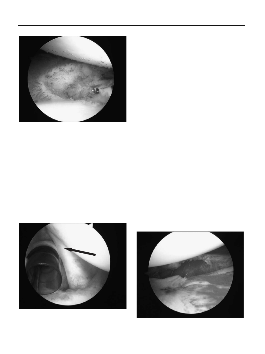
position. The patient is placed in a 60-degree upright
position, with the affected extremity being placed in
approximately 12 pounds of traction-suspension (Fig. 1).
The landmarks of the shoulder are carefully identified
with a marking pen.
The posterior portal is established in the usual
fashion. Sometimes entry into the shoulder through this
portal is difficult because of the restricted joint capsule
and diminished space. Additional traction and rotation
can aid the surgeon in entering the joint.
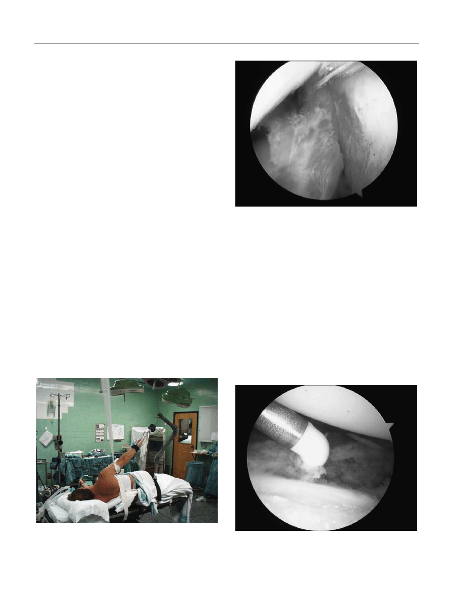
Once the joint is successfully entered, it is common-
place to encounter some early bleeding because of the
synovitis and reactive capsulitis in the joint (Fig. 2). The
control of bleeding needs to be established in the usual
fashion, but is probably even more important in this
particular operative procedure. We employ a pump and
generally start at a 45-mm Hg pressure: this facilitates
either increase or decrease as necessary. Epinephrine can
be used in the irrigating solution. It is imperative to have
hypotensive anesthesia, if medically advisable, with a
systolic pressure below 95 mm Hg. It is imperative to have
adequate flow, which will be established once the anterior
portal is established. Radiofrequency (RF) is used not
only to control the bleeding, but for controlled release.
The anterior portal is then established, either
outside in or inside out. Sometimes difficulties in carrying
out an inside-out method might arise because of the very
limited glenohumeral space. Therefore, even if one is used
to an inside-out method, an outside-in technique might be
necessary.
A bipolar RF device is used for the resection and
hemostasis. Mechanical devices can be used, but the
bipolar RF device is far more versatile in reaching all the
targeted areas in the shoulder. It also has a very
controlled cutting and coagulation mechanism that allows
the surgeon to be more precise. If the surgeon approaches
the axillary nerve and gets too close to it, then the muscle
twitch will alert the surgeon to this risk.
The procedure begins with a release of the rotator
interval, and then working the RF device down the
anterior capsule, staying very close to the labrum, and
attempting to reach the 6-o’clock position, having started
at the 1-o’clock position (Fig. 3). The device is completely
through the capsule when the capsular tissue is seen to
separate while the shoulder joint is under constant
pressure from the irrigating fluid. Approximately 70%
of the time, the underlying muscle tissue is seen, which
also indicates complete capsular resection. A 90-degree
device seems to be the most versatile one to perform the
resection with (Fig. 4). The surgeon can alternate between
cutting when resecting the capsule and coagulation when
FIGURE 1. The patient positioned in the right lateral
decubitus position with the arm in 12 pounds of traction-
suspension.
FIGURE 2. Inflammatory synovitis in the patient’s glenohum-
eral joint.
FIGURE 3. A 90-degree bipolar RF device sectioning the
capsule in proximity to the glenoid.
Sports Med Arthrosc Rev
Volume 15, Number 4, December 2007
Adhesive Capsulitis
r
2007 Lippincott Williams & Wilkins
219
bleeding is encountered. When the 5-0’clock position is
approached, great care is needed in spotting the axillary
nerve, which might come into view. The distension in
the capsule might separate the capsule well enough for
the surgeon to actually visualize it. Under most circum-
stances, a small portion of the tendon of the intra-
articular component of the subscapularis (Fig. 5) will
have to be released. The rationale behind this is to place a
small stress riser in this tendon so that, after the
arthroscopic portion of the procedure, during manipula-
tion, the musculotendinous construct undergoes a con-
trolled stretch rather than a rupture. Approximately 5%
to 7% of the entire subscapularis tendon is cut near its
insertion. This represents about a 3 to 4-mm cut through
the tendon. The intra-articular portion of the subscapu-
laris tendon only represents a small fraction of the entire
subscapularis tendon. The intra-articular pressure needs
to be carefully monitored at this stage because the capsule
is now open and extravasation can occur, along with a
significant amount of fluid loss into the surrounding
tissue. The superior resection of the capsule is then
completed from 11 o’clock to 1 o’clock with the same
anterior portal. The superior release includes the superior
rotator interval and the superior capsule. The biceps and
labrum are not released.
At this stage, the portals are exchanged using
switching sticks, and the resection is visualized anteriorly
and completed posteriorly. The pump pressure is then
lowered to 20 mm Hg, to allow for the visualization of
any unusual bleeding. The posterior capsule is resected
from 11 o’clock to 6 o’clock, again staying very close to
the glenoid. The arthroscopic cannulas and scope are then
removed after a thorough irrigation.
The patient then undergoes a formal manipulation
in the following fashion and order: forward elevation,
followed by external rotation and internal rotation at
0 degrees of adduction. This is followed by extension.
The arm is then taken through another forward elevation
maneuver, and then the external and internal rotation
maneuvers are carried out at 90 degrees of abduction. The
final manipulation that is performed is abduction. The
manipulation is repeated in the above order on as many
occasions as necessary, to achieve as complete an ROM
as is possible.
Surgeons who prefer to manipulate first and
arthroscope after the manipulation do so to visualize
their results, but they usually encounter a significant
amount of bleeding (Fig. 6). It is optional to take a relook
at this stage, but there is no reason to rearthroscope the
shoulder after manipulation.
After the operation, the patient is started on
physical therapy and a home exercise program, both
passive and active-assisted. In some refractory cases,
CPM can be helpful. Pain pumps should be used,
FIGURE 4. Appearance of the capsule after RF resection.
FIGURE 5. The subscapularis tendon with inflammatory
changes. The arrow depicting where a partial release has
been achieved.
FIGURE 6. Capsular tearing after MUA.
Tasto and Elias
Sports Med Arthrosc Rev
Volume 15, Number 4, December 2007
220
r
2007 Lippincott Williams & Wilkins

preferably in the subacromial space for 72 hours, as some
adverse reactions have been reported when they are
placed intra-articularly. We prefer this to an interscalene
block or an indwelling interscalene block. It is imperative
that the patient is followed up very closely, given support
during this most difficult period, and also monitored for
home-therapy progress.
SUMMARY
In summary, adhesive capsulitis is a fairly common
shoulder problem. It can be debilitating for patients, and
most cases take 12 to 24 months to show improvement.
It occurs mainly in diabetic women in their fifth decade
of life.
34
Most patients can be successfully treated with
nonoperatively; however, good results in the refractory
cases can be obtained with arthroscopic capsular release
followed by controlled MUA.
REFERENCES
1. Bernard M, Christian G, Jurg H. Frozen shoulder: MR arthro-
graphic findings. Musculoskelet Imaging. 1945;22:4.
2. Lundberg BJ. The frozen shoulder. Clinical and radiographical
observations. The effect of manipulation under general anesthesia.
Structure and glycosaminoglycan content of the joint capsule. Local
bone metabolism. Acta Orthop Scand Suppl. 1969;119:1–59.
3. Harryman DT II, Lazarus MD. The stiff shoulder. In: Rockwood
CA Jr, Matsen FA III, eds. The Shoulder. Philadelphia: WB
Saunders; 2004:1121–1172.
4. Janda DH, Hawkins RJ. Shoulder manipulation in patients with
adhesive capsulitis and diabetes mellitus: a clinical note. J Shoulder
Elbow Surg.
1993;2:36–38.
5. Bunker FE, Anthony PP. The pathology of frozen shoulder. A
Dupuytren-like disease. JBJS. 1995;Br 77:638–677.
6. Thierry D. Adhesive capsulitis. Emedicine. 2005;11:7.
7. Omer M, Valerie P. Review of the Frozen Shoulder. IAOM-US
newsletter 21.
8. Lori S, Norman C. Adhesive capsulitis: a sticky issue. AAFP.
1999;59:1843–1852.
9. Binder AI, Bulgen DY, Hazleman BL, et al. Frozen shoulder: an
arthrographic and radionuclear scan assessment. Ann Rheum Dis.
1984;43:365–369.
10. Manton Geoffrey L, Schweitzer ME, David K. Utility of MR
arthrography in the diagnosis of adhesive capsulitis. Skelet Radiol.
2001;30:6.
11. Nakagawa OY, Kurai G. Recalcitrant chronic adhesive capsulitis of
the shoulder. Role of contracture of the coracohumeral ligament
and rotator interval in pathogenesis and treatment. JBJS.
1989;71:1511–1515.
12. Bunker TD, Reilly J, Baird KS. Expression of growth factors,
cytokines, and matrix metalloproteinases in frozen shoulder. JBJS.
2000;82-B:768–773.
13. Rodeo SA, Hannafin JA, Tom J, et al. Immunolocalization of
cytokines and their receptors in adhesive capsulitis of the shoulder.
J Orthop Res.
1997;15:427–436.
14. Diercks RL, Stevens M. Gentle thawing of the frozen shoulder.
J Shoulder Elbow Surg.
2004;13:499–502.
15. Gary SP, Kenneth S, David MN. Adhesive capsulitis of the
shoulder: a comprehensive study. Orthop J Harvard Med School.
1994;6:32–33.
16. Dudkiewicz I, Oran A, Salai M, et al. Idiopathic adhesive capsulitis:
long-term results of conservative treatment. Isr Med Assoc J.
2004;6:524–526.
17. Rowe CR, Leffert RD. Idiopathic chronic adhesive capsulitis. In:
Rowe CR, ed. The Shoulder. New York: Churchill Livingstone;
1988:155–163.
18. Cameron RI, McMillan J, Kelly IG. Recurrence of a ‘‘primary
frozen shoulder’’: a case report. J Shoulder Elbow Surg. 2000;9:
65–67.
19. Rizk TE, Christopher RP, Pinals RS, et al. Adhesive capsulitis: a
new approach to its management. Arch Phys Med Rehabil. 1983;
64:29–33.
20. Sean G, Anthony A, Andrew G. Idiopathic adhesive capsulitis: a
prospective functional outcome study of nonoperative treatment.
JBJS.
2000;82:1398.
21. Lee PN, Lee M, Haq AM, et al. Periarthritis of the shoulder: trial of
treatments investigated by multivariate analysis. Ann Rheum Dis.
1974;33:116–119.
22. Buchbinder R, Hoving JL, Green S. Short course of prednisolone
for adhesive capsulitis: a randomized, double blind, placebo
controlled trial. Ann Rheum Dis. 2004;63:1460–1469.
23. Carette S, Moffet H, Tardif J, et al. Intraarticular corticosteroids,
supervised physiotherapy, or a combination of the two in the
treatment of adhesive capsulitis of the shoulder: a placebo-
controlled trial. Arthritis Rheum. 2003;48:829–838.
24. Eustace JA, Brophy DP, Gibney RP, et al. Comparison of the
accuracy of steroid placement with clinical outcome in patients with
shoulder symptoms. Ann Rheum Dis. 1997;56:59–63.
25. Kessel L, Bayley I, Young A. The upper limb: the frozen shoulder.
Br J Hosp Med.
1981;25:334, 336–337, 339.
26. Dodenhoff RM,
Levy D,
Wilson
A,
et
al.
Manipulation
under anesthesia for primary frozen shoulder. Effect on early
recovery and return to activity. J Shoulder Elbow Surg. 2000;9:
2–26.
27. Fox A, Board T, Srinivasan MS. Prospective analysis of the
outcome of glenohumeral manipulation for adhesive capsulitis.
JBJS.
2004;British Volume 87-B(suppl II):169.
28. Fox A, Board T, Srinivasan MS. Improvement in shoulder
function following manipulation for adhesive capsulitis: how
long does it last? JBJS. 2005; British Volume 88-B(suppl I):
138–139.
29. Braun RM, West F, Moorey V, et al. Surgical treatment of the
painful shoulder contracture in the stroke patient. J Bone Joint Surg
Am.
1971;53:1307–1312.
30. Ogilvie-Harris DJ, D’Angelo G. Arthroscopic surgery of the
shoulder. Sports Med. 1990;9:120–128.
31. Segmuller HE, Taylor DE, Hogan CS. Arthroscopic treatment of
adhesive capsulitis. Shoulder Elbow Surg. 1995;4:403–408.
32. Warner Jon JP, Answorth A, Paul M. Arthroscopic release for
chronic, refractory adhesive capsulitis of the shoulder. JBJS.
1996;78:1808–1816.
33. Pearsall AW, Holovacs TF, Speer KP. The intra-articular compo-
nent of the subscapularis tendon: anatomic and histological
correlation in reference to surgical release in patients with frozen-
shoulder syndrome. Arthroscopy. 2000;16:236–242.
34. Norlin R. Frozen shoulder-etiology, pathogenesis, and natural
course. Available at www.shoulderdoc.com.
Sports Med Arthrosc Rev
Volume 15, Number 4, December 2007
Adhesive Capsulitis
r
2007 Lippincott Williams & Wilkins
221
Wyszukiwarka
Podobne podstrony:
2006 gene therpay in sport Br J Sports Med
Adhesive capsulitis (2009)
testy egzaminacyjne z anatomii, szablon krażenie med rat 1 2006 2007, Test krążenie (ratownictwo med
ARCH MED SĄD KRYM , 2007, LVII, 399 405
2006 gene therpay in sport Br J Sports Med
Adhesive capsulitis (2009)
20.Cox arthrosis, różne, ►Medycyna-Fizykoterapia,Psychologia(1234) ---------------------------------
25 Appl Rev Lett 91 233108 2007 Nieznany (2)
Pytania egzaminacyjne 2007 Iwona, Rok II, Med. ratunkowa wieku dziecięcego
24 Phys Rev Lett 99 216802 2007
27 Phys Rev B 76 081406R 2007
32 Phys Rev Lett 98 196806 2007
PDOP 2007
więcej podobnych podstron