
Mendelian and Molecular Genetics
The word inheritance usually brings to mind money or property left by a relative who has passed away. But
there’s another type of inheritance that is right under our noses—actually, your nose is part of the
inheritance. Every living organism has characteristics or features that it passes on to its offspring. These
tendencies of heredity are obvious: even a child knows cows give birth only to other cows, and that
children often look like their parents. But, in fact, the specific biological mechanisms that allow parents to
transmit their features to their offspring were an enormous mystery until about 140 years ago. Scientists
back then knew that parents somehow made a tiny copy of themselves inside an egg or a sperm, but they
had no idea what these copies were or how they worked.
Then, in the 1860s, an Austrian monk named Gregor Mendel started breeding peas in his garden. Where
others saw only plants, Mendel looked deeper and found the basic units of heredity we now call genes. If
you remember from the last chapter, genes are the parts of a chromosome that are transcribed to mRNA
and are ultimately translated to the proteins essential to cellular processes. Mendel had no knowledge of
protein synthesis and had never seen a chromosome, but his simple experiments with peas and the laws he
developed to describe the behavior of hereditary—now termed classical genetics—have provided the
foundation for the modern field of molecular genetics, the study of heredity on the molecular level.
For the SAT II Biology, you need a solid understanding of the basic laws and patterns of both classical
genetics and molecular genetics. Questions on genetics can make up anywhere from 14 to 20 percent of the
core of the SAT II Biology. In addition, the “M” section of the Biology E/M test focuses on evolution in terms
of molecular biology, including genetics.
Basis of Inheritance: Meiosis
Mitosis takes a diploid cell and creates a nearly exact copy. Mitosis has two main functions: (1) it leads to the
creation of all of the somatic (body) cells in humans and other living organisms; (2) in organisms that
undergo asexual reproduction, diploid parent cells undergo mitosis to create identical daughter copies of
themselves. Mitosis creates a daughter cell with chromosomes that are identical to the chromosomes in its
parent cell.
But humans and most other complex plants and animals each have a unique set of chromosomes. This
diversity of chromosomes is the result of sexual reproduction, which involves the contribution of the
genetic material from not one, but two parents. During sexual reproduction the father’s haploid sperm cell
and the mother’s haploid ovum (egg) cell fuse to form a single-celled diploid zygote that then divides
billions of times to form a whole individual.
In order for sexual reproduction to take place, however, the parents first need to have haploid sperm or ova,
also called sex cells, germ cells, or gametes. Meiosis is the name for the special type of cell division that
produces gametes.
Process of Meiosis
Unlike the single-cell division of mitosis, meiosis involves two cellular divisions: meiosis I and meiosis II.
Each stage of meiosis runs through the same five stages as discussed in mitosis. During the first round of
division, two intermediate daughter cells are produced. By the end of the second round of meiotic division
(meiosis II), the original diploid (2n) cell has become four haploid (n) daughter cells.
Meiosis I
Meiosis I is quite similar to mitosis. However, there are a number of crucial differences between meiosis I
and mitosis, all of which will be outlined in the discussion of each individual stage below.
INTERPHASE I
Just as in mitosis, the cell undergoes DNA replication during this intermediate phase. After replication, the
cell has a total of 46 chromosomes, each made up of two sister chromatids joined by a centromere.
PROPHASE I
The major distinction between mitosis and meiosis occurs during this phase. In mitotic prophase, the
double-stranded chromosomes line up individually along the spindle. But in meiotic prophase I,
chromosomes line up along the spindle in homologous pairs. Then, in a process called synapsis, the
homologous pairs actually join together and intertwine, forming a tetrad (two chromosomes of two
chromatids each, or four total chromatids). Often this intertwining leads the chromatids of homologous
chromosomes to actually exchange corresponding pieces of DNA, a process called crossing-over or genetic
reassortment. Throughout prophase I, sister chromatids behave as a unit and are identical except for the
region where crossover occurred.
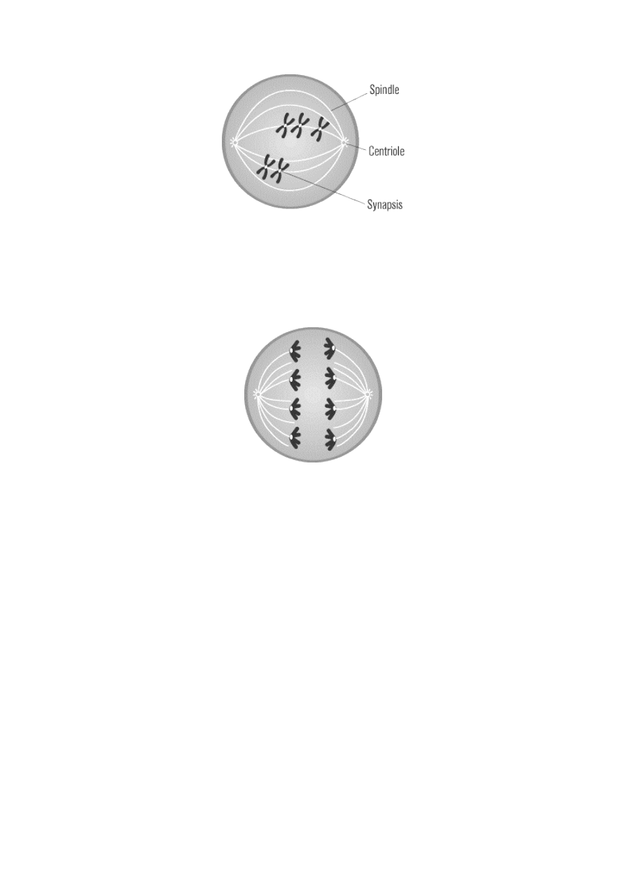
METAPHASE I
After prophase I, the meiotic cell enters metaphase I. During this phase, the nuclear membrane breaks
down, allowing microtubules access to the chromosomes. Still joined at their crossover regions in tetrads,
the homologous pairs of chromosomes, with one maternal and one paternal chromosome in each pair, align
at the center of the cell via microtubules, as in mitotic metaphase. The pairs align in random order.
ANAPHASE I
Anaphase I differs slightly from its mitotic counterpart. In mitotic anaphase, sister chromatids split at their
centromeres and are pulled apart toward opposite poles. In contrast, during anaphase I, the centromeres do
not split: the entire maternal chromosome of a homologous pair is pulled to one end, and the paternal
chromosome is pulled to the other end.
TELOPHASE I
During telophase I, the chromosomes arrive at separate poles and decondense. Nuclear membranes re-form
around them. The cell physically divides, as in mitotic cytokinesis.
THE PRODUCT OF MEIOSIS I
Meiosis I results in two independent cells. One cell contains the maternal homologous pair, with a small
segment of the paternal chromosome from crossover. The other cell contains the paternal homologous pair,
likewise with a small segment of the maternal chromosome. Despite the small region of crossover in the
chromosomes of each cell, the maternal sister chromatids are still quite similar, as are the paternal sister
chromatids. Both cells formed by meiosis I contain a haploid amount of DNA.
The cells produced in meiosis I are different from those produced in mitosis because both haploid members
of the meiotic pair derive from random assortments of either the maternal or paternal chromosomes from
each homologous pair (with the exception of the small crossover sections). In mitosis, the cellular division
separates sister chromatids and results in diploid cells containing one maternal and one paternal copy in
each diploid pair.
Meiosis II
The cells produced by meiosis I quickly enter meiosis II. These cells do not undergo DNA replication before
entering meiosis II. The two cells that move from meiosis I into meiosis II are haploid—each have 23
replicated chromosomes, rather than the 46 that exist in cells entering both mitosis and meiosis I.
Meiotic division II occurs through the familiar phases from meiosis I and mitosis. To distinguish the phases,
they are called prophase II, metaphase II, anaphase II, and telophase II. One important difference between
the events of meiosis I and II is that no further genetic reassortment takes place during prophase II. As a
result, prophase II is much shorter than prophase I. In fact, all of the phases of meiosis II proceed rapidly.
During meiosis II, chromosomes align at the center of the cell in metaphase II exactly the way they do in
mitotic metaphase. In anaphase II, the sister chromatids separate, once again in the same fashion as occurs
in mitotic anaphase. The only difference is that since there was no second round of DNA replication; only
one set of chromosomes exists. When the two cells split at the end of meioisis II, the result is four haploid
cells.
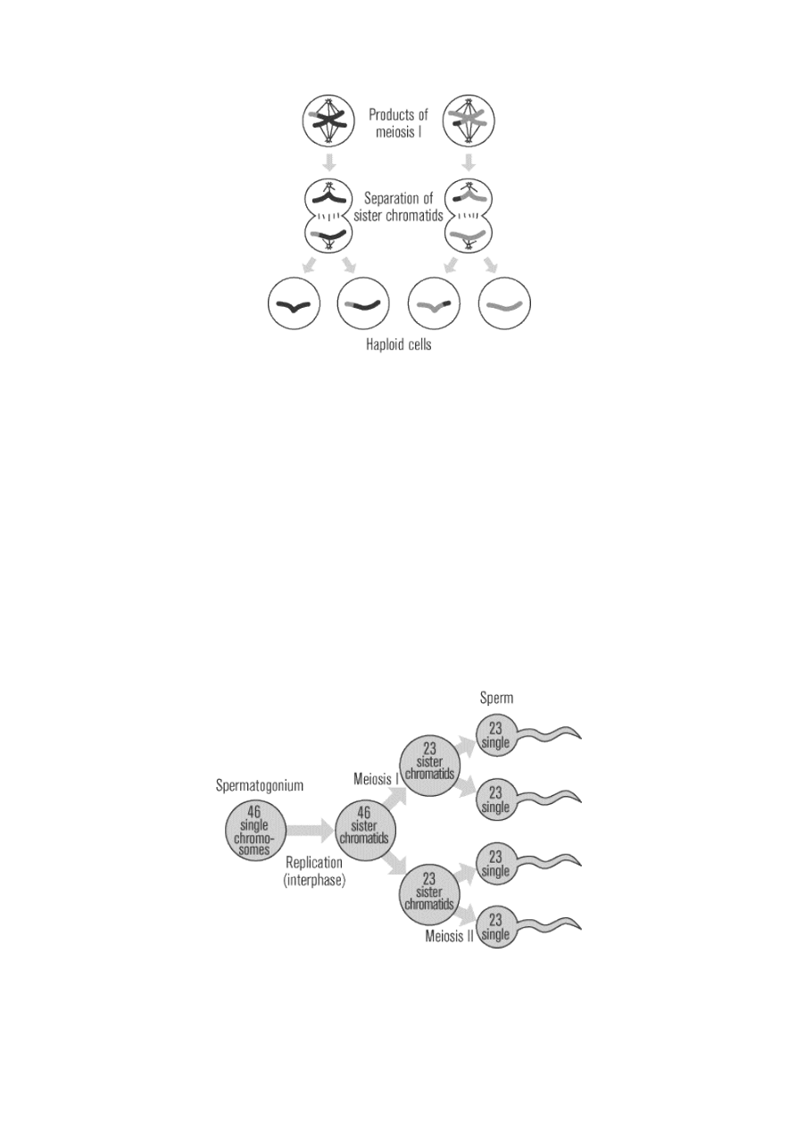
Of the four haploid cells, one cell is composed completely of a maternal homologue, another of a maternal
homologue with a small segment of paternal DNA from crossover in meiosis I, another complete paternal
homologue, and a final paternal homologue with a small segment of maternal DNA from crossover in
meiosis I. These four haploid cells are the gametes, the sperm or egg cells, that fuse together in sexual
reproduction to create new individuals.
Spermatogenesis and Oogenesis
Meiosis, the process by which gametes are formed, can also be called gametogenesis, literally “creation of
gametes.” The specific type of meiosis that forms sperm is called spermatogenesis, while the formation of
egg cells, or ova, is called oogenesis. The most important thing you need to remember about both processes
is that they occur through meiosis, but there are a few specific distinctions between them.
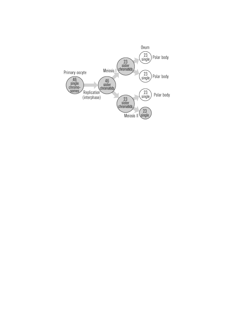
Spermatogenesis
The male testes have tiny tubules containing diploid cells called spermatogonium that mature to become
sperm. The basic function of spermatogenesis is to turn each one of the diploid spermatogonium into four
haploid sperm cells. This quadrupling is accomplished through the meiotic cell division detailed in the last
section. During interphase before meiosis I, the spermatogonium’s 46 single chromosomes are replicated to
form 46 pairs of sister chromatids, which then exchange genetic material through synapsis before the first
meiotic division. In meiosis II, the two daughter cells go through a second division to yield four cells
containing a unique set of 23 single chromosomes that ultimately mature into four sperm cells. Starting at
puberty, a male will produce literally millions of sperm every single day for the rest of his life.
Oogenesis
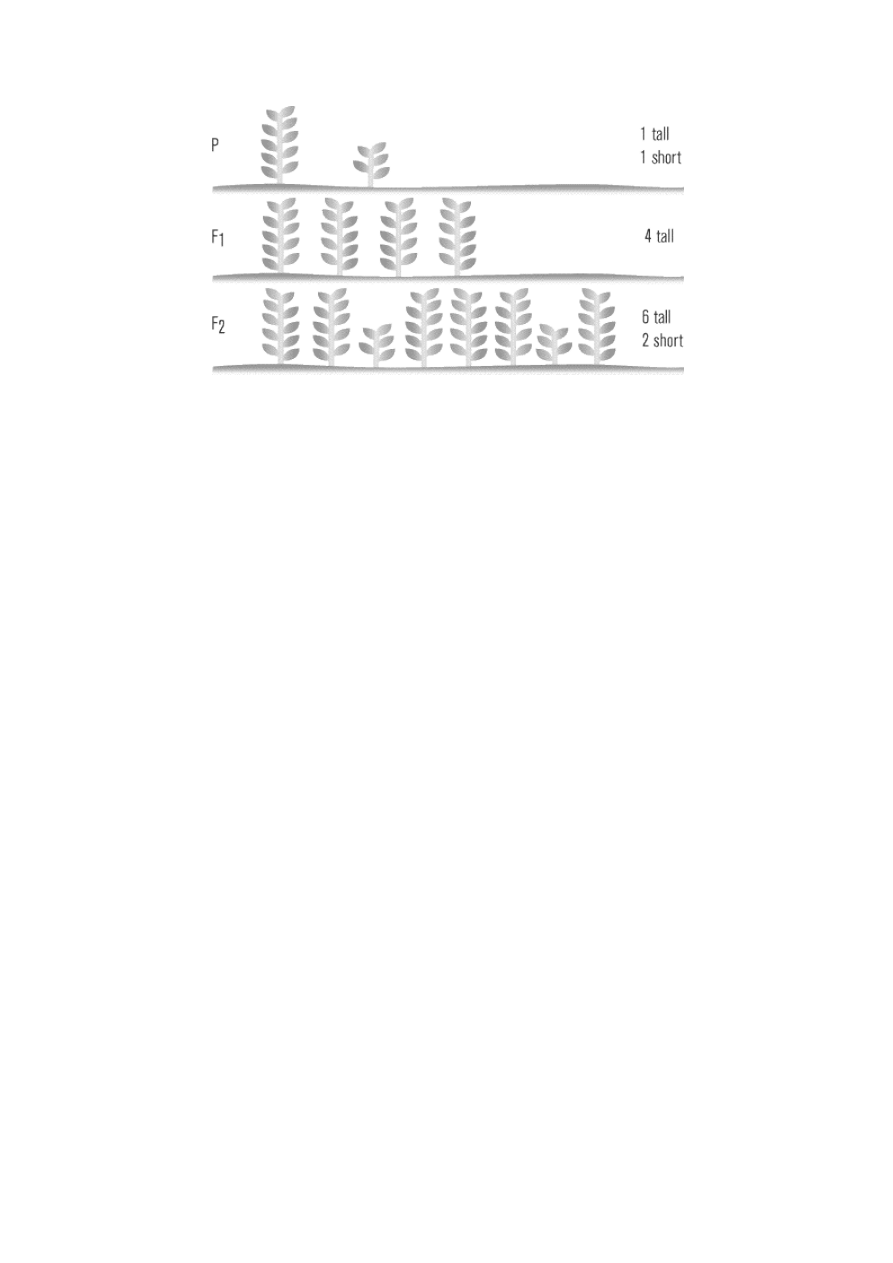
Just like spermatogenesis, oogenesis involves the formation of haploid cells from an original diploid cell,
called a primary oocyte, through meiosis. The female ovaries contain the primary oocytes. There are two
major differences between the male and female production of gametes. First of all, oogenesis only leads to
the production of one final ovum, or egg cell, from each primary oocyte (in contrast to the four sperm that
are generated from every spermatogonium). Of the four daughter cells that are produced when the primary
oocyte divides meiotically, three come out much smaller than the fourth. These smaller cells, called polar
bodies, eventually disintegrate, leaving only the larger ovum as the final product of oogenesis. The
production of one egg cell via oogenesis normally occurs only once a month, from puberty to menopause.
Mendel’s Experiments
Gregor Mendel lived in an Austrian monastery and tended the monastery garden. In 1865, through his
observations of the garden pea plants that grew there, Mendel developed three basic principles that—
although ignored at the time by his scientific colleagues—would later become the foundation for the new
science of genetics.
Every pea plant contains both male and female reproductive parts and will normally reproduce through self-
pollination. Mendel noticed that the self-pollinating pea plants in his garden were true breeding: they all
produced offspring with characteristics identical to their own. Mendel looked at seven different
characteristics, or traits, that showed up in all of the plants. Each of these traits had two contrasting
natures, only one of which would show up in a given true-breeding plant. For example, plant height could be
either short or tall: short, true-breeding plants would only produce short offspring, and tall plants would
only produce tall offspring. At some point, Mendel wondered what would happen if he manually mated
these true-breeding plants with each other—would a tall plant mated with a short plant produce a tall,
medium, or short offspring? Focusing on only one trait at a time, Mendel cross-pollinated plants with each
of the seven contrasting traits and examined their offspring. He called the original true-breeding parents the
P (for parental) generation and called their first set of offspring the F
1
(for “first filial,” from the Latin word
filius, meaning son). The F
1
offspring that result from two parents with different characteristics are also
called hybrids.
Law of Dominance
When Mendel crossed a purebred tall plant with a purebred short plant, all of the offspring in the first
generation (the F
1
generation) were tall. The same thing happened with the other pairs of contrasting traits
he studied: hybrid offspring in the first generation always showed just one of the two forms.
Mendel used the word dominant to describe the form that dominated the phenotype, or physical
appearance, in the F
1
generation. The other form he called recessive, because the characteristic receded
into the background in the F
1
generation. Mendel was the first to realize that hereditary information for two
different forms of a trait can coexist in a single individual, with one form masking the expression of the
other form. This principle, referred to as the law of dominance, provided the basis for Mendel’s subsequent
work.
Law of Segregation
Mendel discovered that mating a tall pea and a short pea would produce an F
1
generation of only tall pea
plants. But, he wondered, were these offspring tall pea plants really identical to their tall parents, or might
they still contain some element of their short parents? To answer this question, Mendel let all seven types of
hybrid F
1
generation plant self-pollinate, producing what he called the F
2
(second filial) generation.
Lo and behold, in each F
2
generation some of the recessive forms of the traits—which had visibly
disappeared in the F
1
generation—reappeared! Approximately one fourth of the F
2
plants exhibited the
recessive characteristic, and three fourths continued to exhibit the dominant form of the trait, like their F
1
parents. This 3:1 ratio of dominant to recessive remained consistent in all of the F
2
offspring.
Mendel came up with a simple but revolutionary explanation for the results he saw in the F
2
generation. He
concluded that within an individual, hereditary information came in paired units, with one unit derived
from each parent. Each simple physical trait, such as stem height, was determined by the combined action
of a single pair of units. Each unit could come in either a dominant form, which he denoted with a capital
letter “A,” or a recessive form, which he denoted with a lowercase “a.” Two units with two possible forms
gave four possible combinations: AA, Aa, aA, and aa; since Aa and aA were equivalent, there were really only
three functional combinations. Because “A” is dominant over “a,” both AA and Aa produced plants with the
same physical characteristics. Only “aa” produced a plant that showed the recessive characteristic.
Mendel realized that the results he saw in the F
2
generation could only be explained if, during the formation
of reproductive cells, paired units are separated at random so that each gamete contains only one of the two
units. This postulate is now known as the law of segregation.
Modern Explanation of Mendel’s Results
With our modern understanding of genes, chromosomes, and cellular reproduction, we can explain the
biological basis of Mendel’s observations and make pretty accurate predictions about the offspring that any
given cross (short for crossbreeding) will produce.
Alleles
Each of the traits that Mendel observed in his pea plants came in one of two varieties; modern science calls
any gene that gives rise to more than one version of the same trait an allele. So, for example, the tall gene
and the short gene are different alleles (variations) of the height gene.
Every somatic cell contains two complete sets of chromosomes, one from each parent. Now you can
understand why homologous chromosomes are similar, but not identical: although they contain the same
genes, they may not contain the same alleles for these genes.
Homozygous and Heterozygous
Going back to Mendel’s plants, we can now say that all of his true-breeding plants contained two of the same
alleles for each of the observed genes. Tall plants in this P generation had two alleles for tallness (TT), and
short P generation plants had two alleles for shortness (tt). Anytime an organism’s two alleles for a specific
trait are identical, that the individual is said to be homozygous (“homo” means same) for that trait.
On the other hand, crossing the tall and short plants to produce F
1
hybrids created a generation of plants
with one tall allele and one short allele (Tt). An organism with two opposing alleles for a single gene is said
to be heterozygous for that trait.
Genotype and Phenotype
Although the P generation of pure-breeding tall plants looked the same as their hybrid F
1
offspring, the P
and F
1
generations did not have identical genetic makeups. The genetic makeup of a certain trait (e.g., TT,
Tt, or tt) is called its genotype, while the physical expression of these traits (e.g., short or tall) is called a
phenotype.
For any given trait, an organism’s genotype will indicate alleles from both parents, while the phenotype only
indicates the allelic form that is physically expressed in that individual. This distinction between genetic
makeup and physical appearance explains the apparent “disappearance” of the recessive alleles in the F
1
generation. Mendel’s results for the F
2
generation can also be reinterpreted in light of these new
distinctions. Mendel’s results showed that 75 percent of the F
2
offspring exhibited the dominant phenotype,
a ratio of 3:1 dominant to recessive. But from a genetic perspective, the breakdown would actually be around
25 percent homozygous dominant (TT), 50 percent heterozygous with a dominant phenotype (Tt), and 25
percent homozygous recessive (tt)—a ratio of 1:2:1.
Punnett Squares
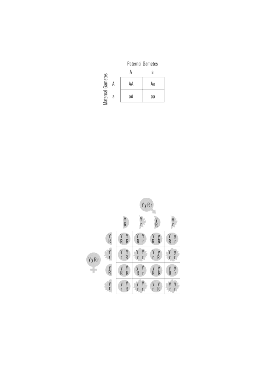
The Punnett square is a convenient graphical method for representing the genotypes of the parental
gametes and all the possible offspring they produce. The Punnett square below shows the mating of two F
1
hybrids (Aa genotype). We call this mating a monohybrid cross, because it involves only one gene.
According to the law of segregation, two possible gametes are formed: A and a. The paternal gametes are
listed as columns across the top of the square, and maternal gametes are listed as rows down the left side of
the square. Combining the gametes in the intersecting boxes provides the genotypes of all possible offspring.
In this case, 25 percent of the F
2
offspring will be AA, 50 percent will be Aa, and 25 percent will be aa. Both
AA and Aa will have the dominant phenotype, giving the 3:1 ratio (75 percent to 25 percent) of dominant to
recessive phenotypes that Mendel observed.
For the SAT II Biology, if you are given the genotypes of two parents, you should be able to predict the
genotypes and phenotypes of their offspring by using a Punnett square.
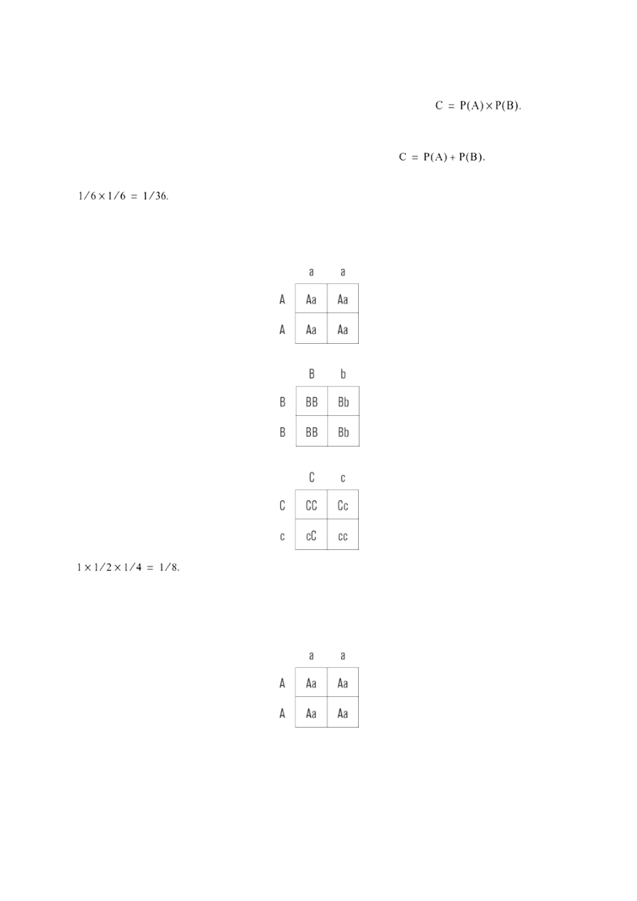
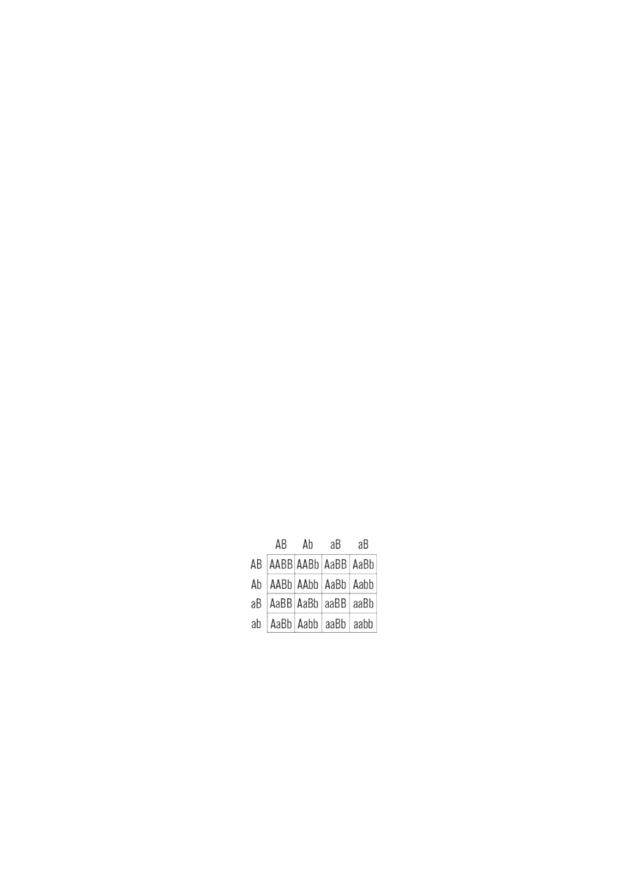
The Law of Independent Assortment
After finishing his monohybrid crosses, Mendel moved on to dihybrid crosses, in which he bred pure,
parental varieties that had two traits distinguishing them from each other. He wanted to determine whether
the inheritance of one trait was connected in any way to the inheritance of the other.
The color and shape of the pea seeds provided two convenient traits to study. The seeds were either yellow
or green, with yellow dominant; in shape, they were either round or wrinkled, with round dominant. Mendel
crossed double dominant (phenotype yellow and round, genotype RRYY) plants with double recessive
(phenotype green and wrinkled, genotype rryy) plants. As expected, the F
1
generation consisted of hybrid
offspring all with the double dominant (round yellow) phenotype and a heterozygous genotype (RrYy). The
key test came in the proportions of different phenotypes in the F
2
generation. If the inheritance of one trait
did not influence the inheritance of the other, then each parent should make equal numbers of the four
possible gametes, and sixteen different genotypes would be equally represented in the offspring. As seen in
the Punnett square below, there should be four different phenotypes (yellow and round, green and round,
yellow and wrinkled, green and wrinkled) occurring in the proportions 9:3:3:1.
Mendel’s phenotype counts of F
2
seeds did indeed show the 9:3:3:1 proportions anticipated in the Punnett
square for the dihybrid cross. From these results, he concluded that the inheritance of one trait was
unrelated to the inheritance of a second trait. The units from any one hereditary pair segregate into the
gametes independently of the segregation of the units from any other pair. This principle is known as the
law of independent assortment.
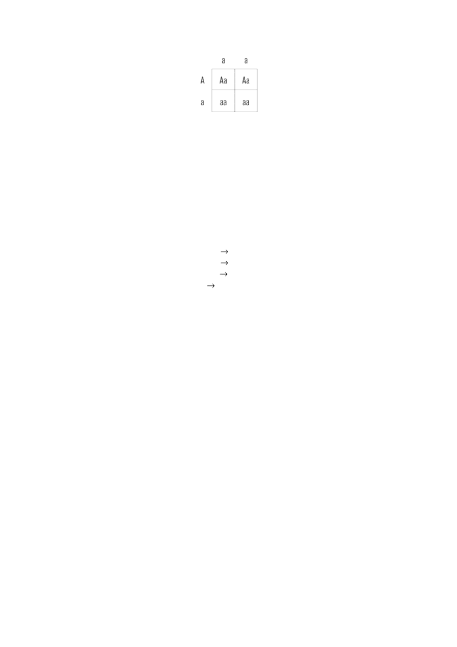
Calculating Probabilities
Drawing Punnett squares is a helpful way to visualize simple genetics problems, but with problems
involving several different genes, it is often easier to use the rules of probability. (A Punnett square for a
three-gene hybrid cross would have 64 squares!) There are two rules of probability that you will need to
solve genetics problems. First, the probability of an outcome that depends on the occurrence of two or more
independent events is obtained by multiplying together the probability of each necessary independent event.
This is the and rule of probability:
If A
and
B must occur in order to bring about outcome C, then the probability of
In contrast, if an outcome depends on the occurrence of any one of several mutually exclusive alternatives,
then the probability of the outcome is obtained by adding together the probabilities of the alternatives. This
is the or rule of probability:
If A
or
B must occur to get outcome C, then the probability of
As an example, we can calculate the probability of getting an 11 when rolling two dice, die A and die B. In
order to roll an 11, we need a 5 and a 6. The probability of rolling a 5 on die A and a 6 on die B is
But we can also roll an 11 with a 6 on die A and a 5 on die B. This is a mutually
exclusive alternative to the first roll we considered; its probability is also
1
/
36
. Since either A5, B6 or A6, B5
gives us a total of 11, the final probability of rolling an 11 using two dice is
1
/
36
+
1
/
36
=
2
/
36
=
1
/
18
.
Moving from gambling to genetics, we can calculate the probability that a cross between genotypes AABBCc
and aaBbCc will produce an offspring with genotype AaBbcc. Taking one gene at a time, the probability of
the Aa combination is a perfect 1, since an AA and aa cross can produce only Aa offspring.
The probability of the Bb combination is
1
/
2
, because the BB and Bb cross will produce Bb offspring 50
percent of the time.
The probability of the cc combination is
1
/
4
, because the Cc and Cc cross gives cc offspring 25 percent of the
time.
Since Aa and Bb and cc must occur to produce our desired outcome, the probability is
Test Crossing (Back Crossing)
A test cross is the means by which a scientist can determine whether an individual with a dominant
phenotype has a homozygous (AA) or heterozygous (Aa) dominant genotype. The test cross involves mating
the individual with the dominant phenotype to an individual with a recessive (aa) phenotype and observing
the offspring produced. If the individual being tested is homozygous dominant, then all offspring will have a
dominant phenotype, since all the offspring will have at least one A allele and the A is dominant.
If the tested individual is heterozygous dominant, then half of the offspring will show the dominant
phenotype, while the other half show the recessive phenotype.
Incomplete Dominance and Codominance
Mendel’s law of dominance is generally true, but there are many exceptions to the law. In some instances,
instead of a heterozygote expressing only one of two alleles, both alleles could be partially expressed. For
example, the flower color of the four o’clock plant is determined by a single gene with two alleles: plants
homozygous for the R
1
allele have red flowers, while plants homozygous for the R
2
allele have white flowers.
If interbred, the heterozygous R
1
R
2
plants have pink flowers. Incomplete dominance is the term used to
describe the situation in which the heterozygote phenotype is intermediate between the two homozygous
phenotypes.
If the heterozygote form simultaneously expresses both alleles fully, then the relationship between the two
alleles is called codominance. An example of codominance appears in human blood type. Blood type is
determined by two alleles, A and B, that code for the presence of antigen A and antigen B on the surface of
red blood cells. Allele A and B are codominant. If only the allele A is present, then only antigen A exists on
the blood cell. If only allele B is present, then only antigen B exists on the blood cell. If both alleles A and B
are present, neither dominates the other and both antigens appear on the red blood cell. A third allele, i, is
recessive: if only it appears, then the blood is of type O. The following is a summary of the genotypes that
result in the four different blood types:
AA and Ai
type A blood
BB and Bi
type B blood
AB and BA
type AB blood
ii
type O blood
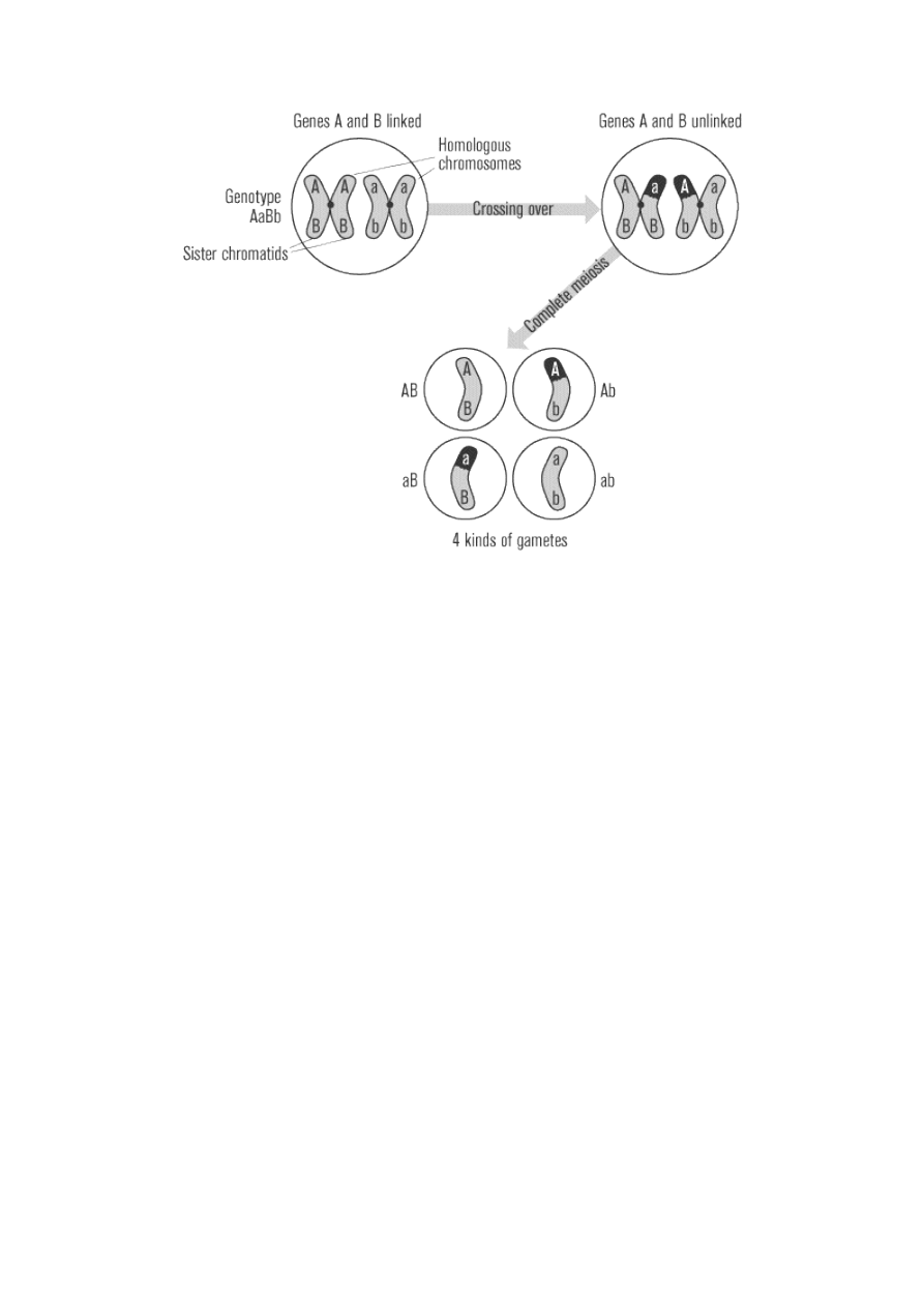
Linkage and Crossing-Over
Fortunately for Mendel, the genes encoding his selected traits did not reside close together on the same
chromosome. If they had, his dihybrid cross results would have been much more confusing, and he might
not have discovered the law of independent assortment. The law of independent assortment holds true as
long as two different genes are on separate chromosomes. When the genes are on separate chromosomes,
the two alleles of one gene (A and a) will segregate into gametes independently of the two alleles of the other
gene (B and b). Equal numbers of four different gametes will result: AB, aB, Ab, ab. But if the two genes are
on the same chromosome, then they will be linked and will segregate together during meoisis, producing
only two kinds of gametes.
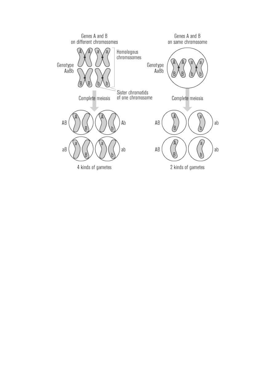
For instance, if the genes for seed shape and seed color were on the same chromosome and a homozygous
double dominant (yellow and round, RRYY) plant was crossed with a homozygous double recessive (green
and wrinkled, rryy), the F
1
hybrid offspring, as usual, would be double heterozygous dominant (yellow and
round, RrYy). However, since in this example the R and Y are linked together on the chromosome inherited
from the dominant parent, with r and y linked together on the other chromosome, only two different
gametes can be formed: RY and ry. Therefore, instead of 16 different genotypes in the F
2
offspring, only
three are possible: RRYY, RrYy, rryy. And instead of four different phenotypes, only the original two will
exist. Notice that the inheritance pattern now resembles that seen in a monohybrid cross, with a 3:1
phenotypic ratio, rather than the 9:3:3:1 ratio expected from the dihybrid cross. If physically linked on a
single chromosome, the round and yellow alleles would segregate together, and the wrinkled and green
alleles would segregate together: no round green seeds or wrinkled yellow seeds would ever appear.
The above explanation, however, neglects the influence of the crossing over of genetic material that occurs
during meiosis. The farther away two genes are from one another, the more likely an exchange point for
crossing over will form between them. At these exchange points, the alleles of one gene switch to the
opposite homologous chromosome, while the other gene alleles remain with their original chromosomes.
When alleles switch places like this, the resulting gametes are called recombinant. In the example above, the
original parental gametes would be RY and ry, while the recombinant gametes would be Ry and rY. Thus
four different kinds of gametes will be formed, instead of only two formed when the genes were linked.
If two genes are extremely close together, crossing over will almost never occur between them, and
recombinant gametes will almost never form. If they are very far apart on the chromosome, crossing over
will almost certainly occur between them, and recombinant gametes will form just as often as if the genes
were on different chromosomes (50 percent of the time). If the genes are at an intermediate distance from
each other, crossing over may sometimes occur between them and sometimes not. Therefore, the percentage
of recombinant gametes (reflected in the percentage of recombinant offspring) correlates with the distance
between two genes on a chromosome. By comparing the recombination rates of multiple different pairs of
genes on the same chromosome, the relative position of each gene along the chromosome can be
determined. This method of ordering genes on a chromosome is called a linkage map.
Mutations
Mutations are errors in the genotype that create new alleles and can result in a variety of genetic disorders.
In order for a mutation to be inherited from one generation to another, it must occur in sex cells, such as
eggs and sperm, rather than in somatic cells. The best way to detect whether a genetic disorder exists is to
use a karyotype, a photograph of the chromosomes from an individual cell, usually lined up in homologous
pairs, according to size.
Autosomal Mutations
Some human genetic illnesses are inherited in a Mendelian fashion. The disease phenotype will have either
a clearly dominant or clearly recessive pattern of inheritance, similar to the traits in Mendel’s peas. Such a
pattern will usually only occur if the disease is caused by an abnormality in a single gene. The mutations that
cause these diseases occur in genes on the autosomal chromosomes, as opposed to sex-linked diseases,
which we cover later in this chapter. (Be careful not to confuse autosomal chromosomes with somatic cells;
autosomal chromosomes are the chromosomes that determine bodily characteristics and exist in all cells,
both sex and somatic.)
Recessive Disorders
A Mendelian genetic illness initially arises as a new mutation that changes a single gene so that it no longer
produces a protein that functions normally. Some mutations, however, result in an allele that produces a
nonfunctional protein. A disease resulting from this sort of mutation will be inherited in a recessive fashion:
the disease phenotype will only appear when both copies of the gene carry the mutation, resulting in a total
absence of the necessary protein. If only one copy of the mutated allele is present, the individual is a
heterozygous carrier, showing no signs of the disease but able to transmit the disease gene to the next
generation. Albinism is an example of a recessive illness, resulting from a mutation in a gene that normally
encodes a protein needed for pigment production in the skin and eyes. The pedigree shown below
diagrams three generations of a hypothetical family affected by albinism.
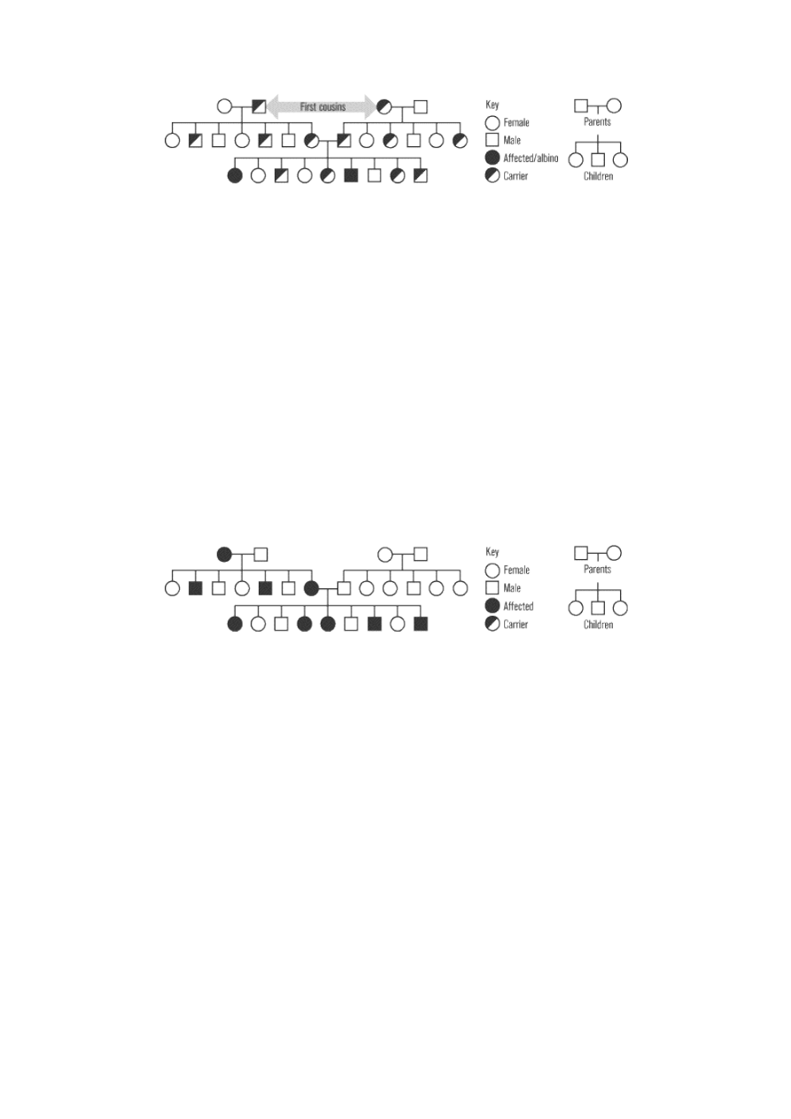
The pedigree demonstrates the characteristic features of autosomal recessive inheritance. The parents of an
affected individual usually show no signs of disease, but both must at least be heterozygous carriers of the
disease gene. Among the offspring of two carriers, 25 percent will have the disease, 50 percent will be
carriers, and 25 percent will be noncarriers. No offspring produced by a carrier and a noncarrier will have
the disease, but 50 percent will be carriers. Although not shown in this pedigree, offspring produced by two
individuals who have the disease in their phenotype, which means both parents are recessive homozygous,
will all develop the disease.
Many recessive illnesses occur with much greater frequency in particular racial or ethnic groups that have a
history of intermarrying within their own community. For example, Tay-Sachs disease is especially common
among people of Eastern European Jewish descent. Other well-known autosomal recessive disorders
include sickle-cell anemia and cystic fibrosis.
Dominant Disorders
Usually, a dominant phenotype results from the presence of at least one normal allele producing a protein
that functions normally. In the case of a dominant genetic -illness, there is a mutation that results in the
production of a protein with an abnormal and harmful action. Only one copy of such an allele is needed to
produce disease, because the presence of the normal allele and protein cannot prevent the harmful action of
the mutant protein. If a recessive mutation is like a car with an engine that cannot start, a dominant
mutation is like a car with an engine that explodes. A spare car will solve the problem in the first case, but
will do nothing to protect the garage in the second case.
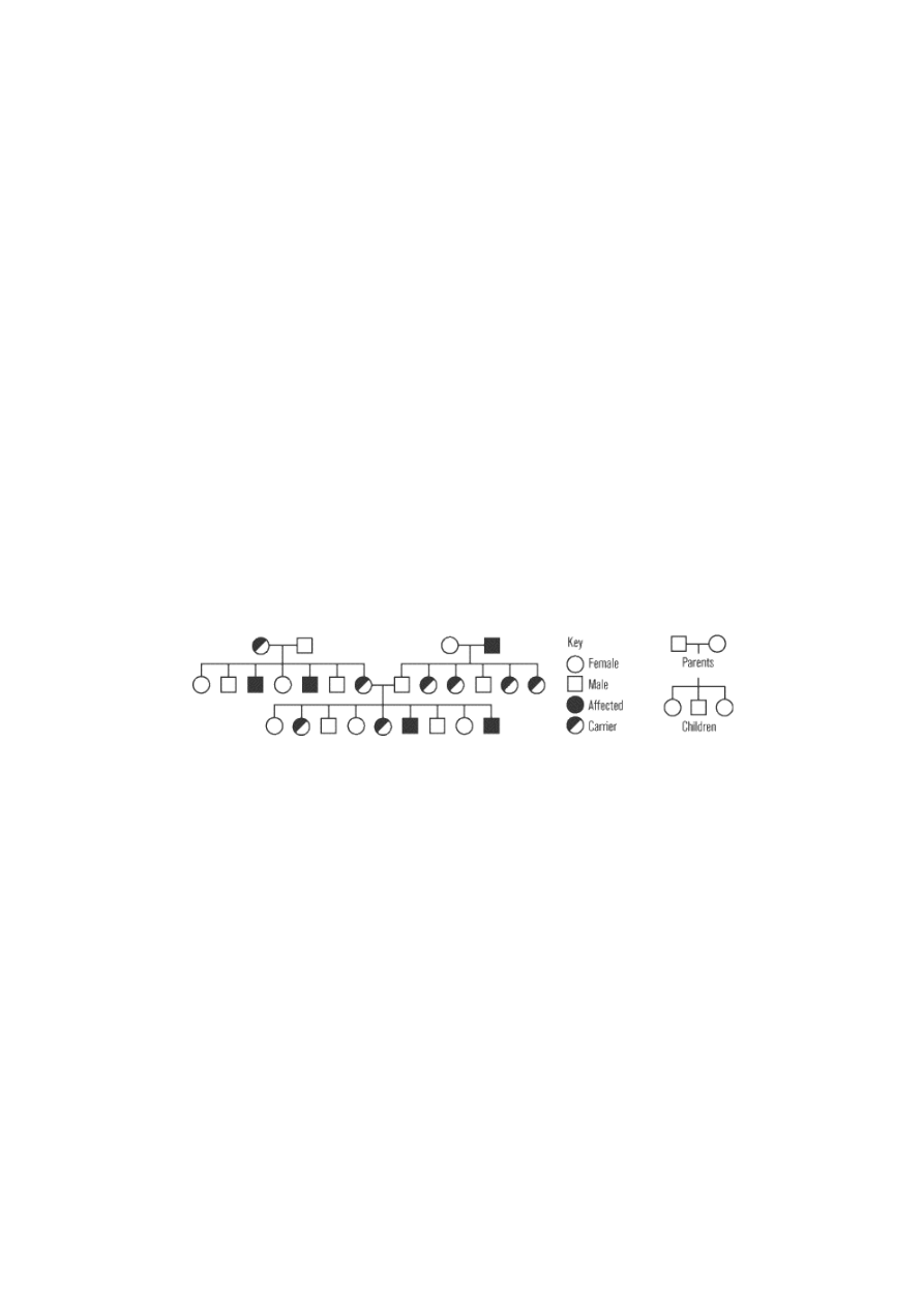
Huntington’s disease, which killed folksinger Woody Guthrie, is a dominant genetic illness. A single mutant
allele produces an abnormal version of the Huntington protein; this abnormal protein accumulates in
particular regions of the brain and gradually kills the brain cells. By middle age, this progressive brain
damage produces severely disturbed physical movements, loss of intellectual functions, and personality
changes. The pedigree shown below diagrams three generations of a hypothetical family with Huntington’s
disease.
This pedigree demonstrates the characteristic features of autosomal dominant inheritance. Notice that all
affected individuals have at least one parent with the disease. Unlike recessive inheritance, there is no such
thing as a carrier: the disease will affect all heterozygous individuals. Among the offspring of an affected
heterozygote and an unaffected person, 50 percent will be affected and 50 percent will be unaffected. None
of the children born to two unaffected individuals will have the disease. (Although not shown in this
pedigree, homozygous dominant mutations often produce very severe cases of the disease, because the
amount of the abnormal protein is doubled and the normal protein is entirely absent.)
Chromosomal Disorders
Recessive and dominant characteristics result from the mutation of a single gene. Some genetic disorders
result from the gain or loss of an entire chromosome. Normally, paired homologous chromosomes separate
from each other during the first division of meiosis. If one pair fails to separate, an event called
nondisjunction, then one daughter cell will receive both chromosomes and the other daughter cell will
receive none. When one of these gametes joins with a normal gamete from the other parent, the resulting
offspring will have either one or three copies of the affected chromosome, rather than the usual two.
Trisomy
A single chromosome contains hundreds to thousands of genes. A zygote with three copies of a chromosome
(trisomy), instead of the usual two, generally cannot survive embryonic development. Chromosome 21 is a
major exception to this rule; individuals with three copies of this small chromosome (trisomy 21) develop
the genetic disorder called Down syndrome. People with Down syndrome show at least mild mental
disabilities and have unusual physical features including a flat face, large tongue, and distinctive creases on
their palms. They are also at a much greater risk for various health problems such as heart defects and early
Alzheimer’s disease.
Monosomy
The absence of one copy of a chromosome (monosomy) causes even more problems than the presence of an
extra copy. Only monosomy of the X chromosome (discussed below) is compatible with life.
Polyploidy
Polyploidy occurs when a failure occurs during the formation of the gametes during meiosis. The gametes
produced in this instance are diploid rather than haploid. If fertilization occurs with these gametes, the
offspring receive an entire extra set of chromosomes. In humans, polyploidy is always fatal, though in many
plants and fish it is not.
Sex Chromosomes and Sex-Linked Traits
Dominant and recessive illnesses occur with equal frequency in males and females. This is because the genes
involved are located on autosomes, which are the same in both genders. Many physical traits, however,
obviously do differ between the two genders. In addition, gender dramatically affects the inheritance of
certain traits and illnesses that have no obvious connection to sexual characteristics.
These sex-linked traits are controlled by genes located on the sex chromosomes. Humans have 46
chromosomes, including 44 autosomes (nonsex chromosomes) and the two sex chromosomes, which can be
either X or Y. The autosomes come in 22 homologous pairs, present in both males and females. Females also
possess a homologous pair of X chromosomes, while males have one X chromosome and one Y chromosome
(the master gene for “maleness” is located on the Y chromosome). All eggs have an X chromosome, so the
sex of a child is determined at the time of fertilization by the type of sperm. If the fertilizing sperm carries an
X chromosome, the child will be female; if it carries a Y chromosome, the child will be male. The X
chromosome is much larger than the tiny Y chromosome, and most of the genes on the X chromosome do
not have a homologous counterpart on the Y.
Genes on autosomes will always be present in two copies: one inherited from the maternal parent, the other
from the paternal parent. The traits controlled by such autosomal genes will be generally unaffected by
gender and will follow Mendelian patterns of inheritance (with the exceptions noted in previous sections).
In contrast, genes on the X chromosome (X-linked genes) are present in two copies in females but only one
copy in males. Female offspring will inherit one copy of an X-linked gene from each parent, but male
offspring must inherit the Y chromosome from their father and therefore always inherit only the maternal
allele of any X-linked gene. For example, color blindness and hemophilia are sex-linked disorders. The
mutated gene that causes these disorders is recessive and exists on the X chromosome. In order for a
female, who is XX, to have a phenotype that is color blind or hemophiliac, both of her parents have to have
the recessive gene. But since males have only one X chromosome inherited from their mother, if their
mother expresses the recessive mutation, that trait will automatically be expressed in the male child’s
phenotype, since the male has no other gene to assert dominance over the recessive mutation.
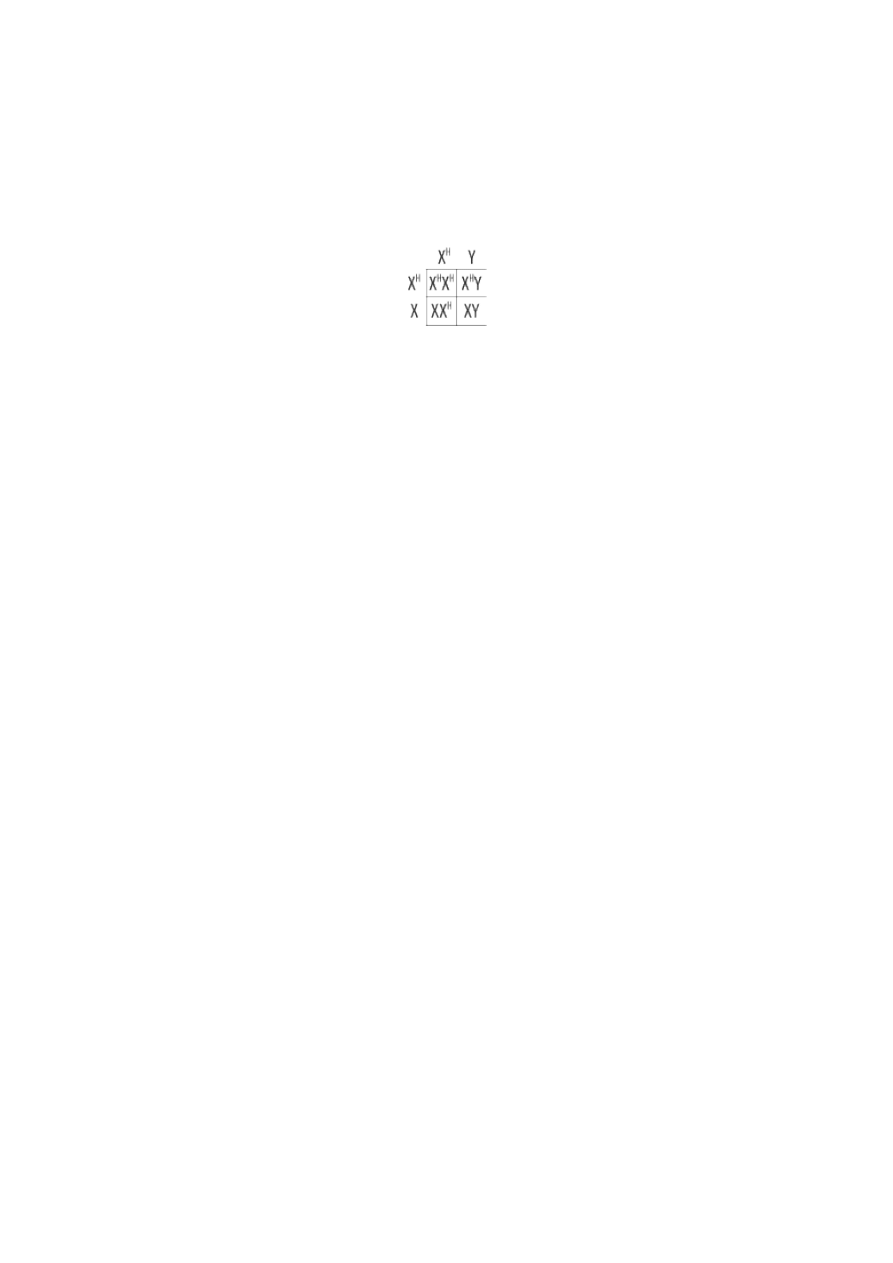
The pedigree shown below diagrams three generations of a hypothetical family affected by hemophilia A.
This pedigree demonstrates many of the characteristic features of X-linked recessive inheritance.
Heterozygous females are carriers who do not express the disease. In contrast, all males with the mutated
allele will express the disease; there are no male carriers. Affected males will transmit the mutated allele to
none of their sons but to all of their daughters, who will then all be carriers. Heterozygous females will
transmit the disease to one-half of their sons, and one-half of their daughters will be carriers. Affected males
generally have an unaffected father and a mother who is a carrier; 50 percent of their maternal uncles will
have the disease.
Review Questions
1.
During which meiotic phase are sister chromatids separated?
(A)
Prophase I
(B)
Metaphase I
(C)
Metaphase II
(D)
Anaphase I
(E)
Anaphase II
2.
One cell that undergoes meiosis will yi eld how many independent cells?
(A)
1
(B)
2
(C)
3
(D)
4
(E)
5

3.
In which generation of a monohybrid cross do all the individuals look the same?
(A)
Parental
(B)
F
1
(C)
F
2
(D)
F
3
(E)
F
4
4. In a monohybrid cross, the two parental genotypes are AA and aa. The genotype of all F
1
plants is Aa. If the
F
1
plants are crossed to get an F
1
generation, what percentage of the F
2
generation will have the dominant
phenotype?
(A)
0%
(B)
25%
(C)
50%
(D)
75%
(E)
100%
5. You are performing a dihybrid cross with two traits encoded by two different genes. These genes are on two
different chromosomes and follow the la w of independent assortment. The two parental genotypes are AABB
and aabb. The genotype of all F
1
plants is Aa Bb. What phenotype ratio will you observe in the F
2
generation?
(A)
8:4:2:1
(B)
1:1:1:1
(C)
4:2:2:1
(D)
1:2:2:1
(E)
9:3:3:1
6. A scientist is studying the inheritance of two traits: stem height and stem color. Tall is dominant over short;
brown is dominant over green. The scientist wants to do a test cross to determine the genotype of a tall, brown
F
1
hybrid individual. What will be the height and color of the other plant he uses in this test cross?
(A)
Tall and brown
(B)
Tall and green
(C)
Short and brown
(D)
Short and green
(E)
It doesn’t matter; any of the above will work.
7.
You are studying the inheritance of stem height and stem color. For height, the tall phenotype is dominant
over short; for color, the brown phenotype is dominant over green. You mate a purebred tall, brown plant to a
purebred short, green plant and all of the F
1
offspring are tall and brown. You then mate two of these F
1
hybrid offspring together, and you count the following phenotypes in their F
2
offspring: 61 tall and brown, 1
tall and green, 2 short and b rown, and 18 short and green. Which of the following is probably true?
(A)
The two genes controlling these traits are sex linked.
(B)
The two genes controlling these traits might be on different chromosomes.
(C)
The two genes controlling these traits are linked with no crossing over.
(D)
The two genes controlling these traits are nearby on the same chromosome.
(E)
The two genes controlling these traits are far apart on the same chromosome.
8.
Which of the following is usually NOT true of an autosomal recessive disease?
(A)
Most people with the disease have parents who do not have the disease.
(B)
An equal number of males and females get the disease.
(C)
If both parents have the disease, 50% of their offspring will have the disease.
(D)
If both parents are heterozygous, 75% of their children will not get the disease.
(E)
The genes for the disease are not located on the sex chromosomes.
9.
A woman who is a heterozygous carrier of the hemophilia allele marries a man who has hemophilia. What
percentage of their female children would you expect to have hemophilia?
(A)
0%
(B)
25%
(C)
50%
(D)
75%
(E)
100%
10. A boy with red -green color blindness (an X -linked recessive condition) has a color -blind father and a mother
who is not color blind. From which parent did the boy inherit his color blindness?
(A)
No wa y to know—it could have been either one.
(B)
His father
(C)
His mother
(D)
It had to come partly from both his mother and father.
(E)
It had nothing to do with either his mother or father.
Explanations
1.
E
In meiosis, sister chromatids separate during anaphase II. During prophase I, sister chromatids are linked at their
centromeres. At metaphase I, each pair of sister chromatids lines up in the middle of the cell together with its
homologous pair of sister chromatids, and only homologous chromosomes separate during anaphase I. During
metaphase II, each pair of sister chromatids again lines up along the metaphase plate, but they do not separate until
anaphase II.
2.
D
Meiosis divides a single diploid cell into four haploid cells. In the case of oogenesis, only one of those daughter cells
goes on to become a fully developed egg cell, leaving three “polar bodies” to disintegrate; the original product, however,
is still four cells.
3.
B
The first (F
1
) generation of offspring from a hybrid cross will all look the same. A hybrid cross involves the mating of a
homozygous dominant (RR) individual with a homozygous recessive (rr) individual, so the parents do not look the same.
In the F
1
generation, all offspring will have genotype Rr and will all show the dominant trait. Other generations will
contain some individuals with the dominant phenotype (RR or Rr) and some with the recessive phenotype (rr) and so will
not all look the same.
4.
D
Approximately 75% of the F
2
generation will exhibit the dominant phenotype. Genotypically, there will be three types of
offspring: 25% homozygous dominant, 50% heterozygous dominant, and 25% homozygous recessive. Only the
homozygous recessive will show the recessive phenotype
—therefore, a total of 75% will have the dominant phenotype.
5.
E
Because these genes are on separate chromosomes and therefore act independently, we can use a Punnett square to
work out the results of this dihybrid cross.
For every sixteen offspring in the F
2
generation, approximately nine will exhibit the dominant phenotype of both genes
(genotype AABB, AABb, AaBB, or AaBb), three will be dominant on gene A only (AAbb or Aabb); three will be dominant
on gene B only (aaBB or aaBb), and one will show a recessive phenotype on both genes (aabb). The result is a
phenotype ratio of 9:3:3:1.
6.
D
In order to determine the genotype of a dominant-appearing individual, the scientist would have to perform a test cross
(also known as a back cross) using a fully recessive individual. In this case, that would be a short and green plant.
7.
D
Given the fact that the expected 3:1 dominant-to-recessive phenotype showed up on the same F
2
offspring plants, it
looks like these genes are linked and were not acting independently of each other. From this information, you can predict
that the two genes controlling these traits are on the same chromosome. Since crossing-over limits the effects of
linkage
—and crossing-over occurs more often when two genes are far apart on the same chromosome— you can
further determine that the two genes must be near each other on the chromosome.
8.
C
The genotype of a person who has an autosomal recessive disease must be homozygous recessive. Therefore, if two
parents have the disease, 100% (not 50 or 75%) of their children should get the disease, since neither parent has
dominant alleles to contribute.
9.
C
50% of the couple’s daughters would be expected to have hemophilia, an X-linked recessive disease. Using a Punnett
square to do the cross of the father (X hemophilia/Y) and the mother (X normal/X hemophilia), you should find that half
of the female children will be homozygous for the X hemophilia gene and have the disease, while half will be
heterozygous and be carriers of the disease.
10.
C
Although the father is color blind, the gene for this condition is located on the X chromosome. The father could not have
given his son the condition, because he only contributes a Y chromosome to his son; therefore, the gene that causes the
disease must have been inherited from the mother.
Wyszukiwarka
Podobne podstrony:
mutacjei naprawacdHuman Molecular Genetics 2
naprDna1rysHuman Molecular Genetics 2
Personality and divorce A genetic analysis
Centre of microbial and plant genetics
naprawaDNAHuman Molecular Genetics 2
Exploring Careers of Biochemistry and Molecular Biology
Primer On Molecular Genetics
Detection and Molecular Characterization of 9000 Year Old Mycobacterium tuberculosis from a Neolithi
Genolevures comparative genomics and molecular evolution of yeast
Clinical aspects and molecular of skin ageing
Personality and divorce A genetic analysis
A Behavioral Genetic Study of the Overlap Between Personality and Parenting
Molecular spectroscopy and structure
Programming Survey Of Genetic Algorithms And Genetic Programming
A Comparison between Genetic Algorithms and Evolutionary Programming based on Cutting Stock Problem
Childhood Experience and the Expression of Genetic Potential
więcej podobnych podstron