Open Life Sci. 2015; 10: 409–416
1 Introduction
Fungal diseases of crops are usually controlled using
resistant cultivars, long rotations, and fumigants,
but mainly by using fungicides. The use of synthetic
fungicides is not an eco-friendly approach as many
are reported to have serious health risks and have been
linked to an increased occurrence of several types of
cancer. Alternative methods to control fungal diseases
have been studied by using compounds derived from
plant sources in an attempt to reduce the use of synthetic
fungicides. Agapanthus africans leaf extracts have
shown good antifungal activity [1]. Plant products have
proved toxic for a large number of fungal and bacterial
pathogens. Soil pathogens such as Pythium sp. could
be controlled by extracts of Larrea tridentata Cov. [2]
while Punica granatum was effective against Fusarium
oxysporum [3]. Allium ursinum [4] flower extract inhibited
mycelial growth of Aspergillus niger, Botrytis cinerea,
Penicillium gladioli, Fusarium oxysporum and Sclerotinia
sclerotiorum. The quality and quantity of biologically
active compounds from Allium species greatly depended
on the target species, the plant organ and harvest time.
Four plant extracts (Adhatoda vasica, Jatropha curcas,
Sapindus emarginatus and Vitex negundo) were able to
control wilt disease of Solanum melogena [5]. Piper betle
was more effective in controlling Fusarium populations
in soil than “carbendazim”, a commercial fungicide
[6]. Extracts of Allium sativum, Coriandrum sativum,
Curcuma longo and Cuminum cyminum possessed a strong
antifungal activity [7]. Forty plants of different families
were tested against Fusarium oxysporum f.sp. cicero, with
Chenopodium ambrosioides having the highest inhibition
[8]. More recently, the antifungal activity of more than
500 plant species has been assessed [9]. Of all plants
tested, only 3% showed a high antifungal activity. Many
authors have also studied the importance of secondary
metabolites in fungal inhibition. The antifungal activity of
Quillaja saponaria extract could be due to the presence of
saponins and phenolic compounds [10]. The relationship
between antifungal activity and total phenolic content
DOI 10.1515/biol-2015-0040
Received March 05, 2014; accepted October 13, 2014
Abstract: The present paper describes the antifungal
activity of some plant extracts on the development of
Fusarium oxysporum f.sp. lycopersici. The best extracts
were selected to be tested as a phytofungicide to control
crop diseases, with the ultimate goal of developing a
green alternative to synthetic fungicides. Using the
conidia germination assay, of the 24 plant extracts
tested, 15 reduced conidia germination and 6 completely
inhibited germination. Extracts of Rivina humulis,
Brassica carinata, Brunfelsia calyicina, Salvia guaranitica
and Punica granatum showed good antifungal activity.
The relationship between total phenolic content (TPC) in
each plant extract tested and the percentage of mycelial
growth inhibition showed a significant correlation (R
2
= 0.69), while no correlation was found between total
flavonoid content (TFC) and percentage mycelial growth
inhibition. Among all extracts tested, Punica granatum
and Salvia guaranitica showed the best inhibitory effect
against Fusarium oxysporum f.sp. lycopersici. Our results
indicate that plant extracts with a good antifungal activity
generally had a high level of total polyphenolic content
and titratable acidity, and low values of pH.
Keywords: Fusarium; plant extract; antifungal activity;
protectant fungicide
Research Article
Open Access
© 2015 Domenico Rongai, et al., licensee De Gruyter Open.
This work is licensed under the Creative Commons Attribution-NonCommercial-NoDerivs 3.0 License.
Domenico Rongai*, Patrizio Pulcini, Barbara Pesce, Filomena Milano
Antifungal activity of some botanical extracts on
Fusarium oxysporum
*Corresponding author: Domenico Rongai, Consiglio per la Ricerca e
la sperimentazione in Agricoltura, Centro di ricerca per l’olivicoltura
e l’industria olearia, viale Petruzzi 75 - 65013 Città Sant’Angelo (PE),
Italy, E-mail: domenico.rongai@entecra.it
Patrizio Pulcini, Barbara Pesce, Filomena Milano, Consiglio per la
ricerca e la sperimentazione in agricoltura, Centro di ricerca per la
patologia vegetale, via C.G Bertero, 22 - 00156 Roma, Italy
Unauthenticated
Download Date | 6/2/17 11:42 AM

410
D. Rongai, et al.
has also been reported [11-14]. Contrasting results are
reported in literature regarding the effect of flavonoids
on antifungal activity. Some authors [11,15] found that
flavonoids were not correlated with antifungal activity
while others [16,17] reported that the inhibition of fungi
was mainly due to flavonoids. The aim of the present work
was to evaluate the antifungal activity of water extracts
of various plant species using phytochemical screening.
Total phenolic and flavonoid content, acidity and pH were
also determined.
2 Experimental procedures
2.1 Plants for extraction and fungus used
Twenty four different plant species from various plant
families were kindly provided by the botanical garden
at “La Sapienza University”, in Rome (Table 1). The fresh
plant material was collected into plastic bags and stored
in a freezer at -20°C. Fusarium oxysporum f. sp. lycopersici
(strain CRA-PAV collection n. ER1372) was used as the
target fungus. The fungus was maintained on potato
dextrose agar (PDA, oxoid cm 0139) and stored at 4°C.
When needed, the isolate was grown for 8 days on PDA in
the dark at 25 ± 2°C. The conidial suspension obtained was
filtered through a double layer of cheesecloth to remove
leaf debris and centrifuged at 2500 r.p.m. for 3 min.
Conidia were than counted and used at a concentration of
5 × 10
4
conidia ml
-1
.
2.2 Preparation of powders and extracts
Fresh material of leaves, bulbs or peel were cut into small
pieces and placed together in the solvent (water). The
heterogeneous mixture was stirred overnight. The material
was sonicated for 3 min (3 s on and 7 s off) and the extract
obtained was then centrifuged at 15 000 (r.p.m.) for 10
minutes and the supernatant filtered through a 0.22 µm
PTFE membrane. The solvent was vacuum evaporated in
a rotatory evaporator, frozen at -80°C for 24 h and finally
freeze dried (-40°C; 7 × 10
-2
mbar) for 2 days. The powder
of the extract obtained was stored in a freezer at -20°C for
further use.
2.3 Antifungal screening
2.3.1 Conidia germination assay
A microtiter plate assay was used to rapidly detect the
antifungal activity of plant extracts. 200 µL of a mixture
containing: 80 µL of conidial suspension, 100 µL of
Czapek Dox Broth and 20 µL of plant extract were pipetted
into each well of the microtitration plate. One plate row
was filled with untreated spore suspension in Czapek Dox
Broth as a positive control. Changes in optical density
following conidial germination were measured 48 h
after inoculation using a microplate reader (Multiscan
– Plus MK II, Labsystems OY, Helsinki, Finland) at a
wavelength of 405 nm. Conidial germination 24 hours
after inoculation was assessed by mounting 10 µl samples
on a glass slide and counting the number of germinated
spores on a gridded square hemocytometer at 4 × 10
-2
mm
2
.
The percentage germination recorded for the eight wells
was averaged. The test was repeated three times.
2.3.2 Mycelial growth inhibition assay
The inhibitory effect of extracts of plant species reported
in Table 1 were also tested using cultures in Petri dishes.
200 mg of each powder plant extract were added to 9.8
mL of Potato Dextrose Agar (PDA) and subsequently put
into sterile 50 mm diameter Petri plates. In addition, a
plate containing a specific standard fungicide (Marisan
50 PB, Dicloran 60%, SIAPA s.r.l., Milano, Italy) was used
at the recommended concentration to serve as a negative
control to determine the effectiveness of the extracts by
comparison. PDA with sterile water served as the control.
Antifungal activity tests were performed by placing 5 mm
mycelial agar discs cut from the actively growing margin of
8 days old F. oxysporum colony in the centre of each plate.
Four replicates for each species extract were used and the
whole experiment was repeated three times. Radial growth
was measured each day, starting 4 days after incubation in
the dark at 25°C, until the 6
th
day. The percentage growth
inhibition of each extract was calculated by the formula:
% inhibition = [growth in control - growth in sample/
growth in control] × 100.
2.4 Determination of total phenolic and fla-
vonoids content
Total phenolic content of all plant extracts was determined
by the Folin-Ciocalteu method [18]. 20 µL of each extract
solution were transferred into separate tubes, which were
then added with 1.58 mL of ultra-pure water. 100 µL of the
Folin-Ciocalteu reagent was then added to the mixture,
mixed well and left for 8 min. After that, 300 µL of 2%
sodium carbonate was added, tubes were uncapped and
shaken two seconds on a vortex and left in the dark for 1
h at room temperature. Measurement was conducted on
Unauthenticated
Download Date | 6/2/17 11:42 AM

Antifungal activity of some botanical extracts on Fusarium oxysporum
411
a spectrophotometer (Varian Cary 100 Conc UV-Vis) at λ
= 760 nm against a ultra-pure water as blank. Gallic acid
was used as a standard phenolic compound to make the
calibration curve that ranges between 0 to 500 mg/L (r
2
=
0.9913). The results are expressed as milligrams of gallic
acid equivalent per gram of dry weight (mg GAE/g dw) of
lyophilized plant extract.
Flavonoid content was estimated using the AlCl
3
method [19]. 0.5 mL of each extract was taken, and 1.5 mL
of methanol was added. 0.1 mL of 10% AlCl
3
and then 0.1
mL of 1M Potassium acetate was added to the reaction
solution. The volume of the solution was made up to 5 ml
with distilled water and the reaction mixture was incubated
at room temperature for 30 minutes. Absorbance was read
at 415 nm at the UV-Vis spectrophotometer. A calibration
curve was generated, using Rutin as a standard flavonoid
compound, from 5 to 100 mg/L (r
2
= 0.9969). Total flavonoid
content was expressed as Rutin Equivalent (mg/L) of the
extract. The experiment was repeated twice.
2.5 Acidity and pH analysis
Acidity was determined by titration with a 0.01 N alkaline
sodium hydroxide solution. Phenolphthalein (1%) was
used as the indicator (2 drops in 20 mL of each sample
before starting the analysis). Sodium hydroxide was
added dropwise with constant swirling until the solution
turned pink throughout. The volume of base required to
reach the equivalence point was used to calculate the
acidity of the extracts expressed in meq NaOH/g. The pH
value of each extract was determined with a Hamilton pH
electrode sensor. All measurements were repeated twice
within a period of 10 days.
2.6 Statistical analysis
A randomized experimental design was used. Statistical
analysis ANOVA was carried out and mean values
compared by Fisher’s protected LSD test at P ≤ 0.05.
SigmaPlot version SPW10 and Sigma Stat version 3.5 were
used to create graphics.
Table 1. Plant species used in the experiments.
Genus
Species
Family
Common name
Parts used
Allium
sativum
Alliaceae
Garlic
Bulb
Allium
triquetrum
Alliaceae
Angled onion
Leaves
Antholyza
aethiopica
Iridaceae
Cobra lily
Leaves
Arctium
lappa
Asteraceae
Greater burdock
Leaves
Boehmeria
nivea
Urticaceae
Ramie
Leaves
Brassica
carinata
Brassicaceae
Ethiopian mustard
Seeds
Brunfelsia
calycina
Solanaceae
Yesterday-today-tomorrow
Leaves
Campsis
radicans
Bignoniaceae
Trumpet vine
Leaves
Celtis
glabrata
Cannabaceae
Hackberry
Leaves
Citrus
limon
Rutaceae
Limon
Leaves
Coffea
arabica
Rubiaceae
Coffea arabica
Leaves
Conium
maculatum
Apiaceae
Poison Hemlock
Leaves
Cycas
revoluta
Cycadaceae
Sago cycad
Fruit
Lavandula
multifida
Lamiaceae
Fernleaf lavender
Leaves
Mallotus
japonicus
Euphorbiaceae
Japanese mallotus
Leaves
Petrea
volubilis
Verbenaceae
Sandpaper vine
Leaves
Philodendron
crassinervium
Araceae
Thick-nerved Philodendron
Leaves
Polygonatum
odoratum
Asparagaceae
Angular Solomon’s seal
Leaves
Punica
granatum
Lythraceae
Pomegranate
Peel
Rivina
humilis
Phytolaccaceae
Pigeonberry
Leaves
Salvia
guaranitica
Lamiaceae
Anise-scented sage
Leaves
Strelitzia
reginae
Strelitziaceae
Bird of paradise
Leaves
Taraxacum
officinale
Asteraceae
Dandelion
Leaves
Yucca
elephantipes
Asparagaceae
Giant yucca
Leaves
Unauthenticated
Download Date | 6/2/17 11:42 AM

412
D. Rongai, et al.
3 Results
3.1 Conidia germination assays
Analysis of optical density and conidia germination showed
a high correlation between difference of absorbance
(DA) at 48 h and the percentage of conidia germination
observed after 24 h (Table 2). Plant extracts with a DA <
0.04 showed no or very low conidia germination. The
extracts with a DA ≤ 0.3 showed a percentage higher than
70; when the DA was > 0.4, the percentage rose over 80.
In the present assay, out of the 24 plant extracts tested, 15
were able to reduce conidia germination and 6 completely
inhibited germination.
3.2 Mycelial growth inhibition assays
The extracts of R. humulis, B. carinata, B. calyicina and S.
guaranitica showed a high antifungal activity: 4 days after
inoculation, the mycelial growth was 33.6, 33.3, 28.2 and
26.0 mm respectively. In contrast, the nontreated control
had mycelial growth of 42.9 mm (Table 3). The highest
antifungal activity was recorded in Punica granatum
extract, where the radial growth was 16.7 mm, even lower
than with Marisan 50 PB, the synthetic fungicide (17.8
mm), though the data are not statistically significant.
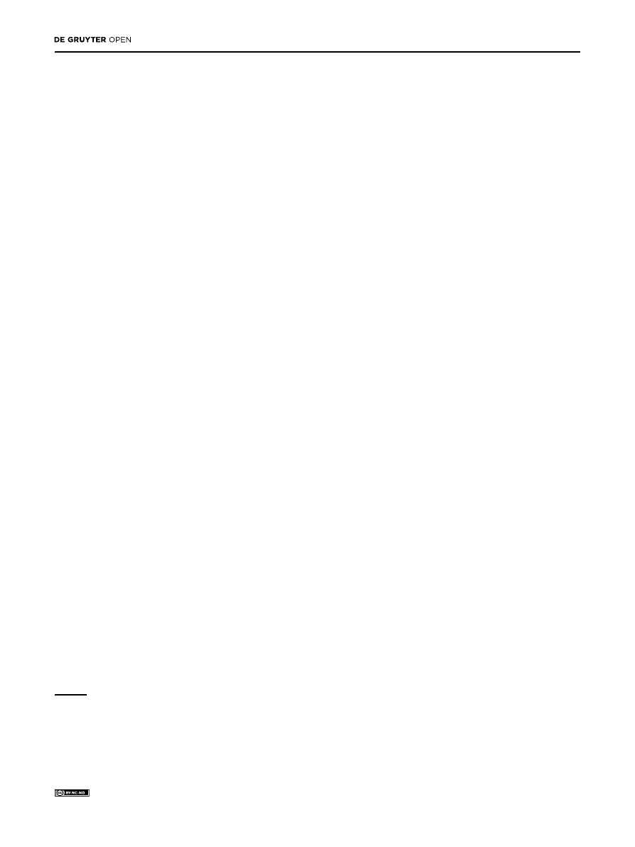
Fungal growth inhibition (Fig. 1) showed that from the 5
th
day the mycelium covered the whole control plates. The
percentage of inhibition over control in P. granatum was
62.77 (4
th
day), 48.53 (5
th
day), 42.71 (6
th
day). These data
were followed by S. guaranitica extract 39.35; 23.35, 12.74,
B. carinata 33.14, 26, 14.7 and B. calycina 31.7, 27.62, 22.06.
3.3 Total phenolic and flavonoids content,
acidity and PH analysis
Total phenolic content (TPC) and flavonoid content
(TFC) varied widely among plant extracts (Table 3). TPC,
expressed as mg GAE/g DW, ranged from 8.29 mg GAE/g
DW in C. radicans to 542 mg GAE/g DW in P. granatum.
TFC, expressed as mg RE/g DW, ranged from 2.71 in P.
crassinervium to 102.76 in P. granatum.
There was a significant correlation (R
2
= 0.69)
between TPC content in each plant extract tested and
Table 2. Difference of absorbance and percentage of conidia germination of 24 plant extracts. Values are the mean of four replications.
Means in the same column followed by same letter are not statistically different at P = 0.05 according to the Fisher LSD Method.
Plant species
Difference of absorbance 48 h after inoculation
Percentage of conidia germination 24 h after
inoculation
Control
0.41a
80a
Allium sativum
0.00d
0e
Allium triquetrum
0.04d
5e
Antholyza aethiopica
0.44a
≥ 80a
Arctium lappa
0.03d
15d
Boehmeria nivea
0.45a
≥ 80a
Brassica carinata
0.01d
0e
Brunfelsia calycina
0.01d
0e
Campsis radicans
0.41a
75a
Celtis glabrata
0.00d
5e
Citrus limon
0.00d
10de
Coffea arabica
0.04d
5e
Conium maculatum
0.53a
75a
Cycas revoluta
0.02d
10de
Lavandula multifida
0.29b
70ab
Mallotus japonicus
0.93a
≥ 80a
Petrea volubilis
0.00d
0e
Philodendron crassinervium 0.33b
70ab
Polygonatum odoratum
0.47a
≥ 80a
Punica granatum
0.03d
0e
Rivina humilis
0.00d
5e
Salvia guaranitica
0.04d
0e
Strelitzia reginae
0.50a
≥ 80a
Taraxacum officinale
0.26b
55cb
Yucca elephantipes
0.10c
65b
Unauthenticated
Download Date | 6/2/17 11:42 AM
Antifungal activity of some botanical extracts on Fusarium oxysporum
413
Table 3. Effect on mycelia growth of F. oxysporum (four days after inoculation) and TPC, TFC, total acid, and pH of some plant extracts tested
at a concentration of 1%.
Plant species
Mycelia growth
TPC
TFC
Acidity
pH
mm
mgGAE/g DW
mgRE/g DW
meq NaOH/g
No treated (positive control) 42.9
a
-
-
-
-
Fungicide (negative control) 17.9
i
-
-
-
-
Allium sativum
34.7
ed
10.14
5.43
0.120
6.57
Allium triquetrum
42.2
a
29.85
15.12
0.248
6.27
Antholyza aethiopica
*
43.29
21.14
0.284
5.78
Arctium lappa
40.1
b
59.85
7.02
0.284
5.70
Boehmeria nivea
*
35.86
9.84
0.212
6.28
Brassica carinata
33.4
e
129.57
31.53
0.540
5.98
Brunfelsia calycina
29.2
g
136.71
10.94
0.344
5.28
Campsis radicans
*
8.29
1.53
0.300
6.24
Celtis glabrata
41.0
b
93.14
17.69
0.104
8.10
Citrus limon
40.0
b
133.57
36.71
0.280
6.08
Coffea arabica
38.5
c
106.57
33.02
0.220
5.82
Conium maculatum
*
33.28
9.69
0.192
6.23
Cycas revoluta
42.1
a
50.14
11.43
0.152
6.18
Lavandula multifida
40.7
b
95.10
14.30
0.916
4.75
Mallotus japonicus
38.1
c
193.70
54.87
0.700
5.15
Petrea volubilis
36.5
d
187.71
59.94
0.416
5.66
Philodendron crassinervium *
13.42
2.71
0.220
6.5
Polygonatum odoratum
*
46.88
21.28
1.240
5.6
Punica granatum
16.7
i
542.50
102.76
1.376
4.08
Rivina humilis
33.6
e
87.28
10.51
0.292
5.79
Salvia guaranitica
26.0
h
210.28
44.20
0.480
5.94
Strelitzia reginae
*
27.86
8.35
0.200
6.57
Taraxacum officinale
41.1
b
84.85
15.79
0.148
6.93
Yucca elephantipes
*
52.70
16.61
0.588
5.66
*=Mycelial growth is greater than control. Values are the mean of four replications. Means in the same column followed by same letter are
not statistically different at P = 0.05 according to the Fisher LSD Method.
Figure 1. Percentage of mycelial inhibition of F. oxysporum f.sp lycopersici observed with water extracts of some plant species tested at
concentration of 1%. From the 5
th
day the mycelium covered the whole plate in the non-treated plates. Abbreviations: Allium sativum (A.s.),
Allium triquetrum (A.t.), Arctium lappa (A.l.), Brassica carinata (B.c.), Salvia guaranitica (S.g.), Celtis glabrata (C.g.), Mallotus japonicas
(M.j.), Coffea arabica (C.a.), Taraxacum officinale (T.o.), Cycas revoluta (C.r.), Petrea volubilis (P.v.), Punica granatum (P.g.), Rivina humulis
(R.h.), Brunfelsia calycina (Bru.c.), Citrus limon (C.l.), Lavandula multifida (L.m.)
Unauthenticated
Download Date | 6/2/17 11:42 AM
414
D. Rongai, et al.
the percentage of mycelial growth inhibition (Fig. 2a). In
most of the extracts tested, TPC is positively correlated
with antifungal activity, except for A. sativum and M.
japonicus (Table 3). The extracts of A. sativum showed a
significant antifungal activity (31.9 mm) but low values
of phenolic content (10.14 mg GAE/g DW), while M.
japonicus showed a low antifungal activity (38.1 mm)
but high value of phenolic content (193.7 mg GAE/g DW).
No significant correlation (R
2
= 0.45) was found between
flavonoid compounds and the percentage of mycelial
growth inhibition (Fig. 2b). We did not find any direct
relationship with the inhibitory effect for titratable
acidity and pH values in our experiment. However,
the extracts with the highest inhibitory effects (Punica
granatum, Salvia guanaritica, Brassica carinata and
Brunfelsia calycina) have high values of TPC and acidity,
and low values of pH (Table 3).
4 Discussion
Conidia germination assays of plant extracts that show
low values of DA, have a very low percentage of conidia
germination. In extracts with DA values close to zero,
there is no germination.
Fourteen plant extracts were able to reduce the radial
growth of F. oxysporum f.sp. lycopersici, compared to the
non-treated control. The efficiency of Rivina humulis
could be due to the presence of alkaloids, flavonoids and
resin, that are known to be bioactive compounds against
bacterial and fungi [20]. Brassica carinata has a high
content of glucosinolates that, after enzymatic-catalysed
hydrolysis, produce cytotoxic compounds with antifungal
activity. The mechanisms of action are still not clear, but
the S-containing compounds, such as carbon disulfide,
dimethyl disulfide, dimethyl sulfide and methanethiol
produced during degradation of glucosinolates, could
have an important role in suppression of fungi. Brunfelsia
calycina belongs to the Solanaceae, a family with a very
good source of alkaloids, flavonoids, saponins, tannins,
and glycosides. Salvia guaranitica extract has a high
sesquiterpene content [21] which occurs as hydrocarbons
or in oxygenated forms. Sesquiterpenes are considered
by some authors [22] to significantly inhibit mycelial
growth and spore germination of F. oxysporum, and this
antifungal activity is based on the permeability of the
cellular walls of fungi. It is also known that bioactivity
of sesquiterpenes is mainly due to their reactions with
–SH group of amino acids, proteins and enzymes.
Moreover, in the Lythraceae some phenolic compounds,
like punicalagin and ellagic acid, may be responsible for
inhibiting fungal mycelial growth. There is no significant
difference between the inhibitory effects of Punica
granatum and the standard fungicide (Marisan 50PB)
at the 4
th
and 5
th
day, while at the 6
th
day the percentage
of inhibition in P. granatum is significantly higher than
with the fungicide. This may be because the antifungal
compounds of the pomegranate extract, although they
are natural, are more persistent than those contained in
the chemical fungicide.
The clear positive correlation found between TPC
and antifungal activity could be due to the water used
in the extraction, a polar solvent able to extract many
polyphenol compounds from the plants. From chemical
analysis, the antifungal activity could be due, at least
partly, to the presence of polyphenol compounds that are
usually the major antifungal compounds of most plant
Figure 2. Linear correlation between the total phenol content (TPC) and mycelial growth inhibition (a); and between total flavonoid content
(TFC) and mycelial growth inhibition (b).
Unauthenticated
Download Date | 6/2/17 11:42 AM

Antifungal activity of some botanical extracts on Fusarium oxysporum
415
extracts. The high antifungal activity of Zizyphus spina-
christ extract is due to phenolic compounds [12,14,23].
These bioactive polyphenol compounds, singly or in
combination, interfere with the life process of fungi by
binding their protein molecules, acting as chelating
agents, altering structural component synthesis,
weakening or destroying the permeability barrier of the
cell membrane and changing the physiological status of
the cells.
In our study we found no correlation between
flavonoids and percentage of mycelial growth inhibition.
These findings are in general agreement with some
previous studies. However, the effects of flavonoids
on phytopatogenic fungi are not well documented. A
few studies indicate that flavonoids are not correlated
with antifungal activity [11] or can stimulate spore
germination [15], while other authors found that extracts
of some plant species are able to inhibit fungi and
bacteria and that their ability is mainly due to flavonoids
[16,17]. Only 2 flavonoids, pisatin and medicarpin, have
been shown to be active against F. oxysporum. The lack
of relationship that we find between TFC and antifungal
activity could be due to the absence of pisatin and
medicarpin in the extracts tested, or to the predominance
of flavonoids that can stimulate fungal growth. These
results are in agreement with [24] and [25] who found
that inhibition effects were higher when polyphenols
were in combination with organic acids. Similarly, pH
may have, in general, a great impact on the antimicrobial
activity of various phenolic compounds [26].
We have identified extracts from six plant
species belonging to six different families (Alliaceae,
Brassicaceae, Lythraceae, Lamiaceae, Solanaceae
and Verbenaceae), showing a good level of antifungal
activity against F. oxysporum f.sp. lycopersici, completely
inhibiting conidial germination. Punica granatum and
Salvia guaranitica seem to have the best inhibitory effect.
Plant extracts with a good antifungal activity generally
have high concentrations of total polyphenolic content
and high levels of titratable acidity. Further studies are
needed with the aim of purifying and characterizing the
polyphenolic compounds of the plant species tested and
of promoting their use in agriculture to reduce fungicide
applications. This work allowed us to select the best
extract which may be used as a phytofungicide to control
crop diseases, with the ultimate goal of developing a
green alternative to synthetic fungicides.
Conflict of interest: Authors declare nothing to disclose.
References
[1] Tegegne G., Pretoriusa J.C., Swart W.J., Antifungal properties
of Agapanthus africanus L. extracts against plant pathogens,
Crop Protection, 2008, 27, 1052-1060.
[2] Lira S.R.H., Balvantin G.G.F., Hernández C.F.D., Gamboa A.R.,
Jasso de Rodríguez D., Jiménez D.F., Evaluation of resin content
and the antifungal effect of Larrea tridentate (Sesse and Moc.
Ex D.C.) Coville extracts from two Mexican deserts against
Pythium sp. Pringsh, Mexican J. Phytopath., 2003, 21, 97-101.
[3] Osorio E., Flores M., Hernández D., Ventura J. Rodríguez R.,
Aguilar C.N., Biological efficiency of polyphenolic extracts from
pecan nuts shell (Carya illinoensis), pomegranate usk (Punica
granatum) and creosote bush leaves (Larrea tridentate Cov.)
against plant pathogenic fungi, Ind. Crops Products, 2010, 31,
153-157.
[4] Pârvu M., Pârvu A.E., Vlase L., Roşca-Casian O., Pârvu O.,
Antifungal properties of Allium ursinum L. ethanol extract, J.
Med. Plant Research, 2011, 5, 2041-2046.
[5] Siva N., Ganesan S., Banumathy N., Muthuchelian K.,
Antifungal Effect of Leaf Extract of Some Medicinal Plant
Against Fusarium oxysporum Causing Wilt Disease of Solanum
melogena L., Ethnobotanical Leaflets, 2008, 12, 156-163.
[6] Singha I.M., Kakoty Y., Unni B.G., Kalita M.C., Das J., Naglot
A., Wann S.B., Singh L., Control of Fusarium wilt of tomato
caused by Fusarium oxysporum f. sp. lycopersici using leaf
extract of Piper betle L.: a preliminary study, World J. Microbiol.
Biotechnol., 2011, 27, 2583-2589.
[7] LalithaV., Kiran B., Raveesha K.A., Antifungal and antibacterial
potentiality of six essential oils extracted from plant source,
Int. J. Eng. Sci. Techn., 2011, 3, 3029-3038.
[8] Minz S., Samuel C.O.,& Tripathi S.C., The effect of Plant Extract
on the Growth of Wilt Causing Fungi Fusarium oxysporum, J.
Pharm. Bio. Sci., 2012, 4, 13-16.
[9] Rongai D., Milano F., Sciò E., Inhibitory Effect of Plant Extracts
on Conidial Germination of the Phytopathogenic Fungus
Fusarium oxysporum, Am. J. Plant Science, 2012, 3, 1693-1698.
[10] Ribera A., Cotoras M., Zúñiga G. E., Effect of extracts from in
vitro-grown shots of Quillaja saponaria Mol. on Botrytis cinerea
Pers, World J. Microbiol. Biotechnol., 2008, 24, 1803-1811.
[11] Stanković M.S., Stefanović O., Čomić L., Topuzović M.,
Radojević I., Solujić S., Antimicrobial activity, total phenolic
content and flavonoid concentrations of Teucrium species,
Cent. Eur. J. Biol., 2012, 7, 664-671.
[12] Anis Z., Sulaiman O., Hashim R., Mehdi S.H., Ghalib R.M.,
Radical scavenging activity, total phenol content and antifungal
activity of Cinnamomum iners Wood, Iranica J. Energy &
Environ., 2012, 3, 74-78.
[13] Mahboubia M., Khamechianb T., Kazempoura N., Total Phenolic
and Flavonoids Content, Antioxidant and Antimicrobial
Activities of Azilia eryngioides Extracts, J. Biol. Active Prod.
Nat., 2012, 2, 144-150.
[14] El-Khateeb A.Y., Elsherbiny E.A., Tadros L.K., Ali S.M.,
Hamed H.B., Phytochemical analysis and antifungal activity
of fruit leaves extracts on the mycelial growth of fungal
plant pathogens, J. Plant Pathol. Microbiol., 2013, 4, 199.
doi:10.4172/2157-7471.1000199.
[15] Ruan Y., Kotraiah V., Straney D.C., Flavonoids stimulate spore
germination in Fusarium solani pathogenic on legumes in a
Unauthenticated
Download Date | 6/2/17 11:42 AM

416
D. Rongai, et al.
manner sensitive to inhibitors of cAMP-dependent protein
kinase, Mol. Plant Micr. Interactions, 1995, 8, 929-938.
[16] Rhouma A., Daoud H.B., Ghanmi S., Salah H.B., Romdhane M.,
Demak M., Antimicrobial activities of leaf extracts of Pistacia
and Schinus species against some plant pathogenic fungi and
bacteria, J. Plant Path., 2009, 91, 339-345.
[17] El Hadrami A., Adam L.R., Daayf F., Biocontrol treatments confer
protection against Verticillium dahliae infection in potato
by inducing anti-microbial metabolites, Mol. Plant Microbe
Interact., 2011, 24, 328–335.
[18] Slinkard K., Singleton V.L., Total phenol analyses: Automation
and Comparison with Manual Methods, Am. J. Enol. Vitic., 1977,
28, 49-55.
[19] Nongalleima K., Dey A., Deb L., Singh C.B., Thongam B., Devi
H.S., Devi S.I., Endophytic Fungus Isolated From Zingiber
zerumbet (L.) Sm. Inhibits Free Radicals And Cyclooxygenase
Activity, Int. J. Pharm. Tech. Res., 2013, 5, 301-307.
[20] Avita E.J., Phytochemical screening and bioactivity assay of
selected South Indian phytolaccaceae, J. Nat. Life Sci., 2013, 1,
26-30.
[21] Carrer R.P., Vanderlinde R., Dutra S., Marcon A., Echeverrigaray
S., Essential oil variotion among Brazilian accessions of Salvia
guaranitica L., Flavour Fragr. J., 2007, 22, 430-435.
[22] Abdelgaleil S.A.M., Badawy M.E.I., SuganumaT., Kitahara K.,
Antifungal and biochemical effects of pseudoguaianolide
sesquiterpenes isolated from Ambrosia maritima L., Afr. J.
Microbiol. Res., 2011, 5, 3385-3392.
[23] Ifesan B.O.T., Fashakin J.F., Ebosele F., Oyerinde A.S.,
Antioxidant and Antimicrobial Properties of Selected Plant
Leaves., Eur. J. Med. Plants, 2013, 3, 465-473.
[24] Orak H. H., Demi̇rci̇ A. Ş., Gümüş T., Antibacterial and antifungal
activity of pomegranate (Punica granatum L. cv.) peel, Electron.
J. Environ. Agric. Food Chem., 2011, 10, 1958-1969.
[25] Oliveira C.E.V., Stamford T.L.M., Neto N.J.G., Souza E.L.,
Inhibition of Staphylococcus aureus in broth and meat broth
using synergies of phenolics and organic acids, Int. J. Food
Microbiol., 2010, 137, 312-316.
[26] Pimia R.P., Nohynek L., Schmidlin S.H., Kahkonen M.,
Heinonen M., Riihinen K.M., Caldentey O.K.M., Berry phenolics
selectively inhibit the growth of intestinal pathogens, J. Appl.
Microbiol., 2005, 98, 991–1000.
Unauthenticated
Download Date | 6/2/17 11:42 AM
Wyszukiwarka
Podobne podstrony:
[Open Life Sciences] Biological activity of new flavonoid from Hieracium pilosella L
[Open Life Sciences] Genetic diversity and population structure of wild pear (Pyrus pyraster (L ) Bu
89 1268 1281 Tool Life and Tool Quality Summary of the Activities of the ICFG Subgroup
Modulation of antifungal activity
Psychology and Cognitive Science A H Maslow A Theory of Human Motivation
antinoceptive activity of the novel fentanyl analogue iso carfentanil in rats jpn j pharmacol 84 188
Antioxidant and antimicrobial activity of extracts
Anticancer activity of Rubia cordifolia
Antibacterial Activity of Isothiocyanates, Active Principles in Armoracia Rusticana Roots
Becker The quantity and quality of life and the evolution of world inequality
In Vitro Anticancer Activity of Ethanolic Extract
AT2H Science Advanced Concepts of Hinduism
Cytotoxic Properties of Some Medicinal Plant Extracts
Elsevier Science The Future of the Financial Exchanges
Influence Of Magnetic Field On Two Phase Flow Convective Boiling Of Some Refrigerant Mixtures
Cytotoxic Activities of Extracts of Medicinal Plants
Chemical Composition and in Vitro Antifungal Activity Screening
Assessment of proliferative activity of thyroid Hürthle
więcej podobnych podstron