
From micro to nano contacts in biological attachment devices
Eduard Arzt, Stanislav Gorb, and Ralph Spolenak
doi:10.1073/pnas.1534701100
2003;100;10603-10606; originally published online Sep 5, 2003;
PNAS
This information is current as of September 2006.
& Services
Online Information
www.pnas.org/cgi/content/full/100/19/10603
etc., can be found at:
High-resolution figures, a citation map, links to PubMed and Google Scholar,
References
www.pnas.org/cgi/content/full/100/19/10603#BIBL
This article cites 20 articles, 3 of which you can access for free at:
www.pnas.org/cgi/content/full/100/19/10603#otherarticles
This article has been cited by other articles:
E-mail Alerts
at the top right corner of the article or
Receive free email alerts when new articles cite this article - sign up in the box
Rights & Permissions
www.pnas.org/misc/rightperm.shtml
To reproduce this article in part (figures, tables) or in entirety, see:
Reprints
www.pnas.org/misc/reprints.shtml
To order reprints, see:
Notes:
From micro to nano contacts in biological
attachment devices
Eduard Arzt
†‡
, Stanislav Gorb
†§
, and Ralph Spolenak
†
†
Max Planck Institute for Metals Research, Heisenbergstrasse 3, 70569 Stuttgart, Germany; and
§
Biological Microtribology Group, Max Planck Institute of
Developmental Biology, Spemannstrasse 35, 72076 Tu¨bingen, Germany
Communicated by Walter L. Brown, Lehigh University, Bethlehem, PA, July 25, 2003 (received for review December 19, 2002)
Animals with widely varying body weight, such as flies, spiders,
and geckos, can adhere to and move along vertical walls and even
ceilings. This ability is caused by very efficient attachment mech-
anisms in which patterned surface structures interact with the
profile of the substrate. An extensive microscopic study has shown
a strong inverse scaling effect in these attachment devices.
Whereas
m dimensions of the terminal elements of the setae are
sufficient for flies and beetles, geckos must resort to sub-
m
devices to ensure adhesion. This general trend is quantitatively
explained by applying the principles of contact mechanics, accord-
ing to which splitting up the contact into finer subcontacts in-
creases adhesion. This principle is widely spread in design of
natural adhesive systems and may also be transferred into practical
applications.
walking
兩 adhesion 兩 locomotion 兩 legs 兩 insects
A
ttachment structures have independently developed several
times in animal evolution (1, 2). Setose or hairy systems of
various animal groups, such as insects, spiders, and lizards
contain surfaces covered by fine patterns of protuberances of
different origin. These highly specialized structures are not
restricted to one particular area of the leg and may be located on
different derivatives of the tarsus and pretarsus (3). Even among
insects, the protuberances belong to different types: represen-
tatives of the Coleoptera and Dermaptera have setae with
sockets providing additional mobility of setae, whereas repre-
sentatives of Diptera have setae without sockets (acanthae).
Setae range in their length from several millimeters to a few
micrometers (4).
Despite
⬎300 years of studies on hairy attachment systems,
there is still a debate concerning the attachment mechanism of
animals walking on smooth walls or ceilings. Different hypoth-
eses have been proposed to explain the mechanism of attach-
ment: sticking fluid, microsuckers, and electrostatic forces (5).
Based on experimental data, some of these theories have been
rejected, and adhesion has been attributed to a combination of
molecular interactions and capillary attractive forces mediated
by secretions (6) or purely van der Waals interactions (7).
Because some animals produce secretory fluids (insects) (8–10)
in the contact area, whereas others do not (spiders, geckos) (11,
12), one can expect different basic physical forces contributing
to the overall adhesion. Recently, strong evidence has been
presented (13) that the adhesion of gecko setae is caused by van
der Waals interaction, rejecting mechanisms relying on capillary
adhesion. Elements of contact mechanics have also been applied
to this problem (13, 14); it was predicted that arrays with smaller
setae endings should result in greater adhesive strength. In the
present study, we combine an extensive microscopical study
¶
of
biological surface devices with the theory of contact mechanics
based on molecular adhesion. We will show that the scaling of the
surface protuberances, for animals differing in weight by 6 orders
of magnitude, can be quantitatively explained by this approach.
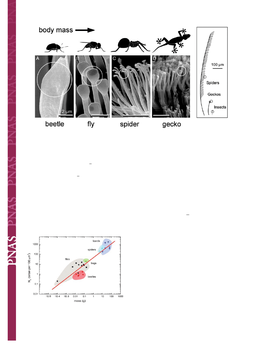
The setae of animals studied
储
are finely structured down to the
m and sub-m levels. The diameters of their spatula-like
terminal elements (Fig. 1) have been measured to range from 0.2
to 5.0
m. Comparative structural data clearly show that the
areal density N
A
of these terminal elements strongly increases
with increasing body mass m (Fig. 2). It is remarkable that a
single master curve exists for the different species; it is given by
log
䡠N
A
(m
⫺2
)
⫽ 13.8 ⫹ 0.699䡠log䡠m(kg), R ⫽ 0.919. Downscaling
of the contact elements, which results in a multiplication of the
number of single contact, thus appears to be a strong design
principle.
We want to interpret these findings in light of theoretical
contact mechanics. Consider a geometry in which a seta termi-
nates in a hemispherical shape. In the purely elastic case, the
diameter d of the area of contact with a flat substrate is given by
the Hertz equation:
d
3
⫽
12RF
E*
,
[1]
where R is the radius of the hemisphere, E* an average plain
strain modulus, and F the compressive contact load.
The Hertz theory has been extended to include surface
attraction effects by Johnson, Kendall, and Roberts (15). Their
result for the diameter of the contact area is
d
3
⫽
12R
E* 兵
F
⫹ 3
R␥ ⫹ 关6R␥F ⫹ 共3R␥兲
2
兴
1/2
其,
[2]
where
␥ is the adhesion energy per area. One of the conse-
quences of their analysis is the prediction of a finite pull-off force
given by
F
C
⫽
3
2
R
␥.
[3]
Let us apply this formalism to the attachment pad of the fly
Eristalis (16). Assume first that no hairy structure is present and
that R
⫽ 100
m is given by the radius of the complete surface
of the attachment organ. To support the weight (80 mg) of the
fly when hanging from the ceiling on one leg, an adhesion energy
in excess of 1 J
兾m
2
would be required according to Eq. 3; this is
clearly unrealistic for van der Waals interaction forces.
‡
To whom correspondence should be addressed. E-mail: arzt@mf.mpg.de.
¶
Electron microscopy and data analysis were performed as follows. Animals were fixed in
70% ethanol. Some pieces of materials were dehydrated in ethanol and critical-point
dried. Pieces of the material were fractured with a razor blade. All preparations were
critical-point dried, mounted on holders, sputter-coated with gold-palladium (10 nm), and
examined in a Hitachi S-800 scanning electron microscope at 20 kV. Measurements of
structures were made on digital pictures with
ANALYSIS 2.1
image analysis software (Soft-
Imaging Software, Mu¨nster, Germany).
储
The following animals with attachment devices were used for analysis: spiders (Cupiennius
salei, and Aphonopelma seemanni), insects (Calliphora erythrocephala, Drosophila mela-
nogaster, Lucilia caesar, Platycheirus angustatus, Sphaerophoria scripta, Episyrpus baltea-
tus, Eristalis pertinax, Myathropa florea, Volucella pellucens, Cantharis fusca, Leptinotarsa
decemlineata, Gastrophysa viridula, Chrysolina fastuosa, Phyllobius pomaceus, and Rhod-
nius prolixus), and geckos (Tarentola mauritanica, Phelsuma madagascariensis, Tarentola
mauritanica, Anolis maynardi, and Gecko gekko).
© 2003 by The National Academy of Sciences of the USA
www.pnas.org
兾cgi兾doi兾10.1073兾pnas.1534701100
PNAS
兩 September 16, 2003 兩 vol. 100 兩 no. 19 兩 10603–10606
APPLIED
PHYSICAL
SCIENCES
EVOLUTION
However, in reality, the animal takes advantage of an impor-
tant consequence of contact theory: inspection of Eq. 3 shows
that the adhesion force is proportional to a linear dimension of
the contact; therefore, by splitting up the contact into n sub-
contacts (setae), each with radius R
兾公n (self-similar scaling),
the total adhesion force is increased to
F
⬘
C
⫽
冑
n
䡠F
C
.
[4]
Van der Waals forces (with typical adhesion energies in the
range from 50 to 10 mJ
兾m
2
) now create sufficient attachment
strength, provided the number of setae is of order 10
3
to 10
4
per
fly pulvillus. This is indeed in accordance with our microscopic
observations (Fig. 1).
It is interesting that such an approach allows some simple
predictions to be made about scaling of attachment devices
between small and large animals: for dimensionality reasons, the
weight of the animal increases more rapidly than the area of the
foot-to-substrate contact. This has to be compensated by a
simultaneous increase in the setal density. It is shown in the
Appendix that two simple cases can be considered:
(i) For self-similar scaling, the contact radius of the terminal
elements is linearly related to their size. The setal areal density
N
A
is then expected to scale as
N
A
⫽ 4
2
m
2/3
,
[5]
where
⫽ (2kp
2/3
2/3
g
兾3
␥) is a geometry-insensitive param-
eter,
is the average mass density, g is the gravitational
acceleration, k is a ‘‘safety’’ factor, and p is a shape factor.
(ii) For curvature invariance, the contact radius R is assumed
to be independent of seta size. This leads to
N
A
⫽
R
m
1/3
.
[6]
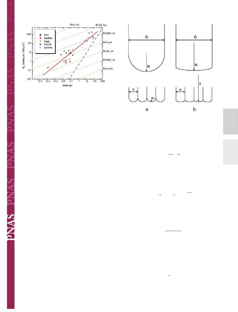
Lines of these slopes are included in Fig. 3 and can now be
compared with the experimental data. It is striking that the
assumption of self-similarity (Eq. 5), leading to a slope of 2
兾3,
explains the scaling, from the fruit fly to the gecko, very well.
Also a value for
can be extracted; the best fit is obtained for
⫽ 3.8 ⫻ 10
6
m
⫺1
䡠kg
⫺1/3
.
Closer inspection of Fig. 3 reveals some additional subtleties.
Whereas the relationship between body mass and density of
single contacts is well borne out for the complete sample of
animals from different evolutionary lineages (red line), the slope
appears to be lower within each lineage and approaches the value
1
兾3 predicted for curvature invariance (green lines). Especially
within lineages of beetles and flies this modified dependence
renders an improved description of the data. Using the above
value for
, values of contact radius R can be extracted from the
data: R
⬇ 1.6
m for the flies (excluding the ultra-light fruit fly)
and R
⬇ 0.3
m for the lizards. It may be suggested that in
heavier animals within a given lineage adhesion is improved by
increasing the seta density slightly at a given radius of curvature
of the terminal elements. In lineages with much larger body
mass, both the seta diameter and the radius of curvature have to
Fig. 1.
Terminal elements (circles) in animals with hairy design of attachment pads. Note that heavier animals exhibit finer adhesion structures.
Fig. 2.
Dependence of the terminal element density (N
A
) of the attachment
pads on the body mass (m) in hairy-pad systems of diverse animal groups
(log
䡠N
A
(m
⫺2
)
⫽ 13.8 ⫹ 0.699䡠log䡠m(kg), R ⫽ 0.919).
10604
兩 www.pnas.org兾cgi兾doi兾10.1073兾pnas.1534701100
Arzt et al.
be reduced. The curvature-invariant mechanism of increasing
adhesion has a natural limit: For curvature invariance the setae
density can only be increased until the seta diameter approaches
the contact diameter for a given curvature R. This limit of
maximum contact can approximately be calculated by the equa-
tions given here; as indicated by a blue line in Fig. 3, it lies well
outside the biological regime.
An additional advantage of the patterned surfaces is the
reliability of contact on various surface profiles and the in-
creased tolerance to defects at individual contacts (17). In the
real situation, failure of some microcontacts because of dust
particles or mechanical damage of single seta would minimally
influence contact adhesion. In the case of a solitary contact, even
a little damage of the contact caused by the presence of dirt or
surface asperities will immediately lead to contact breakage.
The present approach, of course, neglects several additional
contributions, such as the secretion of sticky fluids (18, 19);
however, we note that capillary forces scale in the same way as
the Johnson–Kendall–Roberts force (Eq. 3) so that the scaling
behavior would remain largely unchanged. Also, biological
materials in general and attachment systems in particular (20)
exhibit pronounced viscoelastic effects, which requires a more
complete treatment of the problem using viscoelastic contact
mechanics [e.g., Johnson and Greenwood (21)]. In the light of
these simplifications, it may be surprising that this approach
reproduces the scaling of the attachment devices from the fruit
fly to the gecko, covering 6 orders of magnitude in body mass.
This finding suggests that contact splitting is the overriding
design principle. Overall, it comes as no surprise that the
concepts of contact theory are reflected in the evolutionary
design of biological attachment systems.
Appendix: Derivation of the Scaling Relations
We want to derive the dependence of seta density on animal
mass, as required by contact theory. The mass m is given by
m
⫽ D
3
p,
[A1]
where D is a size parameter that we choose to be the total
apparent contact diameter (i.e., size of one pulvillus);
is the
average mass density, and p is a dimensionless shape factor. The
adhesion force necessary to support this weight is written as
F
W
⫽ k䡠mg,
[A2]
where g is the gravitational acceleration and k is a safety factor.
Consider a pulvillus with n contacts (setae) of diameter s. The
areal density of setae can be expressed as
N
A
⫽
n
D
2
⫽
1
s
2
.
[A3]
For the calculation of the adhesion force, two cases are
distinguished (Fig. 4).
Self-Similarity.
If the contract radius R scales with seta diameter
s (i.e., R
⫽ s兾2 for hemispherical shape), then we obtain for the
Johnson–Kendall–Roberts adhesion force
F
C
⫽ n䡠
3
4
s
␥ ⫽
3
4
D
2
␥
冑
N
A
.
[A4]
Equating Eqs. A4 and A2 yields
N
A
⫽ 4
2
m
2/3
,
[A5]
where
⫽
2kp
2/3
2/3
g
3
␥
,
[A6]
which we assume to be, to first order, independent of the animal
and its size. Eq. A5 predicts plots of log
䡠N
A
vs. log
䡠m lines of slope
2
兾3 (Fig. 3).
Curvature Invariance.
If the contact radius R is fixed and does not
scale with seta diameter, the adhesion force is approximately
given by
F
C
⫽
3
2
R
␥n.
[A7]
Equating Eqs. A7 and A2 leads to
Fig. 3.
Interpretation of Fig. 2 in light of contact theory. A fit to all data (red
line) gives a slope of
⬇2兾3, corresponding to the self-similarity criterion.
Within each lineage, a lower slope of
⬇1兾3 is found, suggesting curvature
invariance of the contacts with radius R (green lines). The approximate limit
for such attachment devices (limit of maximum contact) is shown as a blue line.
Fig. 4.
Two cases of contact scaling. (a) Self-similarity: contact radius R scales
with contact size s. (b) Curvature invariance: contact radius is independent of
contact size.
Arzt et al.
PNAS
兩 September 16, 2003 兩 vol. 100 兩 no. 19 兩 10605
APPLIED
PHYSICAL
SCIENCES
EVOLUTION
N
A
⫽
m
1/3
R
,
[A8]
which corresponds to lines of slope 1
兾3 in Fig. 3.
We gratefully acknowledge stimulating discussions with K. L. Johnson
(Cambridge University, Cambridge, U.K.) and H. Gao, A. Wanner, and
U. Wegst (Max Planck Institute for Metals Research). Support from
members of the Electron Microscopy Unit team (H. Schwarz and J.
Berger) at the Max Planck Institute of Developmental Biology is
gratefully acknowledged. Parts of this work were supported by Federal
Ministry of Science of Germany (Bundesministerium fu¨r Bildung,
Wissenschaft, Forschung und Technologie) Grant BioFuture 0311851
(to S.G.) and a Deutsche Forschungsgemeinschaft Leibniz Award
(to E.A.).
1. Breidbach, O. (1980) Mikrokosmos 69, 200–201.
2. Schliemann, H. (1983) Funkt. Biol. Med. 2, 169–177.
3. Beutel, R. & Gorb, S. N. (2001) J. Zool. Sys. Evol. Res. 39, 177–207.
4. Gorb, S. N. (2001) Attachment Devices of Insect Cuticle (Kluwer, Dordrecht,
The Netherlands), pp. 1–305.
5. Gillett, J. D. & Wigglesworth, V. B. (1932) Proc. R. Soc. London Ser. B 111,
364–376.
6. Stork, N. E. (1980) J. Exp. Biol. 88, 91–107.
7. Autumn, K., Liang, Y. A., Hsieh, S. T., Zesch, W., Wai, P. C., Kenny, T. W.,
Fearing, R. & Full, R. J. (2000) Nature 405, 681–685.
8. Bauchhenss, E. (1979) Zoomorphologie 93, 99–123.
9. Walker, G., Yule, A. B. & Ratcliffe, J. (1985) J. Zool. (London) 205, 297–307.
10. Gorb, S. N. (1998) J. Insect Physiol. 44, 1053–1061.
11. Homann, H. (1957) Naturwissenschaften 44, 318–319.
12. Hiller, U. (1968) Z. Morphol. Tiere 62, 307–362.
13. Autumn, K., Sitti, M., Liang, Y. C. A., Peattie, A. M., Hansen, W. R., Sponberg,
S., Kenny, T. W., Fearing, R., Israelachvili, J. N. & Full, R. J. (2002) Proc. Natl.
Acad. Sci. USA 99, 12252–12256.
14. Arzt, E., Enders, S. & Gorb, S. (2002) Z. Metallkde. 93, 345–353.
15. Johnson, K. L., Kendall, K. & Roberts, A. D. (1971) Proc. R. Soc. London Ser.
A 324, 301–320.
16. Gorb, S., Gorb, E. & Kastner, V. (2001) J. Exp. Biol. 204, 1421–1431.
17. Challet, D. & Johnson, N. F. (2002) Phys. Rev. Lett. 89, 028701.
18. Eisner, T. & Aneshansley, D. J. (1983) Ann. Entomol. Soc. Am. 76, 295–298.
19. Attygalle, A. B., Aneshansley, D. J., Meinwald, J. & Eisner, T. (2000) Zoology
103,
1–6.
20. Gorb, S. N., Jiao, Y. & Scherge, M. (2000) J. Comp. Physiol. A 186,
821–831.
21. Johnson, K. L. & Greenwood, J. A. (1997) J. Colloid Interface Sci. 192, 326–333.
10606
兩 www.pnas.org兾cgi兾doi兾10.1073兾pnas.1534701100
Arzt et al.
Wyszukiwarka
Podobne podstrony:
Guide to the properties and uses of detergents in biology and biochemistry
How to cut Mini and Micro SIM to Nano SIM
Adaptive Filters in MATLAB From Novice to Expert
Notto R Thelle Buddhism and Christianity in Japan From Conflict to Dialogue, 1854 1899, 1987
Guide to the properties and uses of detergents in biology and biochemistry
Far Infrared Energy Distributions of Active Galaxies in the Local Universe and Beyond From ISO to H
ebooksclub org Women and Race in Contemporary U S Writing From Faulkner to Morrison American Literat
A Sarong in my Backpack Adventures from Munich to Pushkar
Adaptive Filters in MATLAB From Novice to Expert
From drain to gain in capture fisheries rents a synthesis study
1980 From Latin to Romance in Sound Charts
William Weber From miscellany to homogenity in Concert Programming
2004 Variation and Morphosyntactic Change in Greek From Clitics to Affixes Palgrave Studies in Langu
Full Toroidal Variable Drive Transmission Systems in Mechanical Hybrid Systems From Formula 1 to Ro
Warning to Japan Norway and the United States From the Cetaceans (DOLPHIN S CONTACTS)
Andrei Marmor Social Conventions From Language to Law Princeton Monographs in Philosophy 2009
Armstrong; From Huponoia to Paranoia On the Secular Co optation of Homeric Religion in Vico, Feuerba
John N Duvall Race and the White Identity in Southern Fiction From Faulkner to Morrison
History of Race Relations in the America from Slavery to Pre doc
więcej podobnych podstron