
REVIEW
Extracellular NAD and ATP: Partners in immune
cell modulation
Friedrich Haag
&
Sahil Adriouch
&
Anette Braß
&
Caroline Jung
&
Sina Möller
&
Felix Scheuplein
&
Peter Bannas
&
Michel Seman
&
Friedrich Koch-Nolte
Received: 8 February 2006 / Accepted: 22 October 2006 / Published online: 9 January 2007
# Springer Science + Business Media B.V. 2007
Abstract Extracellular NAD and ATP exert multiple, par-
tially overlapping effects on immune cells. Catabolism of both
nucleotides by extracellular enzymes keeps extracellular
concentrations low under steady-state conditions and gener-
ates metabolites that are themselves signal transducers. ATP
and its metabolites signal through purinergic P2 and P1
receptors, whereas extracellular NAD exerts its effects by
serving as a substrate for ADP-ribosyltransferases (ARTs) and
NAD glycohydrolases/ADPR cyclases like CD38 and
CD157. Both nucleotides activate the P2X7 purinoceptor,
although by different mechanisms and with different charac-
teristics. While ATP activates P2X7 directly as a soluble
ligand, activation via NAD occurs by ART-dependent ADP-
ribosylation of cell surface proteins, providing an immobilised
ligand. P2X7 activation by either route leads to phosphati-
dylserine exposure, shedding of CD62L, and ultimately to
cell death. Activation by ATP requires high micromolar con-
centrations of nucleotide and is readily reversible, whereas
NAD-dependent stimulation begins at low micromolar con-
centrations and is more stable. Under conditions of cell stress
or inflammation, ATP and NAD are released into the extra-
cellular space from intracellular stores by lytic and non-lytic
mechanisms, and may serve as
“danger signals” to alert the
immune response to tissue damage. Since ART expression is
limited to naïve/resting T cells, P2X7-mediated NAD-induced
cell death (NICD) specifically targets this cell population. In
inflamed tissue, NICD may inhibit bystander activation of
unprimed T cells, reducing the risk of autoimmunity. In
draining lymph nodes, NICD may eliminate regulatory T cells
or provide space for the preferential expansion of primed
cells, and thus help to augment an immune response.
Key words ADP-ribosylation . ADP-Ribosyltransferases .
apoptosis . ATP. ectoenzymes . extracellular purines .
NAD . posttranslational protein modification
Abbreviations
ADP
adenosine diphosphate
ADPR
Adenosine diphosphate ribose
AMP
Adenosine monophosphate
ART
ADP-ribosyltransferase
ATP
Adenosine triphosphate
E-NPP
Ecto-nucleotide pyrophosphatase/
phosphodiesterase
E-NTPD
Ecto-nucleoside triphosphate
diphosphohydrolase
FoxP3
Forkhead box P3
NAADP
Nicotinic acid adenine dinucleotide phosphate
NAD
Nicotinamide adenine dinucleotide
NADP
Nicotinamide adenine dinucleotide phosphate
NICD
NAD-induced cell death
PARP
Poly(ADP-ribose) polymerase
PS
Phosphatidyl serine
ATP and NAD in the extracellular compartment:
From their release to the induction of specific signalling
ATP and NAD+ are classic intracellular metabolites with
center-stage roles in energy metabolism and electron
transfer. In recent years, it has become evident that these
Purinergic Signalling (2007) 3:71
–81
DOI 10.1007/s11302-006-9038-7
F. Haag (
*)
:
S. Adriouch
:
A. Braß
:
C. Jung
:
S. Möller
:
F. Scheuplein
:
P. Bannas
:
F. Koch-Nolte
Institute of Immunology, University Hospital,
Martinistr. 52, 20246 Hamburg, Germany
e-mail: haag@uke.uni-hamburg.de
F. Haag
:
S. Adriouch
:
M. Seman
INSERM U519- EA1556, Faculté de Médecine et de Pharmacie,
Université de Rouen,
F-76183 Rouen Cedex, France
purine nucleotides play important roles also in the
extracellular environment, i.e., as substrates for a flurry
of nucleotide-metabolising ectoenzymes, and, in the case
of ATP, also as a ligand for cell surface receptors (Figs.
and
).
Biosynthesis of NAD presumably takes place in several
locations in the cell [
]. Under physiological conditions
most (more than 70%) of the cellular NAD content is
stored and is utilised in the mitochondria primarily for
metabolic purposes. In the cytoplasm and in the nucleus
NAD serves cell signalling functions, as a precursor for
calcium mobilising metabolites and as a substrate for two
families of nuclear enzymes, i.e., poly-ADP-ribosyl
polymerases (PARPs) and the sirtuin (homologues of
the yeast
“silent mating type information regulation 2”
(Sir2) gene) family of NAD-dependant lysine deacetylases,
both of which play important roles in coordinating DNA
repair, regulating transcription levels and controlling pro-
gression towards apoptosis [
]. Under pathophysiologi-
cal conditions, such as ischemia, oxidative stress or
DNA-damaging agents, cells release their mitochondrial
NAD content to the cytoplasm and the nucleus by still
unknown mechanisms [
]. It is not surprising, then, that
NAD plays an essential role in the cellular response to
stress.
Similarly, following the induction of cellular stress part
of this cellular content of NAD and ATP may be released
into the extracellular space. This may occur by several
mechanisms involving active exocytosis or diffusion
through transmembrane transporters in living cells or
passive leakage across the membrane in dying cells [
–
].
Of note, release of purines by injured or dying cells has
recently been suggested to serve as a
“danger signal” that
may alert the immune system to tissue damage [
].
Immune modulation by extracellular ATP
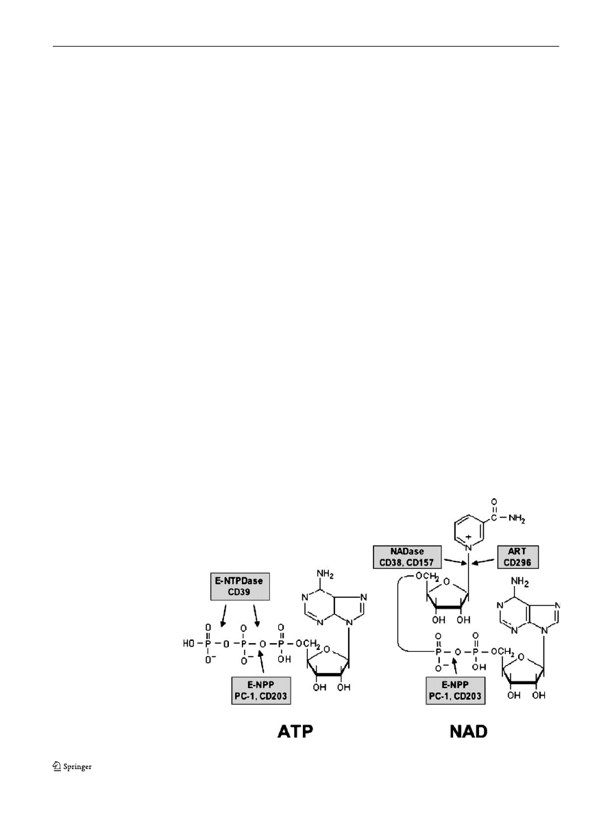
Once released, extracellular ATP and NAD can be degraded
into further metabolites such as ADP, AMP or adenosine by
extracellular enzymes, i.e., ecto-nucleoside triphosphate
diphosphohydrolase (E-NTPDs), ecto-nucleotide pyrophos-
phatase/phosphodiesterase (E-NPPs), and the ecto-5
′-nucle-
otidase CD73 (Figs.
). ATP or its by-products activate
different members of the purinoceptor family of receptors.
Purinoceptors comprise adenosine-sensitive P1 receptors
(A1, A2a, A2b, and A3) and P2 receptors, which are
activated by ATP, ADP, UTP, UDP or UDP-glucose [
] (and NAD, see note added in proof). P2 receptors are
further divided into two groups: the G protein-coupled
seven-transmembrane P2Y receptors (P2Y1, -2, -4, -6, -11,
-12, -13, -14), and the P2X ligand-gated ion channels
(P2X1-7) [
]. Triggering of purinoceptors by their
ligands regulates important physiological functions such as
platelet aggregation, local regulation of blood pressure,
modulation of cardiac functions in ischemic conditions or
regulation of the development of inflammation [
,
,
Regulation of immune functions by ATP and its
metabolites has been reviewed elsewhere [
]. ATP can
in principle transmit signals through several different
receptors, including the complete P2X family and a
subgroup of P2Y receptors (P2Y1, 2, 11, 12, 13) [
These receptors differ greatly in their relative sensitivities to
ATP, with EC50s in the nanomolar (P2Y), low micromolar
Fig. 1 Chemical structure of
ATP and NAD, and sites of
cleavage by different
ecto-enzymes
72
Purinergic Signalling (2007) 3:71
–81
(most P2X) or high micromolar (P2X7) ranges [
]. The
situation is further complicated by the fact that extracellular
ATP is rapidly metabolised, and its break-down products,
notably ADP and adenosine, have signalling functions of
their own through different receptors. Both pro- and anti-
inflammatory effects of ATP on immune cells have been
reported, depending on the cell type and the available
concentration of ATP. In general, P2X7, which requires
high ATP concentrations acting for a short time, mediates
mainly pro-inflammatory effects, such as the processing
and release of interleukin- (IL-) 1 and IL-18 [
], in
dendritic cells and macrophages, and induces cell death in
T cells. Activation of P2X7 also stimulates the production
of tumor necrosis factor alpha (TNFa) in microglial cells
[
]. Low concentrations of ATP present during the
maturation of DCs reduce their capacity to induce Th1-
typical responses in primed T cells [
,
]. These anti-
inflammatory effects may be due either to direct action on
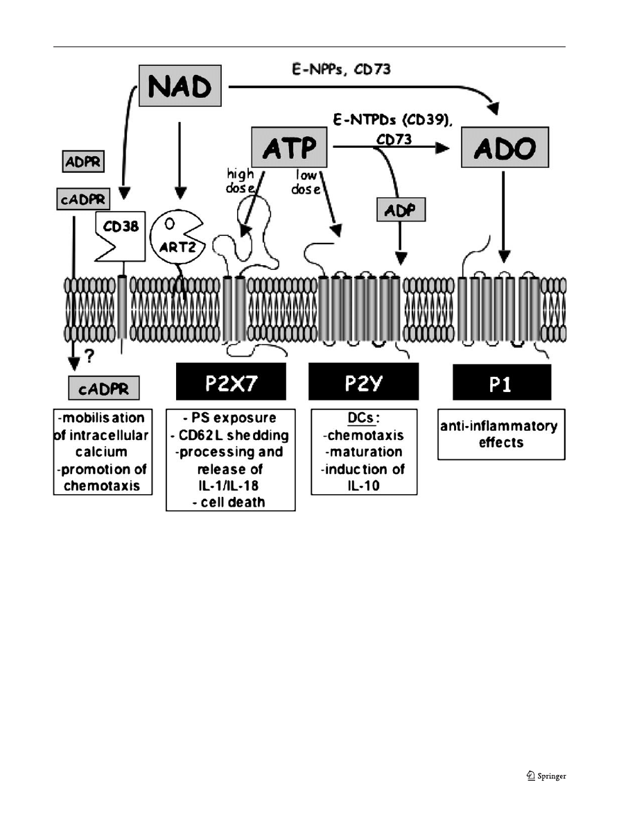
Fig. 2 Action of extracellular ATP and NAD and their metabolites on
different cell surface receptors. Extracellular ATP present in high,
intermediate, or low concentrations can activate P2X7, other P2X, or
P2Y receptors, respectively, or is hydrolysed by the sequential action
of ecto-nucleoside triphosphate diphosphohydrolases (E-NTPDs) such
as CD39 and ecto-5
′-nucleotidase (CD73) to ADP and adenosine
(ADO). For clarity, P2X receptors other than P2X7 are not shown,
since their presence on immune cells is not well documented. ADP can
act on P2Y receptors, and adenosine can activate G protein-coupled P1
receptors. Extracellular NAD serves as a substrate for cell-surface
ADP-ribosyltransferases (ART2), or is hydrolyzed to ADPribose by
CD38. CD38 can also synthesise cyclic ADP-ribose, a known
intracellular calcium mobilising agent. It is not known how cADPR
gains access to the intracellular compartment. NAD (and ATP) may
also be hydrolysed by ecto-nucleotide pyrophosphatase/phosphodies-
terases (E-NPPs) to AMP, which in turn is hydrolysed by CD73 to
adenosine. See text for details
Purinergic Signalling (2007) 3:71
–81
73
some P2Y receptors like P2Y11, or by adenosine signalling
through P1 receptors (Figs.
and
Immune modulation by extracellular NAD
Similar to ATP, NAD is also degraded in the extracellular
compartment, giving rise to the generation of metabolites
like cyclic ADP-ribose or adenosine that are active signal
transducers (Fig.
). In contrast to ATP, signalling through
intact NAD does not involve specific membrane receptors
(see note added in proof). Nevertheless, NAD may regulate
cellular functions through two known enzyme families. The
first family, comprising CD38 and the functionally related
CD157 enzyme, possesses NAD-glycohydrolase and ADP-
ribose cyclase activities. They catalyse cleavage of NAD
into ADP-ribose or cyclic ADP-ribose and nicotinamide
[
], as well as the transglycosidation of NADP and
nicotinic acid to yield NAADP [
]. Cyclic ADP-ribose
and NAADP are newly recognised second messenger
molecules, which trigger calcium release from IP3-inde-
pendent intracellular stores, and which may thus play
important regulatory roles [
]. However, it is contro-
versial whether these second messengers are generated by
extracellular CD38 and are then translocated to the cytosol
by hitherto unknown mechanisms, or whether they are
generated from intracellular NAD by an intracellular
isoform of CD38. CD38 may also be involved in the
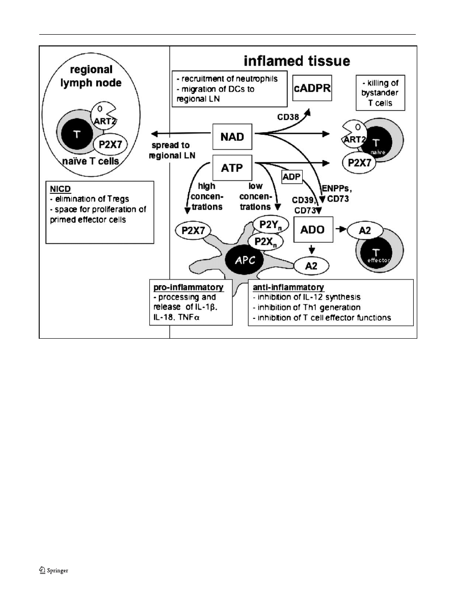
Fig. 3 Hypothetical scheme of the interplay of purine sensors during
an immune response. ATP and NAD are released locally at sites of
tissue injury or inflammation. At high concentrations, ATP acts on the
P2X7R receptor to exert pro-inflammatory effects on antigen present-
ing cells or to kill T cells; at low concentrations it acts on other P2
receptors to downregulate the initiation of Th1 responses. NAD is used
by ARTs on T cells to activate P2X7, or by CD38 to generate cyclic
ADP-ribose. It is conceivable that NAD may exert distant effects by
reaching lymph nodes draining inflammatory sites in physiologically
relevant concentrations. Both ATP and NAD are degraded by
metabolising enyzmes to yield other signalling molecules, notably
adenosine (ADO), which exerts predominantly anti-inflammatory
effects through P1 receptors of the A2-subfamily. See text for details
74
Purinergic Signalling (2007) 3:71
–81

regulation of immune functions by limiting the substrate
availability for ADP-ribosyltransferases (see below) [
].
Mice lacking the CD38 glycohydrolase/ADP-ribosyl cy-
clase show an impaired antibody response to T-cell
dependent antigens [
], which may be due to a defect
in the migratory capacity of dendritic cells [
The second family of enzymes mediating signalling by
NAD comprises the mono(ADP-ribosyl)transferases
(ARTs), which are structurally related to ADP-ribosylating
bacterial toxins. These enzymes catalyse a posttranslational
modification of proteins by transferring the ADP-ribose
moiety from NAD to specific amino acids, e.g., arginine
residues, on target proteins [
]. This family contains five
known mammalian members, ART1-ART5, which are GPI-
anchored membrane proteins (ART1-ART4) or secreted
enzymes (ART5) [
]. Human ART1 was recently assigned
the CD number CD296 [
]; it is expressed by activated
granulocytes as well as by skeletal muscle, heart, and
epithelial cells [
]. ART4 has been identified as the
carrier of the Dombrock blood group alloantigens and was
recently assigned the CD number CD297 [
]. ART4 is
expressed prominently by erythrocytes and at lower levels
also on monocytes and splenic macrophages. Only ART1,
ART2 and ART5 have been shown so far to possess
arginine-specific activity, while ART3 and ART4 may have
acquired a new target specificity. Akin to the well-known
phosphorylation reaction, posttranslational protein modifi-
cation by ADP-ribosylation regulates (inhibits or activates)
the functions of target proteins [
]. The ART enzyme
family members thus represent new players in the epige-
netic regulation of protein functions.
It has been shown that ART2, like many other GPI-
anchored proteins, is segregated into specialised cholester-
ol- and ganglioside-enriched microdomains on the cell
surface termed lipid rafts [
]. Localisation into lipid rafts
is important for the activity of ART2, presumably by
focussing it on its target molecules. Indeed, substantial
fractions of two known non-GPI-anchored target proteins of
ART2, i.e., LFA-1 and P2X7, may also be recruited into
lipid rafts [
].
Purine sensors on cells of the immune system
Cells of the immune system express a variety of purine
sensors on their surfaces, either as purinoreceptors or as
ecto-enzymes that metabolise purine nucleotides (Fig.
).
For many of the molecules, a detailed expression analysis is
still hampered by a lack of suitable antibodies.
The only ATP-sensitive purinoreceptor that has been
positively identified on peripheral T cells to date is P2X7.
In these cells, P2X7 mediates ATP- and NAD-dependent
phosphatidyl serine (PS) exposure, CD62L shedding, and
ultimately cell death [
,
]. Of the P2Y receptors, P2Y6
and P2Y14 have been described on T cells [
,
], but
these receptors are sensitive to UDP and UDP-glucose,
respectively.
P2X7 is also expressed on antigen-presenting cells,
including dendritic cells and macrophages, where it
mediates release of the non-classically secreted cytokines
IL-1
β and IL-18 [
,
], and promotes phagosome/
lysosome fusion [
–
]. P2X7 is not expressed on resting
B cells in the mouse, but in the human has been identified
on a subset of chronic B-cell lymphomas (B-CLL) [
Immature dendritic cells also express the P2Y11 receptor
[
]. This receptor, which is sensitive to nanomolar
concentrations of ATP (see note added in proof), has been
implicated in several responses of DCs to ATP. Low doses
of ATP synergise with other stimuli like TNFa or LPS to
enhance DC maturation, but the net effect is to reduce the
production of IL-12p70 and increase the production of IL-
10 [
]. As is the case for the anti-inflammatory A2
subgroup of P1 receptors (see below), stimulation of P2Y11
causes an elevation of intracellular cAMP in DCs, which
mediates its effects on DC maturation [
]. Using a
different biochemical pathway, P2Y11 also inhibits the
migratory response of immature DCs to chemotactic
gradients, causing these cells to remain longer at sites of
tissue damage [
The G protein-coupled P1 receptors fall into two
functional groups, which serve to lower (A1 and A3
receptors) or to increase (A2a and A2b receptors) intracel-
lular levels of cyclic AMP (cAMP). A1/A3 receptors are
expressed on immature DCs, where they induce calcium
flux and promote chemotaxis [
,
]. A2a/b receptors
down-regulate the production of IL-12 in LPS-matured
DCs and thus inhibit the differentiation of naive CD4+ T
cells towards a Th1 phenotype. T cells also express A2a
receptors. Stimulation of these receptors by adenosine
inhibits TCR-mediated T cell proliferation and upregulation
of the IL-2 receptor, as well as most of the effector
functions of cytotoxic T cells [
,
]. The A2a/b receptors
are the most prominent P1 receptors on immune cells, and
are responsible for the dominant anti-inflammatory effects
of adenosine on the immune system (recently reviewed in
[
]). Nucleotide-metabolising enzymes are widely distrib-
uted among cells of the immune system. Ecto-ATPase and
5
′-nucleotidase activities, which are sufficient to convert
ATP into adenosine, are found on many lymphocytes and
antigen presenting cells. It is worth noting that ecto-
adenylate kinase, the enzyme catalysing the reverse
pathway, i.e., the generation of ATP from extracellular
adenosine, is also present on the surface of lymphocytes
[
]. Adenosine can also be generated from extracellular
NAD by the sequential action of E-NPPs and 5
′-nucleotid-
ase. Although the expression of E-NPPs on immune cells
Purinergic Signalling (2007) 3:71
–81
75

has not been studied in detail, it is of note that E-NPP1, also
known as PC-1, was originally identified as a marker for
plasma cells. Finally, immune cells express NAD-depen-
dent NADase and ADP-ribosyltransferase activities. In the
mouse, the CD38 NADase/ADPR-cyclase is expressed on
B cells and other antigen-presenting cells, and on activated,
but not resting T cells [
,
]. Conversely, the ART2 ADP-
ribosyltransferases are expressed on resting mouse T cells,
but not on activated ones or on antigen-presenting cells.
The biological functions of ARTs and their substrate,
NAD, in immune regulation
Extracellular NAD selectively induces apoptosis of mouse
T cells (both CD4+ and CD8+), but not of B cells [
].
Interestingly, apoptosis is observed with NAD concentra-
tions as low as 1 micromolar. Furthermore, sensitivity to
NAD is dependent on the activation state of lymphocytes.
Indeed, in vitro stimulation of T cells with mitogens prior to
NAD incubation results in relative insensitivity to NAD-
induced apoptosis. Similarly, in vivo-activated cells, present
in freshly isolated T-cell preparations, do not respond to
NAD treatment, resulting in enrichment of CD44high,
CD62Llow activated /memory T cells in the surviving
fraction. Thus, NAD selectively induces apoptosis of naive
mouse T lymphocytes [
]. It has recently been shown that
sensitivity to NAD-induced cell death is especially high in
CD4+/CD25+ regulatory T cells expressing the transcrip-
tion factor forkhead box P3 (FoxP3) [
What is the molecular mechanism underlying NAD-
induced apoptosis? Apart from NAD, none of the structur-
ally related molecules tested (i.e., nucleosides, nucleotides
or products of NAD metabolism) induced apoptosis in the
micromolar range. Therefore, NAD must induce apoptosis
through direct interaction with membrane proteins like
ARTs, which are able to use extracellular NAD. Consistent
with this interpretation, ART2 knock-out mice are com-
pletely resistant to NAD-induced apoptosis [
]. However,
although C57BL/6 mice express high levels of ART2, T
lymphocytes from this strain are relatively resistant to the
effects of NAD. Therefore, ART2.2 is required, but not
sufficient to account for NAD-induced apoptosis, and
another essential factor must be involved in the process [
].
This downstream effector was identified by pharmaco-
logical studies [
]. Etheno-NAD, an NAD analogue
modified in the adenine moiety, can be used as a substrate
by ART2 resulting in etheno-ADP-ribosylation of target
proteins [
]. However, etheno-NAD, like the NAD
analogues NHD or NGD, was unable to induce apoptosis.
Furthermore, pre-treatment with etheno-NAD, NHD or
NGD prevented subsequent NAD-induced apoptosis. These
data implied that the downstream effector was sensitive to
the modification of NAD in the adenine group, a property
known to hold true for purinoceptors. As adenosine itself
was unable to trigger apoptosis of T lymphocytes, it was
poorly conceivable that the effector belonged to the P1
purinoceptors. Conversely, the fact that a high dose of ATP
is able to induce apoptosis of T cells suggested that P2
purinoceptors could be involved. Within the P2 receptor
family, P2X7 was a good candidate, because, firstly, P2X7
is expressed on T lymphocytes and, secondly, because
P2X7 triggering is known to induce apoptosis [
Several lines of evidence demonstrated that P2X7 does
indeed mediate NAD-induced apoptosis as a consequence
of ADP-ribosylation of membrane proteins by ART2.
Foremost, pharmacological studies showed that the P2X7
inhibitors KN-62 and oATP block NAD-induced apoptosis.
Furthermore NAD, like ATP, was able to induce other
known effects characteristic of P2X7 activation, such as the
formation of non-selective membrane pores permeable to
large molecules up to 900 Da, the exposition of PS on the
outer leaflet of the cell membrane, and the shedding of
CD62L. Moreover NAD, like ATP, induced calcium uptake
that could be inhibited by the P2X7 inhibitor KN-62.
Finally, the generation of antibodies directed against mouse
P2X7 allowed us to show that P2X7 is ADP-ribosylated in
the presence of NAD and is, therefore, directly targeted by
ART2 [
].
Further confirmation of the involvement of P2X7 in
NAD-induced apoptosis was brought by examination of the
gene coding for P2X7 in C57BL/6 mice. Cloning and
sequencing of the P2rx7 gene showed that these mice
harbour an inactivating mutation (P451L) within the
cytoplasmic domain of the receptor. Transfection studies
in HEK cells showed that the P451L mutation severely
affects P2X7 functions like pore formation and calcium flux
in response to ATP in comparison to the wild type [
]. It
seemed likely, therefore, that the P451L mutation in P2X7
accounted for the resistance to NAD-induced apoptosis in T
lymphocytes from C57BL/6 mice. The P451L mutation in
the mouse is reminiscent of a naturally occurring E496A
mutation in the cytoplasmic domain of the human P2X7,
which similarly affects the function of the receptor [
].
Collectively, these studies identify ADP-ribosylation of
P2X7 as an alternative way of activating P2X7 in T
lymphocytes [
]. Although P2X7 has been implicated in
important cellular functions, it is not fully clear how it is
activated in vivo. Near millimolar concentrations of
exogenous ATP are required to elicit P2X7 activation in
vitro [
]. In this context, ART2-catalysed ADP-
ribosylation, requiring only micromolar concentrations of
NAD, represents an appealing alternative pathway for the
activation of P2X7. NAD-dependent activation of P2X7
76
Purinergic Signalling (2007) 3:71
–81

constitutes a novel mechanism for inducing T lymphocyte
apoptosis. It is of note that this mechanism only affects
resting T lymphocytes [
], consistent with the observation
that ART2 is shed from the cell surface by the action of a
metalloproteinase following T-cell activation [
]. Other
known apoptosis-triggering pathways in T lymphocytes
affect either immature T cells during their development in
the thymus, or activated mature T lymphocytes, a process
known as AICD (activation-induced cell death). NAD-induced
apoptosis or NICD (NAD-induced cell death) is the first
apoptosis pathway described affecting naive T lymphocytes.
P2X7 is also expressed on macrophages and dendritic
cells. Recent studies have underlined the central role of
P2X7 in inflammation by controlling IL-1
β maturation and
release [
]. Furthermore, P2X7 may play a role in the
elimination of intracellular pathogens such as mycobacteria
or Chlamydia by promoting phagosome-lysosome fusion
[
–
]. To date no clear evidence of ART expression on
these cell types has been found. It is also not known yet
whether P2X7 can be activated by soluble ARTs or by an
ART present on the surface of an apposing cell. It thus
presently remains unclear whether NAD-dependent activa-
tion of P2X7 can also occur on these cells.
Activation of P2X7 by ATP or via NAD-dependent
ADP-ribosylation shows important differences. First of all,
activation via NAD begins at substrate concentrations around
1
μM and increases in a dose-dependent manner. By contrast,
ATP-mediated activation does not occur below a certain
threshold concentration, which is dependent on the cell type
and usually lies above 100
μM. Importantly, brief treatment
with NAD leads to long lasting activation of P2X7, while
treatment with ATP in the same conditions produces reversible
effects [
,
]. These characteristics are compatible with the
notion of a covalently bound (immobilised ADP-ribose in the
case of NAD-mediated activation) versus a soluble (ATP) ligand.
Endogenous sources of NAD and ATP
Under normal conditions the concentrations of ATP and
NAD in body fluids are below the concentrations required
to induce P2X7-dependent apoptosis. The concentration of
NAD for instance is reported to be maintained between 0.1
to 0.5
μM in the serum of non-treated animals [
]. It is
therefore plausible that extracellular nucleotides are re-
leased from intracellular sources, where they are present in
higher concentrations. As mentioned above, three situations
may lead to the release of nucleotides from intracellular
compartments: (1) liberation of intracellular contents by
dying cells, (2) exocytosis of nucleotide-containing gran-
ules and (3) diffusion of these molecules towards the
extracellular space across membrane channels.
Experiments in vitro demonstrated that cell lysates
contain amounts of NAD sufficient to induce ART2-
dependent apoptosis of T lymphocytes [
]. These data
suggest indirectly that NAD may be released in vivo by
dead cells resulting from accidental tissue damage or
consequent to the activity of the immune system itself.
Notably, T cells from ART2
−/− mice were not affected in
these experiments, indicating that
— at least at the
concentrations employed
— the lysates did not contain
sufficient quantities of ATP to trigger the P2X7 receptor.
This is consistent with the observation that activation of
P2X7 occurs at low doses of NAD, but requires a threshold
concentration of ATP [
,
The hypothesis that purines are actively secreted from
living cells and function as neurotransmitters was first
formulated by G. Burnstock in 1972 [
]. Since then,
many groups have confirmed that ATP, together with other
neurotransmitters, is actively released from pre-synaptic
vesicles of so-called
“purinergic” nerves [
]. Recently, a
similar mechanism has also been described for the release
of NAD. According to this report, NAD can be released in
combination with ATP and other neurotransmitters from
stimulated postganglionic nerve terminals connected to
blood vessels and urinary bladder [
]. Outside the nervous
system, other mechanisms can account for the release of
purine nucleotides. For example, both NAD and ATP have
been reported to be transported through Connexin43 gap-
junction hemi-channels [
]. Evidence is accumulating
that these channels preferentially open under conditions of
metabolic or mechanical stress [
,
]. In vitro, release of
purine nucleotides can be induced by moderate osmotic
shocks, application of shear forces and following mechan-
ical stress. In all these cases, release of purines appears to
be an active process that follows the increase of
intracellular calcium concentration [
]. Finally,
extracellular ATP itself may be a signal for the release
of further ATP through activation of the P2X7 purino-
receptor [
].
Evidence is accumulating linking the release of purine
nucleotides into the extracellular compartment with inflam-
mation or cellular stress. If purine nucleotides are prefer-
entially released into the extracellular compartment under
conditions of cellular stress, it is conceivable that the
massive recruitment of polymorphonuclear neutrophils and
macrophages into sites of inflammation, which results in
oxidative stress, tissue damage and massive neutrophil
death, may lead to the release of NAD by both lytic and
non-lytic mechanisms. In this context it is interesting to
note that the only direct demonstration to date of ADP-
ribosylation occurring in vivo using endogenous NAD as a
substrate stems from an inflammatory environment. In
humans, the defensin HNP-1 has been reported to be
Purinergic Signalling (2007) 3:71
–81
77

ADP-ribosylated in the bronchoalveolar fluid of smokers
[
]. This suggests that NAD might be released in
situations leading to chronic airway inflammation.
The regulation of purinergic signalling
by nucleotide-catabolising enzymes
The deleterious effect of the ADP-ribosylation of P2X7
receptors on naive T lymphocytes raises the question of
whether this phenomenon also occurs under physiological
conditions. In in vitro experiments, more than 70% of naive
T lymphocytes are susceptible to NICD [
]. Obvious-
ly, this does not reflect the situation in a normal mouse. In
living organisms, a flurry of nucleotide-catabolising
enzymes tightly regulate the concentration of extracellular
NAD and ATP. The steady-state concentration of NAD in
biological fluids thus results from the equilibrium between
its release from intracellular stores and its degradation by
NAD-catabolising enzymes.
In principle, extracellular NAD may be hydrolysed by
the CD38 and CD157 NAD-glycohydrolases or by phos-
phodiesterases (E-NPPs) present on the membrane of
several cells as well as in soluble form in biological fluids.
CD38 is the major NAD-glycohydrolase/ADP-ribosyl
cyclase present in the extracellular compartment [
].
Experiments in vitro point to a major role of CD38 in
controlling the level of ADP-ribosylation on the surface of
T cells [
]. In these experiments, the presence of CD38,
which is highly expressed by B cells, and at lower level on
activated T cells, greatly impaired the level of ADP-
riboyslation detected on the surface of T cells upon
treatment with NAD. Magnetic depletion of B cells from
cell preparations and/or the use of ara-F-NAD to selectively
block CD38 activity, greatly enhanced detectable ADP-
ribosylation. In line with these findings, cells prepared from
CD38
−/− deficient mice showed increased apparent ADP-
ribosyl transferase activity in vitro. These observations
underline the important role of the CD38 NADase in
regulating ADP-ribosylation reactions by limiting the
concentration of available extracellular NAD. However, the
situation may be more complex in vivo, where other NAD-
metabolising enzymes such as phosphodiesterases, which are
expressed for instance on vascular endothelium, may also play
a role to control the level of NAD in body fluids.
In a similar fashion, it has been shown that the CD39
ecto-nucleotidase is the major determinant for the regula-
tion of extracellular ATP levels in blood. CD39 is also
highly expressed on Langerhans cells of the skin, where it
prevents hyperreactivity of these cells to ATP released from
keratinocytes, for instance during injury by topically
applied irritant chemicals [
]. Interestingly, CD39-defi-
cient mice also show reduced T-cell dependent immune
responses to antigens applied to the skin, pointing to as yet
undefined modulating effects of ATP during the generation
of these responses [
].
The biological significance of NAD-mediated signalling
Although several target proteins for ART2 have been
identified, the activation of the P2X7 purinoreceptor is
presently the best-studied example of the functional con-
sequences of mono-ADP-ribosylation. NAD-dependent,
ART-mediated ADP-ribosylation represents an alternate
mechanism of P2X7 activation that differs from
“classical”
activation by ATP in important aspects.
What may be the biological significance of NAD-
mediated activation of P2X7? Importantly, it focuses
P2X7 activation on a special population of cells, i.e., those
that express both P2X7 and ART2. This is essentially the
population of naive or resting T lymphocytes, since ART2
expression is limited to T cells, and activation of these cells
leads to the proteolytic cleavage of ART2 by metal-
loproteases and its shedding from the cell surface as an
active enzyme [
]. It is not yet clear whether the ART2
enzyme liberated from activated T cells is capable of ADP-
ribosylating other target proteins that may be soluble or on
the surface of other cells. Cells that have shed their ART2,
however, are resistant to NAD-mediated activation of P2X7
and thus insensitive to NICD.
For these cells NAD-dependent P2X7 activation pro-
vides a mechanism for signalling through P2X7 that
requires only low concentrations of extracellular nucleotide,
which may be more easily attainable in vivo than the high
concentrations required for ATP-mediated signalling.
The precise role of NICD in vivo remains elusive.
However, based on the available data, one can speculate
that NICD is a mechanism to focus immune reactivity onto
appropriate targets, i.e., pathogens causing tissue damage,
while at the same time protecting against auto-immune
reactions. According to this hypothesis, NAD is preferen-
tially released at sites of tissue damage and inflammation,
and would act locally to eliminate regulatory T cells, while
sparing antigen-specific effector cells. This would result in
an augmentation of the immune response at a site of
infection. In addition, NICD would affect naïve T cells
present in the local environment, thereby limiting the
unwanted activation of bystander cells. Finally, it is
conceivable that NAD might gain access to the draining
lymph nodes in concentrations sufficient to elicit a
cytotoxic effect. Here it would also act to augment the
immune response. Besides killing regulatory T cells present
in the lymph node, NAD could eliminate a fraction of naive
T lymphocytes, thus creating space for the expansion of
activated and memory lymphocytes.
78
Purinergic Signalling (2007) 3:71
–81

Conclusions
Extracellular nucleotides such as ATP and NAD are ideally
suited as extracellular signal transmitters, since they can be
rapidly mobilised from intracellular stores and their action
is rapidly terminated by degradation through nucleotide
catabolising enzymes. ATP and NAD are preferentially
released from intracellular stores in conditions of cell stress
or inflammation, and thus may function as classical
“danger
signals
” to alert the immune response. A high degree of
plasticity is attained by their capacity to signal through
different receptors at different concentrations, as well as by
their degradation to metabolites that by themselves are
capable of signal transmission through other receptors.
Consequently, the net effect of signalling in a given
microenvironment will be critically dependent on the
locally available nucleotide concentration and the particular
constellation of nucleotide receptors and nucleotide-utilis-
ing enzymes present. Understanding the intricacies of the
network of purine sensors on the surface of immune cells
remains a major challenge that will ultimately lead to the
development of new possibilities for pharmacological
modulation of immune responses.
Note added in proof
A recent study shows that P2Y11 can be
activated by micromolar concentrations of ecto-NAD. Moreschi I et al
(2006) Extracellular NAD+ is an agonist of the human P2Y11
purinergic receptor in human granulocytes. J Biol Chem 281:31419
–29.
Acknowledgements
This work was supported by DFG grant
No310/6-1 to FKN and FH, and grants from the Ligue Nationale
Contre le Cancer, the Association pour la Recherche sur le Cancer,
and the Ministère de la Recherche to MS. SA was a recipient of
stipends from the Fondation pour la Recherche Medicale and the
INSERM/DFG.
References
1. Berger F, Lau C, Dahlmann M et al (2005) Subcellular
compartmentation and differential catalytic properties of the three
human nicotinamide mononucleotide adenylyltransferase iso-
forms. J Biol Chem 280:36334
–36341
2. D
’Amours D, Desnoyers S, D’Silva I et al (1999) Poly(ADP-
ribosyl)ation reactions in the regulation of nuclear functions.
Biochem J 342:249
–268
3. Herceg Z, Wang ZQ (2001) Functions of poly(ADP-ribose)
polymerase (PARP) in DNA repair, genomic integrity and cell
death. Mutat Res 477:97
–110
4. Di Lisa F, Ziegler M (2001) Pathophysiological relevance of
mitochondria in NAD(+) metabolism. FEBS Lett 492:4
–8
5. Schwiebert EM, Zsembery A (2003) Extracellular ATP as a
signaling molecule for epithelial cells. Biochim Biophys Acta
1615:7
–32
6. Bruzzone S, Guida L, Zocchi E et al (2001) Connexin 43 hemi
channels mediate Ca2+-regulated transmembrane NAD+ fluxes in
intact cells. Faseb J 15:10
–12
7. Smyth LM, Bobalova J, Mendoza MG et al (2004) Release of
beta-nicotinamide adenine dinucleotide upon stimulation of
postganglionic nerve terminals in blood vessels and urinary
bladder. J Biol Chem 279:48893
–48903
8. la Sala A, Ferrari D, Di Virgilio F et al (2003) Alerting and tuning
the immune response by extracellular nucleotides. J Leukoc Biol
73:339
–343
9. Hanley PJ, Musset B, Renigunta V et al (2004) Extracellular ATP
induces oscillations of intracellular Ca2+ and membrane potential
and promotes transcription of IL-6 in macrophages. Proc Natl
Acad Sci U S A 101:9479
–9484
10. Di Virgilio F (2005) Purinergic mechanism in the immune system: a
signal of danger for dendritic cells. Purinergic Signalling 1:205
–209
11. Ralevic V, Burnstock G (1998) Receptors for purines and
pyrimidines. Pharmacol Rev 50:413
–492
12. Dubyak GR (2003) Knock-out mice reveal tissue-specific roles of
P2Y receptor subtypes in different epithelia. Mol Pharmacol
63:773
–776
13. Fredholm BB, Abbracchio MP, Burnstock G et al (1997) Towards
a revised nomenclature for P1 and P2 receptors. Trends Pharmacol
Sci 18:79
–82
14. Khakh BS, Burnstock G, Kennedy C et al (2001) International
union of pharmacology. XXIV. Current status of the nomenclature
and properties of P2X receptors and their subunits. Pharmacol
Rev 53:107
–118
15. Burnstock G, Williams M (2000) P2 purinergic receptors:
modulation of cell function and therapeutic potential. J Pharmacol
Exp Ther 295, 862
–869
16. Bodin P, Burnstock G (2001) Purinergic signalling: ATP release.
Neurochem Res 26:959
–969
17. Ferrari D, Chiozzi P, Falzoni S et al (1997) Purinergic modulation
of interleukin-1 beta release from microglial cells stimulated with
bacterial endotoxin. J Exp Med 185:579
–582
18. Mehta VB, Hart J, Wewers MD (2001) ATP-stimulated release of
interleukin (IL)-1beta and IL-18 requires priming by lipopolysac-
charide and is independent of caspase-1 cleavage. J Biol Chem
276:3820
–3826
19. Suzuki T, Hide I, Ido K et al (2004) Production and release of
neuroprotective tumor necrosis factor by P2X7 receptor-activated
microglia. J Neurosci 24:1
–7
20. la Sala A, Sebastiani S, Ferrari D et al (2002) Dendritic cells
exposed to extracellular adenosine triphosphate acquire the
migratory properties of mature cells and show a reduced capacity
to attract type 1 T lymphocytes. Blood 99:1715
–1722
21. la Sala A, Ferrari D, Corinti S et al (2001) Extracellular ATP
induces a distorted maturation of dendritic cells and inhibits their
capacity to initiate Th1 responses. J Immunol 166:1611
–1617
22. Berthelier V, Tixier JM, Muller-Steffner H et al (1998) Human
CD38 is an authentic NAD(P)+ glycohydrolase. Biochem J
330:1383
–1390
23. Aarhus R, Graeff RM, Dickey DM et al (1995) ADP-ribosyl
cyclase and CD38 catalyze the synthesis of a calcium-mobilizing
metabolite from NADP. J Biol Chem 270:30327
–30333
24. Guse AH (2000) Cyclic ADP-ribose. J Mol Med 78:26
–35
25. De Flora A, Franco L, Guida L et al (1998) Ectocellular CD38-
catalyzed synthesis and intracellular Ca(2+)
— mobilizing activity
of cyclic ADP-ribose. Cell Biochem Biophys 28:45
–62
26. Krebs C, Adriouch S, Braasch F et al (2005) CD38 controls ADP-
ribosyltransferase-2-catalyzed ADP-ribosylation of T cell surface
proteins. J Immunol 174:3298
–3305
27. Cockayne DA, Muchamuel T, Grimaldi JC et al (1998) Mice
deficient for the ecto-nicotinamide adenine dinucleotide glycohy-
drolase CD38 exhibit altered humoral immune responses. Blood
92:1324
–1333
28. Partida-Sanchez S, Goodrich S, Kusser K et al (2004) Regulation
of dendritic cell trafficking by the ADP-ribosyl cyclase CD38:
impact on the development of humoral immunity. Immunity
20:279
–291
Purinergic Signalling (2007) 3:71
–81
79

29. Koch-Nolte F, Petersen D, Balasubramanian S et al (1996) Mouse
T cell membrane proteins Rt6-1 and Rt6-2 are arginine/protein
mono(ADPribosyl)transferases and share secondary structure
motifs with ADP-ribosylating bacterial toxins. J Biol Chem
271:7686
–7693
30. Glowacki G, Braren R, Firner K et al (2002) The family of toxin-
related ecto-ADP-ribosyltransferases in humans and the mouse.
Protein Sci 11:1657
–1670
31. Koch-Nolte F, Glowacki G, Bannas P et al (2005) Use of genetic
immunization to raise antibodies recognizing toxin-related cell
surface ADP-ribosyltransferases in native conformation. Cell
Immunol 236:66
–71
32. Yadollahi-Farsani M, Kefalas P, Saxty BA et al (1999) Poly-
morphic forms of human ADP-ribosyltransferase-1 differences in
their catalytic activities revealed by labeling of membrane-
associated substrates. Eur J Biochem 262:342
–348
33. Zolkiewska A, Moss J (1993) Integrin alpha 7 as substrate
for a glycosylphosphatidylinositol-anchored ADP-ribosyltrans-
ferase on the surface of skeletal muscle cells. J Biol Chem
268:25273
–25276
34. Zhao Z, Gruszczynska-Biegala J, Zolkiewska A (2005) ADP-
ribosylation of integrin alpha7 modulates the binding of integrin
alpha7beta1 to laminin. Biochem J 385:309
–317
35. Parusel I, Kahl S, Braasch F et al (2005) A panel of monoclonal
antibodies recognizing GPI-anchored ADP-ribosyltransferase
ART4, the carrier of the Dombrock blood group antigens. Cell
Immunol 236:59
–65
36. Koch-Nolte F, Reche P, Haag F et al (2001) ADP-ribosyltrans-
ferases: plastic tools for inactivating protein and small molecular
weight targets. J Biotechnol 92:81
–87
37. Bannas P, Adriouch S, Kahl S et al (2005) Activity and
specificity of toxin-related mouse T cell ecto-ADP-ribosyltrans-
ferase ART2.2 depends on its association with lipid rafts.
Blood 105:3663
–3670
38. Di Virgilio F, Chiozzi P, Ferrari D et al (2001) Nucleotide
receptors: an emerging family of regulatory molecules in blood
cells. Blood 97:587
–600
39. Seman M, Adriouch S, Scheuplein F et al (2003) NAD-induced T
cell death: ADP-ribosylation of cell surface proteins by ART2
activates the cytolytic P2X7 purinoceptor. Immunity 19:571
–582
40. Somers GR, Hammet FM, Trute L et al (1998) Expression of the
P2Y6 purinergic receptor in human T cells infiltrating inflamma-
tory bowel disease. Lab Invest 78:1375
–1383
41. Scrivens M, Dickenson JM (2005) Functional expression of the
P2Y14 receptor in murine T-lymphocytes. Br J Pharmacol
146:435
–444
42. MacKenzie A, Wilson HL, Kiss-Toth E et al (2001) Rapid
secretion of interleukin-1beta by microvesicle shedding. Immunity
15:825
–835
43. Lammas DA, Stober C, Harvey CJ et al (1997) ATP-induced
killing of mycobacteria by human macrophages is mediated by
purinergic P2Z(P2X7) receptors. Immunity 7:433
–444
44. Fairbairn IP, Stober CB, Kumararatne DS et al (2001) ATP-
mediated killing of intracellular mycobacteria by macrophages is a
P2X(7)-dependent process inducing bacterial death by phago-
some-lysosome fusion. J Immunol 167:3300
–3307
45. Coutinho-Silva R, Stahl L, Raymond MN et al (2003) Inhibition
of chlamydial infectious activity due to P2X7R-dependent
phospholipase D activation. Immunity 19:403
–412
46. Adinolfi E, Melchiorri L, Falzoni S et al (2002) P2X7 receptor
expression in evolutive and indolent forms of chronic B
lymphocytic leukemia. Blood 99:706
–708
47. Wiley JS, Dao-Ung LP, Gu BJ et al (2002) A loss-of-function
polymorphic mutation in the cytolytic P2X7 receptor gene and
chronic lymphocytic leukaemia: a molecular study. Lancet
359:1114
–1119
48. Wilkin F, Duhant X, Bruyns C et al (2001) The P2Y11 receptor
mediates the ATP-induced maturation of human monocyte-
derived dendritic cells. J Immunol 166:7172
–7177
49. Wilkin F, Stordeur P, Goldman M et al (2002) Extracellular
adenine nucleotides modulate cytokine production by human
monocyte-derived dendritic cells: dual effect on IL-12 and
stimulation of IL-10. Eur J Immunol 32:2409
–2417
50. Schnurr M, Toy T, Stoitzner P et al (2003) ATP gradients
inhibit the migratory capacity of specific human dendritic cell
types: implications for P2Y11 receptor signaling. Blood 102:
613
–620
51. Panther E, Idzko M, Herouy Y et al (2001) Expression and function
of adenosine receptors in human dendritic cells. Faseb J 15:
1963
–1970
52. Panther E, Corinti S, Idzko M et al (2003) Adenosine affects
expression of membrane molecules, cytokine and chemokine
release, and the T-cell stimulatory capacity of human dendritic
cells. Blood 101:3985
–3990
53. Huang S, Apasov S, Koshiba M et al (1997) Role of A2a
extracellular adenosine receptor-mediated signaling in adenosine-
mediated inhibition of T-cell activation and expansion. Blood
90:1600
–1610
54. Koshiba M, Kojima H, Huang S et al (1997) Memory of
extracellular adenosine A2A purinergic receptor-mediated signal-
ing in murine T cells. J Biol Chem 272:25881
–25889
55. Sitkovsky M, Lukashev D (2005) Regulation of immune cells by
local-tissue oxygen tension: HIF1 alpha and adenosine receptors.
Nat Rev Immunol 5:712
–721
56. Yegutkin GG, Henttinen T, Samburski SS et al (2002) The
evidence for two opposite, ATP-generating and ATP-consuming,
extracellular pathways on endothelial and lymphoid cells. Bio-
chem J 367:121
–128
57. Adriouch S, Ohlrogge W, Haag F et al (2001) Rapid induction of
naive T cell apoptosis by ecto-nicotinamide adenine dinucleotide:
requirement for mono(ADP-ribosyl)transferase 2 and a down-
stream effector. J Immunol 167:196
–203
58. Aswad F, Kawamura H, Dennert G (2005) High sensitivity of
CD4+CD25+ regulatory T cells to extracellular metabolites
nicotinamide adenine dinucleotide and ATP: a role for P2X7
receptors. J Immunol 175:3075
–3083
59. Ohlrogge W, Haag F, Lohler J et al (2002) Generation and
characterization of ecto-ADP-ribosyltransferase ART2.1/ART2.2-
deficient mice. Mol Cell Biol 22:7535
–7542
60. Krebs C, Koestner W, Nissen M et al (2003) Flow cytometric and
immunoblot assays for cell surface ADP-ribosylation using a
monoclonal antibody specific for ethenoadenosine. Anal Biochem
314:108
–115
61. Di Virgilio F, Chiozzi P, Falzoni S et al (1998) Cytolytic P2X
purinoceptors. Cell Death Differ 5:191
–199
62. Di Virgilio F (1998) ATP as a death factor. Biofactors 8: 301
–
303
63. Adriouch S, Dox C, Welge V et al. (2002) Cutting edge: a natural
P451L mutation in the cytoplasmic domain impairs the function of
the mouse P2X7 receptor. J Immunol 169:4108
–4112
64. Gu BJ, Zhang W, Worthington RA et al (2001) A Glu-496 to Ala
polymorphism leads to loss of function of the human P2X7
receptor. J Biol Chem 276:11135
–11142
65. Surprenant A, Rassendren F, Kawashima E et al (1996) The
cytolytic P2Z receptor for extracellular ATP identified as a P2X
receptor (P2X7). Science 272:735
–738
66. North RA, Surprenant A (2000) Pharmacology of cloned P2X
receptors. Annu Rev Pharmacol Toxicol 40:563
–580
67. Kahl S, Nissen M, Girisch R et al (2000) Metalloprotease-
mediated shedding of enzymatically active mouse ecto-ADP-
ribosyltransferase ART2.2 upon T cell activation. J Immunol
165:4463
–4469
80
Purinergic Signalling (2007) 3:71
–81

68. Davies CA, Perrett D, Zhang Z et al (1999) Simultaneous analysis
of nitrite, nitrate and the nicotinamide nucleotides by capillary
electrophoresis: application to biochemical studies and human
extracellular fluids. Electrophoresis 20:2111
–2117
69. O
’Reilly T, Niven DF (2003) Levels of nicotinamide adenine
dinucleotide in extracellular body fluids of pigs may be growth-
limiting for Actinobacillus pleuropneumoniae and Haemophilus
parasuis. Can J Vet Res 67:229
–231
70. Burnstock G (1972) Purinergic nerves. Pharmacol Rev 24:509
–581
71. Stout CE, Costantin JL, Naus CC et al (2002) Intercellular
calcium signaling in astrocytes via ATP release through connexin
hemichannels. J Biol Chem 277:10482
–10488
72. Contreras JE, Sanchez HA, Eugenin EA et al (2002) Metabolic
inhibition induces opening of unapposed connexin 43 gap junction
hemichannels and reduces gap junctional communication in
cortical astrocytes in culture. Proc Natl Acad Sci U S A 99:495
–500
73. Cherian PP, Siller-Jackson AJ, Gu S et al (2005) Mechanical
strain opens connexin 43 hemichannels in osteocytes: a novel
mechanism for the release of prostaglandin. Mol Biol Cell
16:3100
–3106
74. Boudreault F, Grygorczyk R (2004) Cell swelling-induced ATP
release is tightly dependent on intracellular calcium elevations. J
Physiol 561:499
–513
75. Genetos DC, Geist DJ, Liu D et al (2005) Fluid Shear-Induced
ATP Secretion Mediates Prostaglandin Release in MC3T3-E1
Osteoblasts. J Bone Miner Res 20:41
–49
76. Pellegatti P, Falzoni S, Pinton P et al (2005) A novel recombinant
plasma membrane-targeted luciferase reveals a new pathway for
ATP secretion. Mol Biol Cell 16:3659
–3665
77. Paone G, Wada A, Stevens LA et al (2002) ADP ribosylation of
human neutrophil peptide-1 regulates its biological properties.
Proc Natl Acad Sci U S A 99:8231
–8235
78. Mizumoto N, Kumamoto T, Robson SC et al (2002) CD39 is the
dominant Langerhans cell-associated ecto-NTPDase: modulatory
roles in inflammation and immune responsiveness. Nat Med
8:358
–365
Purinergic Signalling (2007) 3:71
–81
81
Document Outline
- Extracellular NAD and ATP: Partners in immune cell modulation
- Abstract
- ATP and NAD in the extracellular compartment: From their release to the induction of specific signalling
- Immune modulation by extracellular ATP
- Immune modulation by extracellular NAD
- Purine sensors on cells of the immune system
- The biological functions of ARTs and their substrate, NAD, in immune regulation
- Endogenous sources of NAD and ATP
- The regulation of purinergic signalling by nucleotide-catabolising enzymes
- The biological significance of NAD-mediated signalling
- Conclusions
- References
- Abstract
Wyszukiwarka
Podobne podstrony:
Agatha Christie Tommy and Tuppence 02 Partners In Crime
Ebsco Garnefdki Negative life events and depressive symptoms in late life Buffering effects of par
Ebsco Garnefdki Negative life events and depressive symptoms in late life Buffering effects of par
Functional Origins of Religious Concepts Ontological and Strategic Selection in Evolved Minds
2008 4 JUL Emerging and Reemerging Viruses in Dogs and Cats
Angielski tematy Performance appraisal and its role in business 1
Kissoudi P Sport, Politics and International Relations in Twentieth Century
Greenshit go home Greenpeace, Greenland and green colonialism in the Arctic
Ionic liquids as solvents for polymerization processes Progress and challenges Progress in Polymer
Civil Society and Political Theory in the Work of Luhmann
RATIONALITY AND SITUATIONAL LOGIC IN POPPER
Ouellette J Science and Art Converge in Concert Hall Acoustics
2008 5 SEP Practical Applications and New Perspectives in Veterinary Behavior
Mutations in the CgPDR1 and CgERG11 genes in azole resistant C glabrata
business groups and social welfae in emerging markets
Central Bank and its Role in Fi Nieznany
więcej podobnych podstron