
1
Alachlor oxidation by the filamentous fungus Paecilomyces marquandii
Journal of Hazardous Materials 261 (2013) 443
– 450
http://dx.doi.org/10.1016/j.jhazmat.2013.06.064
Mirosława Słaba, Rafał Szewczyk, Milena Adela Piątek & Jerzy Długoński*
*Department of Industrial Microbiology and Biotechnology, Faculty of Biology and Environmental Protection,
University of Łódź, Banacha 12/16, 90-
237 Łódź, Poland, tel:+48-42-6354465, fax: +48-42-6655818, e-mail: jdlugo@biol.uni.lodz.pl
Keywords: alachlor, biooxidation, biotransformation, byproducts identification, Paecilomyces marquandii,
Abstract: Alachlor, a popular chloroacetanilide herbicide, can be a potential health risk factor. Soil microorganisms are primarily responsible for
conversion and migration of alachlor in natural environment, but knowledge concerning alachlor biodegradation is not complete. Therefore, we
studied the ability of Paecilomyces marquandii, soil fungus tolerant to heavy metals, to eliminate alachlor and proposed a new pathway of its
transformation. After 7 days of incubation only 3.3% of alachlor was detected from an initial concentration 50 mg L
-1
and 20.1% from a concentration
100 mg L
-1
. The qualitative IDA LC-MS analysis showed the presence of ten metabolites. All of them were dechlorinated mainly through oxidation,
but also reductive dechlorination was observed. The main route of alachlor conversion progressed via N-acetyl oxidation resulting in the formation
of mono-, di- and trihydroxylated byproducts. N-acetyl oxidation as a dominant route of alachlor metabolism by fungi has not been described so far.
The toxicity of alachlor tested with Artemia franciscana did not increase after treatment with P. marquandii cultures. Paecilomyces marquandii strain
seems to be an interesting model for the research on alachlor conversion by soil microscopic fungi, due to its dechlorination and hydroxylation
ability as well as high tolerance to heavy metals.
1. Introduction
Alachlor [2-chloro-N-2,6-diethylphenyl-N-(methoxymethyl)acetamide]
is a pre-emergence herbicide, widespread all over the world due to
its effectiveness and moderate persistence in environment
compared to other pesticides. Its solubility in water reaching 242 mg
L
-1
, low rate of mineralization and direct application into soil caused
the leaching of alachlor into groundwater and migration in water
environment [1]. This xenobiotic and its metabolites have been
widely detected in rivers, sea, wastewater and drinking water [2].
Harmful effect of alachlor on human health has been proved by
WHO [3]. Environmental Protection Agency (EPA, US) recognized
this herbicide as slightly toxic (3rd class of toxicity). Based on the
long-term animals study, alachlor was classified as a carcinogen of
B2 group by EPA [4-6]. Additionally, alachlor was found to be a
xenoestrogen, which disrupts the normal function of human and
animal hormonal systems, modeling their activity in the way
characteristic for female sexual hormones and it was included into
the group of endocrine disrupting compounds (EDCs). Introduced to
environment they cause a lot of unfavourable changes observed
especially among animals living in marine and inland waters, such
as malformation of sexual features, fertility disruptions and in
consequence dying out of some species of bivalves, amphibians,
fishes and mammalians[7-10]. The occurrence of xenoestrogens in
drinking water can result in a wrong form of sex in the foetal period
of humans and lead to cancer genesis [11-13]. For this reason,
alachlor like most xenoestrogens was included in the European
Union legislation [14] and Polish legislation [15 ] as a hazardous
priority substance, which should be totally eliminated from
environment.
The possibilities of alachlor elimination from water environment and
drinking water by ozonation and advanced oxidation treatment are
being intensively investigated [16]. A combined method of photo-
Fenton and biological oxidation has also been used [17].
Microbiological degradation via cometabolism by different groups of
microorganisms, representing soil microflora is a major way of its
conversion in natural environment [18-19]. White et al. [20] reported
that
microscopic
fungi
are
capable
of
degrading
most
chloroacetanilide herbicides. The genus of Paecilomyces represents
ubiquitous soil fungi, often isolated from heavy metal polluted areas
[21-23]. Its remarkable enzyme activity and degradative abilities
were also documented [24-26] Literature data concerning alachlor
degradation by soil microbial communities or pure microorganism
cultures and their metabolic pathways are limited and usually reveal
only a few byproducts [27-28]. Only Tiedje and Hagedorn, [29] and
Sette et al. [30] documented the degradation of alachlor by pure
cultures
of
soil
fungus
Chaetomium
globosum
and
soil
streptomycetes and proposed a microbial pathway of herbicide
transformation.
In our earlier work the ability of Paecilomyces marquandii to
simultaneously remove alachlor and zinc was estimated [31]. In the
present study we focused on the identification of an alachlor
degradation by-product, proposed a metabolic pathway and checked
the toxicity of alachlor untreated and treated with P. marquandii.
2. Materials and methods
2.1. Chemicals
Ethyl acetate needed for alachlor extraction was purchased from
POCH S.A. (Gliwice, Poland), whereas high purity solvents used
during sample preparation for HPLC analysis were obtained from J.
T. Baker Chemical Co. (Netherlands). Alachlor, PESTANAL
®
,
analytical standard (99.2%) and all the other chemicals were from
Sigma
–Aldrich Chemical Co. (Germany).
2.2. Microorganism
Paecilomyces marquandii S. Hughes, 1951 (basionym: Verticillium
marquandii (Massee, 1898), a filamentous fungus from the collection
of the Department of Industrial Microbiology and Biotechnology,
University of Lodz (identification number: IM 6003) was tested in this
work. This strain was selected from postflotation dumps of non-
ferrous metal works (Silesia, Poland), strongly polluted with heavy
metals [32].
2.3. P. marquandii culture conditions
Ten-day-old spores obtained from ZT agar slants were used to
inoculate 20 ml Sabouraud medium (per liter: 10 g peptone, 20 g
glucose) in 100 ml Erlenmeyer flasks. The cultivation (with conidia
density of 5x10
7
mL
-1
) was carried out on a rotary shaker (160 rpm)
for 24 h at 28
o
C. The preculture (3 ml) was transferred to 17 ml of
fresh medium and incubated for the next 24 h. The homogenous
preculture (15%), prepared as presented above, was introduced into
Sabouraud medium, supplemented with alachlor at 50 and 100 mg
L
-1
concentrations, or without the xenobiotic in the control cultures.
Abiotic controls, containing the medium and alachlor at appropriate
concentrations were also incubated. The cultures were grown for 7
days under standard conditions. Next, mycelia samples were
separated for analyses. Biomass was washed with distilled water
and dry weight was quantified by the
method described by Różalska
et al. [33].
2.4. Alachlor extraction and samples preparation
Samples were prepared according to the method described by Słaba
et al. [31] with some modifications. The cultures were homogenized
with 20 ml ethyl acetate (MISONIX, England) at 4
o
C and with 120 W
power input. After homogenization, the samples were extracted and
this step was repeated with the second portion of ethyl acetate.
Next, the extracts were dried with anhydrous sodium sulfate and
evaporated under reduced pressure at 40
o
C. Evaporated residues
were dissolved in 2 ml of ethyl acetate and 0.2 ml was transferred to
chromatography plates for quantitative and qualitative analys es.
2.5. Cytochrome P-450 inhibition studies
Proadifen (0.1 mM) and 1-aminobenzotriazole (1 mM) were
introduced to 17 ml Sabouraud medium inoculated with 3 ml of
fungal homogenous preculture. After 30 min alachlor (50 mg L
-1
) was
2
added and the samples were incubated and prepared, as described
above. Initial concentration of inhibitors was individually selected for
P. marquandii as the highest dose, not inhibiting fungal growth by
more than 15%. During the samples incubation the inhibitors level
was monitored chromatographically and in the case of depletion it
was supplied to a proper concentration.
2.6. HPLC-MS/MS analysis
Analyses were performed on the Agilent 1200 LC System coupled
with an AB Sciex 3200 QTRAP mass detector equipped with
TurboSpray Ion Source (ESI). The column used was Agilent XDB-
C18, 1.8 µm, 4.6 x 50 mm and a mobile phase was a mixture of: A –
H
2
O+5 mM ammonium formate: B
– ACN+5 mM ammonium
formate.
2.6.1. Quantitative analysis
10 µl of the each tested sample was injected on a column
maintained in isocratic conditions A:B
– 20:80, temperature 30 C
and 500 µl min
-1
flow. The retention time of alachlor was 2.35 min.
MS/MS detection was made in an MRM positive ionization mode.
Optimized MRM pairs for alachlor were: 270.1-238.2 m/z (CE=13)
–
quantifier ion, 270.1-162.3 (CE=25)
– qualifier ion. The other
parameters of the detector were: CUR: 25.00; TEM: 600.00; GS1:
55.00; GS2: 40.00; interface heater: ON; IS: 5500.00; CAD: Medium;
DP: 21.00; EP: 5.50; CEP: 14.00; CXP: 4.00. A standard equation
used for the quantitative analysis showed linearity in the range from
0 to 10 µg mL
−1
of alachlor (r=0.9984).
2.6.2. Qualitative analysis
10 µl of the tested samples was injected on a column with mobile
phase flow 500 µl min
-1
and the temperature set at 30 C. The
following gradient was applied: -2
– 0 min preinjection equilibration
80:20 (A:B); 0
– 2 min 80:20 (A:B); 20:80 (A:B) in 12 min and
maintained until17 min; 17.10 reversed to start conditions 80:20
(A:B) and maintained till the end of the method in 18.00 min. MS/MS
detection was made in an IDA (Information Dependant Acquisition)
mode composed of mixed scan modes and IDA criteria for dynamic
m/z filtering. The method was constructed as follows: Prec 1
(Precursor ion scan), Prec 2, ER (Enhanced Resolution scan), IDA
criteria, EPI (Enhanced Product Ion scan). Precursors were 117.1
m/z (DP=15-25, CE=60-70) working in the range 120-280 m/z and
162.1 m/z (DP=15-25, CE=20-30) working in the range 164-320 m/z.
Both precursors were assigned as markers for potential alachlor
metabolites. ER scan worked in 250 Da/s scan rate (DP=15-25) and
it was used for isotopic distribution studies of molecular ion species.
EPI scan was working in the range 50-320 m/z (DP=15-25, CE=40,
CES=20) and was used for mass spectra collection. The rest of the
MS parameters were the same as in the quantitative method. The
most important IDA criteria used for selective m/z filtering were as
follows: chosen 1 to 2 most intense peaks from the range 117-320
m/z, whose charge state was +1 and exceeded 10000 counts
intensity, excluding former target ions for 30s after 3 occurrences.
Further explanation of the method setup is provided in the results
section.
2.7. Toxicity study
Artemia franciscana (formerly A. salina) Artoxkit M (MicroBioTests,
Inc., Mariakerke, Belgium) was used according to the standard
producer’s procedure. A. franciscana cysts were incubated in
standard saline water for 30 h at 25
o
C at the lightness 3000 lux. The
motile larvae were applied in an acute toxicity test. The cultures of P.
marquandii with or without alachlor (control) were separated by
filtration. The supernatant and abiotic samples containing alachlor at
the same concentration as in biotic samples were extracted with
ethyl acetate two times, dried with anhydrous sulfate and evaporated
under reduced pressure at 40
o
C. Evaporated samples were
dissolved in 0.2 ml ethanol and diluted with saline water to obtain the
same volume as after filtration. Next, appropriate dilutions were
performed. A. franciscana controls with saline water and with the
same volume of ethanol as in the samples were also carried out. All
samples with alachlor and controls were in three replicates and all
tests were performed in triplicate.
2.8. Statistical analysis
All experiments were carried out in triplicate. One-way analysis of
variance (ANOVA) was used to determine the significance of the
differences between the samples. All statistical analyses were
performed using Excel 2000 (Microsoft Corporation, USA).
3. Results and discussion
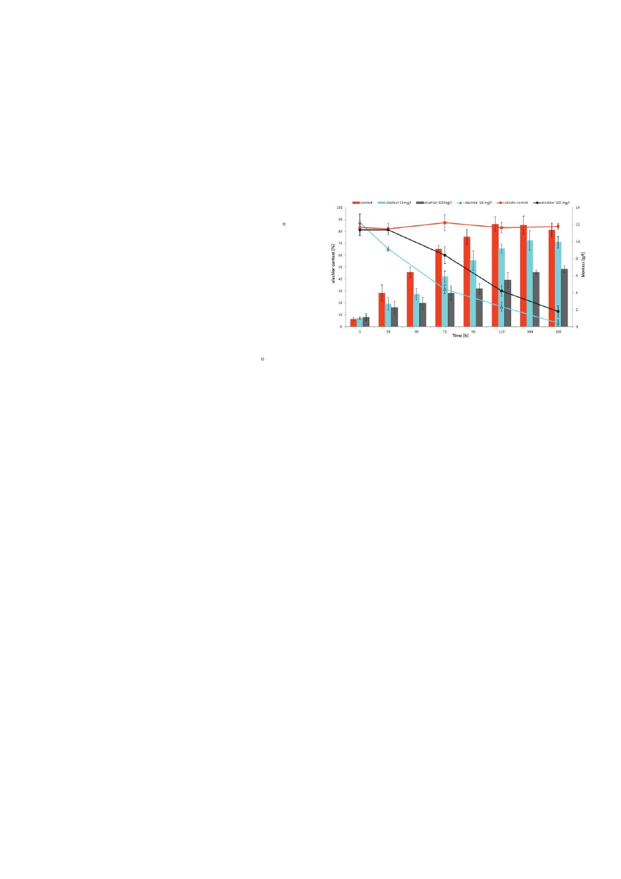
3.1. Growth and removal of alachlor by P. marquandii
The investigated fungus growth and alachlor elimination by P.
marquandii in liquid Sabouraud medium amended with alachlor (50
and 100 mg in 1litre) has been illustrated in Fig.1. Both tested
concentrations of alachlor inhibited growth of the fungus, but from
144 h the difference between the control and the culture with a 50
mg L
-1
dose of the herbicide did not have statistical importance
(Fig.1). Toxic substrate applied at concentration of 100 mg L
-1
repressed significantly fungal growth during the whole time of
incubation (40-60%).
Fig. 1. The fungal growth and alachlor degradation by P. marquandii
cultures on Sabuoraud medium containing alachlor at concentrations
50 and 100 mg L−1.
After 7days of incubation only 3.3 and 20.1% of alachlor added at
the initial concentrations of 50 and 100 mg L
-1
were detected. It was
noteworthy that 70% of the supplied alachlor disappeared as soon
as after 72 h, when xenobiotic was introduced at a dose of 50 mg L
-
1
. Our results are comparable with the results of most papers
investigating microbial transformation of alachlor by streptomycetes,
yeast and filamentous fungi via cometabolism [27,28,29,30,34].
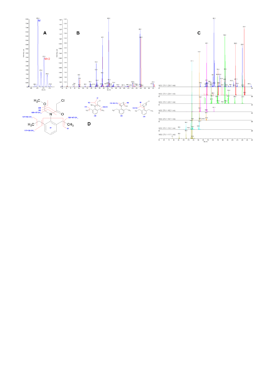
3.2. Qualitative analysis of alachlor biodegradation
Alachlor fragmentation patterns were initially examined for a proper
IDA LC-MS/MS method setup. In the ER scan (Fig. 2 A) a molecular
ions cluster of alachlor showed a typical isotopic distribution for a
compound containing one chlorine atom. Several EPI experiments
were made to collect mass spectra at different collision energies
(CE) from each isotopic form. The most important data from EPI
experiments were collected from molecular ion M and M+2 isotopes,
which revealed the presence of chlorine atoms in the following mass
spectrum fragments: 238 m/z, 224 m/z, 220 m/z, 210 m/z, 208 m/z,
90 m/z and 77 m/z (Fig. 2 B). Ion clusters around 78 m/z and 91 m/z
are typically a result of aromatic ring presence and that is why we
did several MS3 scans to have a deeper insight into the process of
alachlor fragmentation (Fig. 2C). The results of such approach
showed that, while fragments above 162 m/z are mostly a result of
fragmentations and rearrangements of dimethyl ether (C
2
H
6
O) and
chloracetaldehyde (C
2
H
3
ClO) substituents attached to nitrogen atom,
fragments equal to or below 162 m/z come from the fragmentation of
the 2,6-diethyl-N-methylaniline structure (methaniminium ion
–
C
11
H
16
N
+
). As it is shown in Figure 2B and 2C, ion clusters 77-79 m/z
and 90-92 m/z originate from two different types of fragmentation,
but chlorine containing fragments are much stronger than fragments
coming from the ring related structure. Having all data, we provided
the explanation of alachlor mass spectrum in Fig. 2D and chose ions
117.1 m/z and 162.1 m/z as the best markers due to their structure,
intensity and stability for the IDA method applied in metabolite
identification studies.
Samples for qualitative analysis were collected in 0, 24, 72, 120 and
168 h of culturing and included corresponding biotic and abiotic
controls acting as a reference for alachlor metabolites searching. All
samples were prepared in triplicates and examined by the IDA LC-
MS/MS method. Based on mass spectra analysis, we found 10
metabolites. All of them underwent characteristic fragmentation in
the EPI scan confirming the presence the of 2,6-diethyl-N-
methylaniline substructure and absence of the chlorine atom
3
Fig. 2. Mass spectra analysis of alachlor showing their typical
fragmentation pattern.
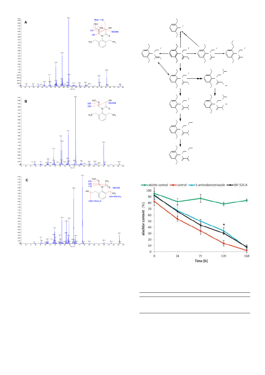
examined in the ER scan. In two cases
– RT=8.2 (M=295.1) and
RT=13.9 (M=264.2) we could not identify the structure further as the
m/z signals were too weak and difficult to interpret. Examples of
mass spectra and their basic interpretation are shown in Fig. 3. The
first two examples show mass spectra coming from the compounds
with a removed chlorine atom as a result of mono- and
dihydroxylation
(Fig.
3A
and
3B,
respectively)
of
the
chloracetaldehyde (C
2
H
3
ClO) substituent of alachlor. Dihydroxylation
on C-terminal of the acetaldehyde substituent of N-(2,6-
diethylphenyl)-2,2-dihydroxy-N[(hydroxymethoxy)methyl]acetamide
can be reasonably explained and confirmed by rearrangements
between neighbour chains, which results in the formation of ion 63
m/z.
In
the
case
of
N-[2-ethyl-6-(2-hydroxyethyl)phenyl]-N-
(methoxymethyl)acetamide (Fig. 3C), the fragmentation pattern of
the 2,6-diethyl-N-methylaniline substructure is a little different as a
result of hydroxylation of one ethyl group at a distal point from the
benzene ring. The most important ions confirming this structure are:
176.2 m/z (C
11
H
14
NO
+
), 158.2 m/z (C
11
H
12
N
+
) formed as a result of
H
2
O loss from ion 176.2 m/z, 143.2 (C
10
H
8
N
+
) formed as a result of
CH
3
loss from ion 158.2 m/z. All the other mass spectra of potential
metabolites were examined and interpreted in a similar way. The
summary of qualitative analysis is presented in Supplementary Data
(Table S1).
Relative intensity of the alachlor metabolites checked in the samples
collected during the culture of P. marquandii revealed that the
majority of them appeared at a relatively low level from 72 h and
reached
their
maximum
at
120-168
h.
N-[2-ethyl-6-(2-
hydroxyethyl)phenyl]-N-(methoxymethyl)acetamide
(6)
appeared
only at 168 h of incubation. Parallel analysis of metabolite peaks
area tendencies and one to each other area ratio in the following
hours of the experiment (Supplementary Data, Fig S1) helped us
formulate the alachlor biodegradation pathway showed in Fig. 4. The
main route of biodegradation starts with oxidative dechlorination
resulting in the formation of N-(2,6-diethylphenyl)-2-hydroxy-N-
(methoxymethyl)acetamide
(5).
This
compound
undergoes
consecutive hydroxylations of terminal carbon atoms of both
dimethyl ether and hydroxyacetaldehyde substituents attached to the
nitrogen atom of the 2,6-diethyl-N-methylaniline substructure.
Oxidation reactions generate various di- or trihydroxy derivatives and
{(2,6-diethylphenyl)[(hydroxymethoxy)methyl]amino}(oxo)acetic acid
(7). On the other hand, this route also leads to methylation of the
hydroxyl group as a next step after full oxidation of terminal carbons
and
formation
of
N-(2,6-diethylphenyl)-N-
[(dihydroxymethoxy)methyl]-2-hydroxy-2-methoxyacetamide
(10).
Other side reactions are probably reductive dechlorinations that start
from N-(2,6-diethylphenyl)-N-(methoxymethyl)acetamide (8)
formation. This derivative was also previously detected by us in P.
marquandii cultures analyzed with GC/MS application [31]. One
route leads to a loss of chloracetaldehyde and methanol from
alachlor and the other one leads to hydroxylation of the ethyl group
attached to a benzene ring after previous dechlorination of the
chloracetaldehyde substituent (6).
Alachlor byproducts have been often identified in bacteria enriched
soil samples or microbial cultures [18,27,28]. Nevertheless, only few
papers presented metabolic pathway of alachlor transformation by
microorganisms. Tiedje and Hagedorn [29] described alachlor
conversion by soil fungus Chaetomium globosum and identified four
metabolites:
2-chloro-2',6'-diethylacetanilide,
2,6-diethyl-N-
(methoxymethyl)aniline, 2,6-diethylaniline and 1-chloroacetyl-2,3-
dihydro-7-ethylindole. Chlorinated and dechlorinated indole and
quinoline derivative compounds were detected as main metabolites
of soil actinomycetes [30]. The metabolic pathway of alachlor
biotransformation, proposed by us, differs from this provided by
Tiedje and Hagedorn [29] and Sette et al. [30]. Alachlor was
metabolized by P. marquandii mainly by hydroxylation. Both
hydroxylation of the N-alkyl group and benzylic hydroxylation of ethyl
side chains occurred, although the first type of reaction dominated.
Our results are in opposition to the findings of Hapeman
–Somich
[35], Pothuluri et al. [27] and Qiang et al. [16] showing that ethyl
chains of alachlor are more susceptible to oxidation. Filamentous
fungus Cunninghamella elegans oxidized alachlor at the benzylic
position [27]. Pothuluri et al [27] noticed a connection between the
preferential hydroxylation of the arylethyl side chain of alachlor and
cytochrome P-450 monooxygenase activity. It was well documented
that the fungus C. elegans can metabolize different xenobiotics with
an involvement of cytochrome P-450 [27,36,37]. Considering such a
possibility, we investigated whether P. marquandii could transform
alachlor in the presence of cytochrome P-450 inhibitors proadifen
(SKF 525-A) and 1-aminobenzotriazole (Fig.5). Although during the
incubation ca. 20% mitigation of alachlor degradation in the
presence of inhibitors was observed, at 168 h the differences
between the control without inhibitors and the cultures supplied with
SKF 525-A and 1-aminonenzotriazole did not exist. The herbicide
elimination under inhibitors pressure was effective, suggesting a
negligible role of cytochrome P-450 in alachlor metabolism. These
results can help to explain the difference in the alachlor
hydroxylation by compared fungi.
3.3. Toxicological study
Xenobiotic
transformations
occurring
via
biooxidation
and
dechlorination often lead to the substrate detoxification [27]. On the
other hand, it is known that mammalian metabolic activation of
alachlor involving monooxygenation and conjugation processes can
result in the formation of toxic and carcinogenic intermediate
4
Fig. 3. Mass spectra and fragmentation patterns of exemplary
alachlor degradation byproducts and alachlor intermediates
originating from P. marquandii cultures detectedby LC
–MS/MS.
metabolites [38-39]. Therefore, we tested abiotic samples and fungal
cultures containing the same concentration of alachlor with the
application of Artemia franciscana toxkit. These crustaceans
inhabiting aquatics environments with different salinity (5 - 250 g L
-1
)
are commonly used in ecotoxicological studies [40].
Studies concerning alachlor toxicity were conducted with P.
marquandii extracts, because the diluted supernatants obtained after
the fungus cultures demonstrated significant toxicity to A.
.franciscana, which was not observed in the case of extracts in the
same dilutions (data not shown). An inhibition of the tested
crustacean motility by extracts diluted in saline water after 24 h
incubation was observed and the results were expressed as effect
concentration, which inhibited nauplii motility by 50% (EC
50
) (Table
1). The 24 h EC
50
of alachlor (corresponding to the initial alachlor
dose) reached
8.17 ± 1.11 and 7.35 ± 2.54 mg L
-1
for abiotic and
biotic samples and did not show a statistically significant difference
(p<0.05). The value of 24 h EC
50
obtained for the alachlor solution
without the extraction procedure was 10.80
± 2.51 mg L
-1
.Generally,
alachlor has moderate toxicity to aquatic invertebrates. EC
50
or LC
50
N
O
CH
3
O
OH
C
H
3
C
H
3
N
O
O
OH
C
H
3
C
H
3
OH
OH
N
O
CH
3
O
OH
C
H
3
C
H
3
OH
N
O
CH
3
O
CH
3
C
H
3
C
H
3
N
O
O
C
H
3
C
H
3
OH
O
O
H
CH
3
N
O
O
O
C
H
3
C
H
3
OH
OH
N
O
O
C
H
3
C
H
3
OH
OH
O
H
O
CH
3
NH
CH
3
C
H
3
C
H
3
N
O
CH
3
O
CH
3
C
H
3
OH
N
O
CH
3
O
Cl
C
H
3
C
H
3
N
O
CH
3
O
H
OH
C
H
3
C
H
3
4
9
2
1
3
5
7
8
10
6
Fig. 4. A proposed pathway of alachlor biotransformation by P.
marquandii.
Fig. 5. Effect of cytochrome P-450 inhibitors on alachlor elimination
by P. mar-quandii.* Significant differences at p < 0.05.
Table 1. The toxicity test of alachlor samples treated and untreated
with P. marquandii.
Samples
24 h EC
50
[mg L
−1
]
Extract of Sabouraud medium after 7 day- incubation (without P.
marquandii inoculation)
8.17
± 1.11
Extract of P. marquandii cultures after 7- day incubation
7.35
± 2.54
Pure alachlor solution (without incubation and extraction)
10.80
± 2.51
of alachlor to Daphnia reached 10-13 mg L
-1
[41-44]. The toxic effect
of pure alachlor solution determined by us was comparable with the
acute toxicity of this herbicide to Daphnia. The results of a toxicity
assay with A. franciscana showed that biotransformation of alachlor
by P. marquandii did not increase its toxicity. Direct ozonation and
O
3
/H
2
O
2
advanced oxidation resulted only in slight mitigation of
alachlor toxicity Qiang et al. [16]. The inhibition of D. magna motility

5
amounted to 23.3 ± 5.8% (after ozonation) and 26.7 ± 11.5%
(advanced process) in comparison to 33.8 ± 5.8 for the untreated
control. The lack of a decrease in alachlor toxicity as a result of
oxidation by P.marquandii could be caused by the compensating
effect of byproducts with lower and higher toxicity. Besides
hydroxylated intermediates, P. marquandii
produced also 2’,6’-
diethyl-N-methylaniline (4). It is a derivative of toxic and carcinogenic
2’,6’-diethylaniline
(DEA),
which
was
reported
in
many
biodegradation studies [18,29,45]. Although extracts originating from
control fungal cultures without alachlor supplementation did not
affect A. franciscana in the tested range of dilutions it cannot be
ruled out that mycelium produces some toxic metabolites under
alachlor exposure, influencing toxkit organisms.
Additionally, some data show that substitution of chlorine by a
hydroxyl group did not affect alachlor toxicity [46]. Although
degradation of this herbicide by P. marquandii did not lead to direct
detoxification, dechlorinated metabolites produced by P. marquandii
can be more easily degraded by other soil microorganisms.
4. Conclusions
The obtained results point to P. marquandii as a new valuable
research model for the study of alachlor degradation, differing from
other microscopic soil fungi. The IDA MS/MS analysis of fungal
cultures extracts resulted in the identification of ten alachlor
metabolites. The pathway of the herbicide degradation involved
mainly dechlorination and oxidation reactions. We did not observe
an increase in toxicity during alachlor biooxidation by P. marquandii
cultures examined with the use of A. franciscana toxikits, but the
appearance of a
2’,6’-diethyl-N-methylaniline derivative requires
more detailed studies to check the safety of potential environmental
applications.
Acknowledgement
This study was supported by the Grant of the National Centre for
Science in Cracow, Poland, No UMO-2011/01/B/NZ9/02898.
References
[1]
B. Lauga, N. Girardin, S. Karama, K. Le Ménach, H. Budzinski, R. Duran,
Removal of alachlor in anoxic soil slurries and related alteration of the active
communities, Environ. Sci. Pollut. Res. 20 (2013) 1089-105.
[2]
T.L. Potter, T.L. Carpenter, Occurrence of alachlor environmental degradation
products in groundwater, Environ. Sci. Technol. 29 (1995) 1557
–63.
[3]
World Health Organization, Guidelines for drinking-water quality. WHO 2006
http://www.who.int/water_sanitation_health/dwq/gdwq3rev/en/index.html
[4]
D.M. Tessier, J.M. Clark, Quantitative assessment of the mutagenic potential of
environmental degradative products of alachlor, J. Agric. Food Chem. 43 (1995)
2504-12.
[5]
L. Fava, P. Bottoni, A. Crobe, E. Funari, Leaching properties of some
degradation products of alachlor and metolachlor, Chemosphere 41 (2000) 1503-
8.
[6]
J.H. Zhu, X.L. Yan, Y. Liu, B. Zhang, Improving alachlor biodegradability by
ferrate oxidation, J. Hazard. Mater. 135 (2006) 94-9.
[7]
G. Daston, J.C. Cook, R.J. Kavlock, Uncertainties for endocrine disrupters: Our
view on progress, Toxicol. Sci. 74 (2003) 245
–52.
[8]
H.S. Kang, M.C. Gye, M.K. Kim, Effects of alachlor on survival and development
of Bombina orientalis (Boulenger) embryos, Bull. Environ. Contam. Toxicol. 74 (
2005) 1199-1206.
[9]
A. Goksoyr, Endocrine disruptors in the marine environment: mechanisms of
toxicity and their influence on reproductive processes in fish, J. Toxicol. Environ.
Health A 69 (2006) 175
–184.
[10]
T.B. Hayes, P. Case, S. Chui, D. Chung, C. Haeffele, K. Haston, M. Lee, V.P.
Mai, Y. Marjuoa, J. Parker, M. Tsui, Pesticide mixtures, endocrine disruption, and
amphibian declines: are we underestimating the impact?, Environ. Health
Perspect. 114 (2006) 40-50.
[11]
O. Osano, W. Admiral, D. Otieno, Developmental disorders in embryos of the
frog Xenopus laevis induced by chloroacetanilide herbicides and their
degradation products, Environ. Toxicol. Chem. 21 (2002) 375
–379.
[12]
S. Snyder, M. Benotti, Endocrine disruptors and pharmaceuticals: implications
for water sustainability, Water Sci. Technol. 61 (2010) 145-54.
[13]
A. Fucic, M. Gamulin, Z. Ferencic, J. Katic, M. Krayer von Krauss, A. Bartonova,
D.F. Merlo, Environmental exposure to xenoestrogens and oestrogen related
cancers: reproductive system, breast, lung, kidney, pancreas, and brain, Environ.
Health A 11 (2012) art. No S9.
[14]
Directive 2008/105/EC of the European Parliament and of the Council of 16
December 2008. Official Journal of the European Union L 348/84, 24.12.2008.
[15]
Decree of the Polish Ministry of the Environment from July 2, 2010. 138/934
02.07.2010.
[16]
Z. Qiang, C. Liu, B. Dong, Y. Zhang, Degradation mechanism of alachlor during
direct ozonation and O
3
/ H
2
O
2
advanced oxidation process, Chemosphere 78
(2010) 517-26.
[17]
M.M.
Ballesteros Martín, J.A. Sánchez Pérez, J.L. García Sánchez, L. Montes
Casas López, I. Oller, S. Malato Rodríguez, Degradation of alachlor
and pyrimethanil by combined photo-Fenton and biological oxidation, J. Haz.
Mat. 155 (2008) 342-9.
[18]
D.M. Stamper, O.H. Tuovinen, Biodegradation of the acetanilide herbicides
alachlor, metolachlor, and propachlor, Crit. Rev. Microbiol. 24 (1998) 1-22.
[19]
J. Xu, M. Yang, J. Dai, H. Cao, C. Pan, X. Qiu, M. Xu, Degradation of acetochlor
by four microbial communities. Bioresour. Technol. 99 (2008) 7797
–802.
[20]
P.M. White, T.L. Potter, A.K. Culbreath, Fungicide dissipation and impact on
metolachlor aerobic soil degradation and soil microbial dynamics, Sci. Total
Environ. 408 (2010) 1393-1402.
[21]
L. Zucconi, C. Ripa, F. Alianiello, A. Benedetti, S. Onofri, Lead resistance,
sorption and accumulation in a Paecilomyces lilacinus strain, Biol. Fert. Soil 37
(2003) 17-22.
[22]
M. Słaba, M. Bizukojć, B. Pałecz, J. Długoński, Kinetic study of toxicity of zinc
and lead ions to the heavy metals accumulating fungus Paecilomyces
marquandii, Bioprocess Biosyst. Eng. 28 (2005) 185-97.
[23]
X. Zeng, J. Tang, H.Yin, X. Liu, P. Jiang, H. Liu, Isolation, identification and
cadmium adsorption of a high cadmium-resistant Paecilomyces lilacinus, African
J. Biotechnol. 9 (2010) 6525-33.
[24]
E. Estévez, M.C. Veiga, C.Kennes, Biofiltration of waste gases with the fungi
Exophiala oligosperma and Paecilomyces variotii, Appl. Microbiol. Biotechnol. 67
(2005) 563-68.
[25]
H. Gradišar, J. Friedrich, I. Križaj, R. Jerala, Similarities and specificities of fungal
keratinolytic proteases: comparison of keratinases of Paecilomyces marquandii
and Doratomyces microsporus to some known proteases, Appl. Environ.
Microbiol. 71 (2005) 3420-6.
[26]
Z. Liu, D. Zhang, Z. Hua, J. Li, G. Du, J. Chen, A newly isolated Paecilomyces
sp. WSH-L07 for laccase production: isolation, identification, and production
enhancement by complex inducement, J. Ind. Microbiol. Biotechnol. 36 (2009)
1315-21.
[27]
J.V. Pothuluri, J.P. Freeman, F.E. Evans, E.T. Moorman, C.R. Cerniglia,
Metabolism of alachlor by the fungus Cunninghamella elegans, J. Agric. Food
Chem. 41 (1993) 483
–8.
[28]
A. Munoz, W.C. Koskinen, L. Cox, M. J. Sadowsky, Biodegradation and
mineralization of metolachlor and alachlor by Candida xestobii, J. Agric. Food
Chem. 59 (2011). 619-27.
[29]
J.M. Tiedje, M.L. Hagedorn, Degradation of alachlor by a soil fungus,
Chaetomium globosum, J. Agric. Food Chem. 23 (1975) 77-81.
[30]
L.D. Sette, L.A. Mendonca Alves da Costa, A.J. Marsaioli, G.P. Manfio,
Biodegradation of alachlor by streptomycetes, Appl. Microbiol. Biotechnol. 64
(2004) 712
–17.
[31]
M. Słaba, R. Szewczyk, P. Bernat, J. Długoński, Simultaneous toxic action zinc
and alachlor resulted in enhancement of zinc uptake by Paecilimyces
marquandii, Sci. Total. Environ. 407 (2009) 4127-33.
[32]
M. Słaba, J. Długoński, Selective recovery of Zn
2+
from waste slag from a metal-
processing plant by microscopic fungus Verticillium marquandii, Biotechnol. Lett.
22 (2000) 1699-1704.
[33]
S. Różalska, R.Szewczyk, J. Długoński, Biodegradation of 4-n-nonylphenol by
the non-ligninolytic filamentous fungus Gliocephalotrichum simplex: A proposal of
a metabolic pathway, J. Hazard. Mater. 180 (2010) 323-31.
[34]
D.R. Shelton, S. Khader, J.S. Karns, B.M. Pogell, Metabolism of twelve
herbicides by Streptomyces, Biodegradation, 7 (1996) 129
–36.
[35]
C.J. Hapeman-Somich, Mineralization of pesticide degradation products. in: L.
Somasundaram, J.R. Coast, (Eds.), Pesticide Transformation Products: Fate and
Significance in the Environment, ACS symposium Series, 459. American
Chemical Society, 1991, pp. 133
–147.
[36]
P. Bernat, J. Długoński, Degradation of tributyltin by the filamentous fungus
Cunninghamella elegans, with involvement of cytochrome P-450. Biotechnol.
Lett. 24 (2002) 1971-74.
[37]
K. Lisowska, J. Długoński, Concurrent corticosteroid and phenanthrene
transformation by filamentous fungus Cunninghamella elegans. J. Steroid
Biochem. Mol. Biol. 85 (2003) 63-69.
[38]
S. Coleman, S. Liu, R. Linderman, E. Hodgson, R.L. Rose, In vitro metabolism of
alachlor by human liver microsomes and human cytochrome P450 isoforms,
Chem. Biol. Interact. 122 (1999) 27
–39.
[39]
B.A. Wetmore, A.D. Mitchell, S.A. Meyer, M.B. Genter, Evidence for site-specific
bioactivation of alachlor in the olfactory mucosa of the Long-Evans rat, Toxicol.
Sci. 49 (1999) 202
–12.
[40]
B.S. Nunes, F.D. Carvalho, L.M. Guilhermino, G. Van Stappen, Use of the genus
Artemia in ecotoxicity testing, Environ. Pollut. 144 (2006) 453
–62.
[41]
Alachlor (Lasso), Herbicide Profile 6/85.
[42]
Alachlor Sigma-Aldrich, Material Safety Data Sheet.
[43]
W.A. Hartman, D.B. Martin, Effects of Four Agricultural Pesticides on Daphnia
pulex, Lemna minor, and Potamogeton pectinatus, Bull. Environ. Contam.
Toxicol. 35 (1985) 646-51.
[44]
H. He, G. Chen, J. Yu, J. He, X.Huang, S. Li, Q. Guo, T. Yu, H. Li, Individual and
joint toxicity of three chloroacetanilide herbicides to freshwater cladoceran
Daphnia carinata, Bull. Environ. Contam. Toxicol. 90 (2013) 344-50.
[45]
C.Z. Chen, C.T. Yan, P.V. Kumar, J.W. Huang, J.F. Jen, Determination of
alachlor and its metabolite 2,6-diethylaniline in microbial culture medium using
online microdialysis enriched-sampling coupled to high-performance liquid
chromatography, J. Agric. Food Chem. 59 (2011) 8078-85.
[46]
J.K. Lee, Degradation of the herbicide alachlor by soil microorganisms. Korean J.
Environ. Agric. 3 (1984) 1-9.
Wyszukiwarka
Podobne podstrony:
Szewczyk, Rafał i inni Intracellular proteome expression during 4 n nonylphenol biodegradation by t
Szewczyk, Rafał; Długoński, Jerzy Mikrobiologiczny rozkład pentachlorofenolu (2007)
Szewczyk, Rafał; Długoński, Jerzy Pentachlorophenol and spent engine oil degradation by Mucor ramos
Szewczyk, Rafał i inni Rapid method for Mycobacterium tuberculosis identification using electrospra
Modelowanie form odzieży dla figur nietypowych Monika Mosionek i Mirosława Szewczyk
Coaching grupowy Praktyczny podręcznik dla liderów, trenerów, doradców i nauczycieli = Grela Joanna,
Zagadnienia bezpieczenstwa informacyjnego w standardzie TETRA V D Rafal Niski Miroslaw Radziwanowski
3 tydzień Wielkanocy, III piątek
24 piątek
32 piątek
23 piątek
2 tydzień Wielkanocy, II piątek
03 piątek
otyłość i cukrzyca dla pielęgniarek na piątek
18 piątek
01 piątek
22 piątek
19 piątek
więcej podobnych podstron