SCAN0129
CHAPTER 5 T Crystalline Lens
93

A

FIGURĘ 5-8
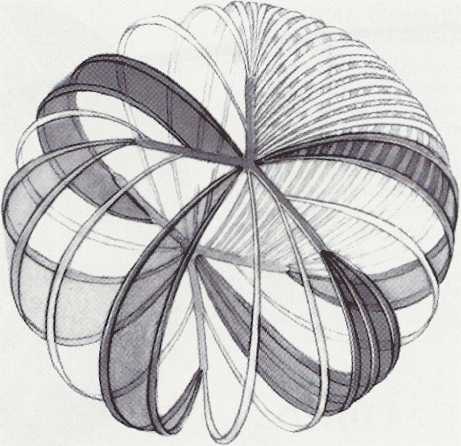
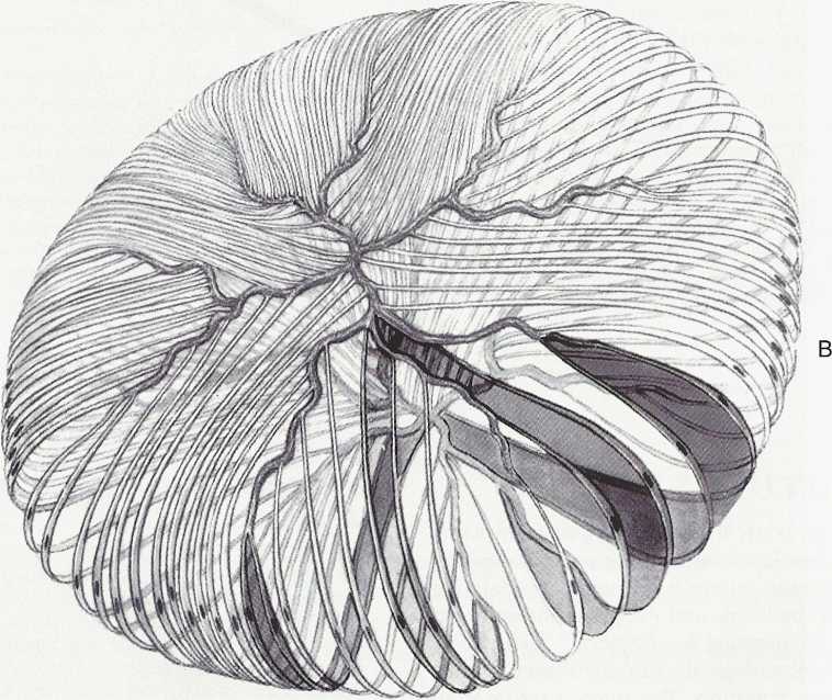
Fetal and adult lenses, showing sutures and arrangement of lens cells. A, Fetal nucleus. Anterior Y suture is at a, and posterior suture is at b. Lens cells are depicted as wide bands. Cells that attach to tips of Y sutures at one pole of lens attach to fork of Y at opposite pole. B, Adult lens cortex. Anterior and posterior organization of sutures is morę complex. Lens cells that arise from tip of a suture branch insert farther anteriorly or posteriorly into a fork at opposite pole. This arrangement conserves shape of lens. In this drawing, for educational purposes, the suture appears to lie in a single piane, but the reader should remember that the suture extends throughout the thickness of the cortex and nucleus to the level of the Y sutures in the fetal nucleus.
Wyszukiwarka
Podobne podstrony:
image002 Figurę I: Distribution and spread of adansonii hybrid in South America. Adapted from Townse
ELECTROMAGNETIC ENERGY AND FORCES In this chapter we will consider the storage and transformation of
119BD CcnA2 CcnD1 Figurę 3.3 : Proliferation and apoptosis of Cd36-nuli bonę marrow MSCs Celi count
SCAN0122 CHAPTER 2 ▼ Cornea and Selera 27FIGURĘ 2-17 Limbus. Limbal conjunctiva (A) is formed by an
SCAN0132 CHAPTER 6 ▼ Aqueous and Yitreous Chambers 111VIT R E O U S CHAMBER The vitreous chamber is
więcej podobnych podstron