SCAN0122
CHAPTER 2 ▼ Cornea and Selera
27

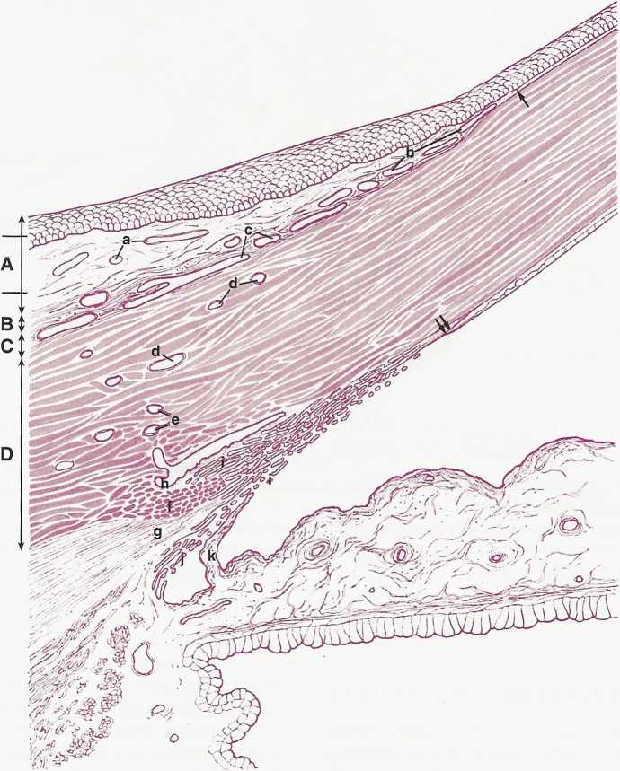
FIGURĘ 2-17
Limbus. Limbal conjunctiva (A) is formed by an epithelium (1) and a loose connective tissue stroma (2). Tenon's capsule (B) forms a thin, poorly defined connective tissue layer over episclera (C). Limbal stroma occupies the area (D) and is composed of ścierał and corneal tissues that merge in this region. Conjunctival stromal vessels are also seen (a). They form peripheral corneal arcades (b), which extend anteriorly to termination of Bowman's layer (arrow). Episcleral vessels (c) are cut in different planes. Vessels forming intrascleral (d) and deep ścierał plexus (e) are shown within limbal stroma. Ścierał spur has coarse and dense coilagen fibers (f). Anterior part of longitudinal portion of eiliary muscle (g) merges with ścierał spur and trabecular meshwork. Lumen of Schlemm's canal (h) and loose tissues of its wali are seen clearly. Sheets of the trabecular meshwork (i) are outer to cords of uveal meshwork (j).
An iris process (k) is seen to arise from iris surface and join trabecular meshwork at level of anterior portion of ścierał spur. Descemet's membranę terminates (double arrows) within anterior portion of the triangle, outlining aqueous outflow system. (From Hogan MJ, Alvarado JA, Weddell JE: Histology of the human eye, Philadelphia, 1971, Saunders.)
Wyszukiwarka
Podobne podstrony:
SCAN0120 CHAPTER 2 ▼ Cornea and Selera 17FIGURĘ 2-10 Summary diagram of corneal stroma. A, Fibroblas
SCAN0132 CHAPTER 6 ▼ Aqueous and Yitreous Chambers 111VIT R E O U S CHAMBER The vitreous chamber is
SCAN0144 CHAPTER 9 ▼ Ocular Adnexa and Lacrimal System 169 Supraorbital artery Lacrimal artery Super
84842 SCAN0124 CHAPTER 3 ▼ Uvea 43 The ciliary body can be divided into two parts: the pars plicata
SCAN0127 CHAPTER 4 ▼ Retina 75 Temporal on retina Nasal Perimetric angles in degre
48206 SCAN0133 CHAPTER 9 T Ocular Adnexa and Lacrimal System 161 CHAPTER 9 T Ocular Adnexa and Lacri
skanowanie0014 (47) • The communicative teaching is marked by an atmosphere of using and working wit
Preterite Preterite The preterite is formed by taking the infmitive, dropping the last two letters,
więcej podobnych podstron