48206 SCAN0133
CHAPTER 9 T Ocular Adnexa and Lacrimal System 161
CHAPTER 9 T Ocular Adnexa and Lacrimal System 161
Palpebral
conjunctiva
Accessory lacrimal gland (of Krause)
Superior tarsal muscle (of Muller)
Accessory lacrimal gland (of Wolfring)
Meibomian
gland
Gland of Moll
Subcutaneous connective tissue
Submuscular areolar layer
Orbicularis oculi muscle
Hair follicle
Riolan’s
muscle
Zeis gland
Tarsal
piąte
Porę of
meibomian gland
Epidermis of skin
Aponeurosis of levator muscle
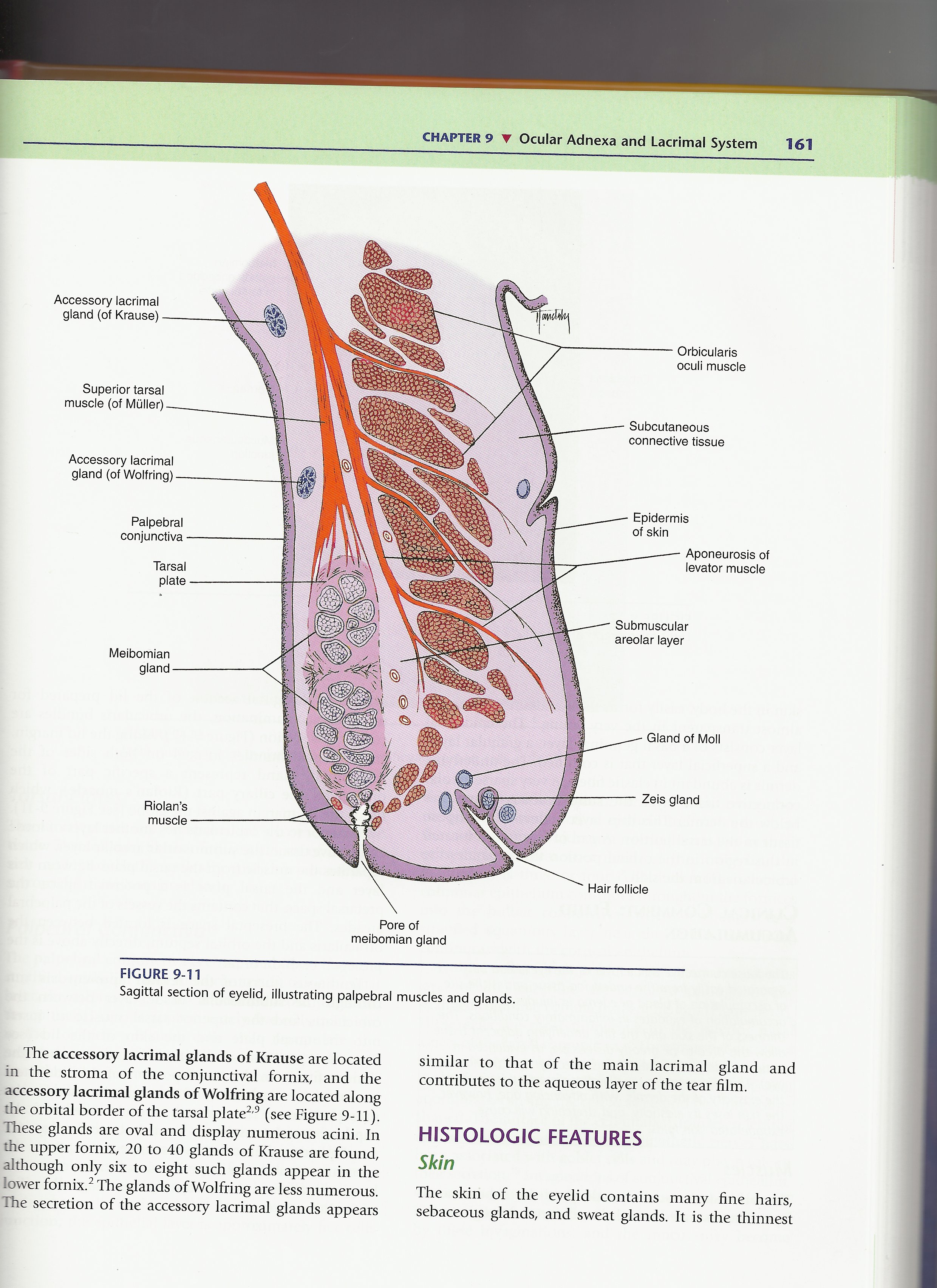
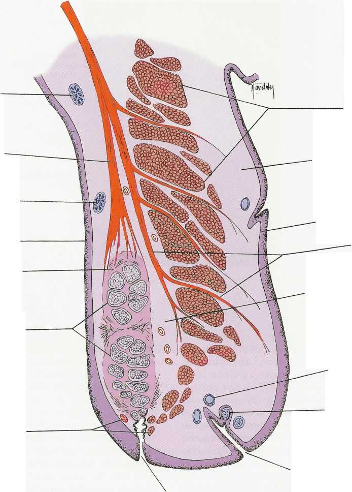
FIGURĘ 9-11
Sagittal section of eyelid, illustrating palpebral muscles and glands.
The accessory lacrimal glands of Krause are located in the stroma of the conjunctival fornix, and the accessory lacrimal glands of Wolfring are located along the orbital border of the tarsal piąte2,9 (see Figurę 9-11). These glands are oval and display numerous acini. In the upper fornbc, 20 to 40 glands of Krause are found, although only six to eight such glands appear in the Iower fornbc.2 The glands of Wolfring are less numerous. The secretion of the accessory lacrimal glands appears similar to that of the main lacrimal gland and contributes to the aąueous layer of the tear film.
HISTOLOGIC FEATURES
Skin
The skin of the eyelid contains many fine hairs, sebaceous glands, and sweat glands. It is the thinnest
Wyszukiwarka
Podobne podstrony:
SCAN0144 CHAPTER 9 ▼ Ocular Adnexa and Lacrimal System 169 Supraorbital artery Lacrimal artery Super
SCAN0122 CHAPTER 2 ▼ Cornea and Selera 27FIGURĘ 2-17 Limbus. Limbal conjunctiva (A) is formed by an
SCAN0129 CHAPTER 5 T Crystalline Lens 93 A FIGURĘ 5-8 Fetal and adult lenses, showing sutures and ar
SCAN0132 CHAPTER 6 ▼ Aqueous and Yitreous Chambers 111VIT R E O U S CHAMBER The vitreous chamber is
SCAN0120 CHAPTER 2 ▼ Cornea and Selera 17FIGURĘ 2-10 Summary diagram of corneal stroma. A, Fibroblas
23476 SCAN0121 CHAPTER 2 T Cornea and Selera 19 brane can be detached easily from posterior stroma.1
więcej podobnych podstron