SCAN0120
CHAPTER 2 ▼ Cornea and Selera
17
FIGURĘ 2-10
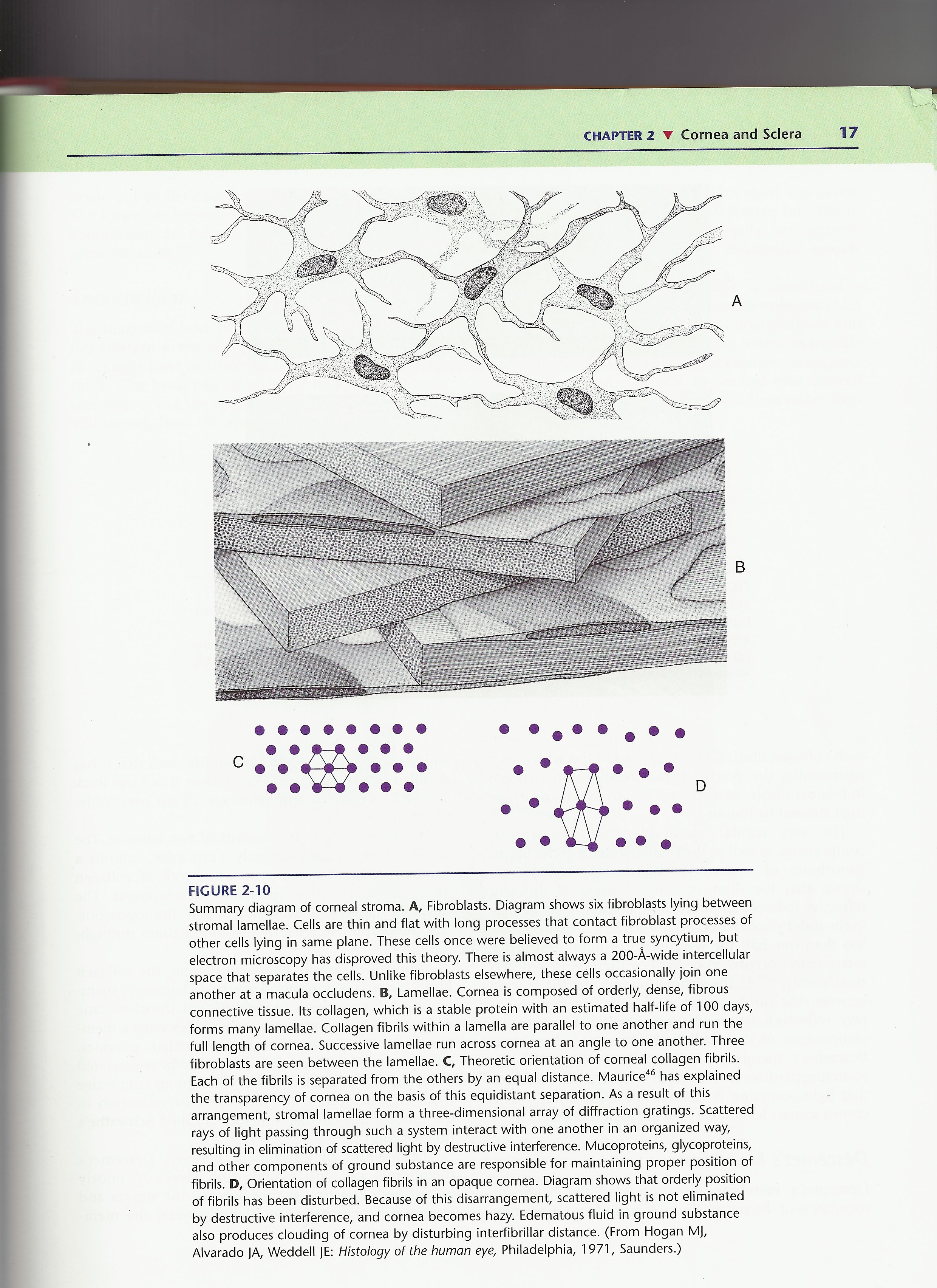
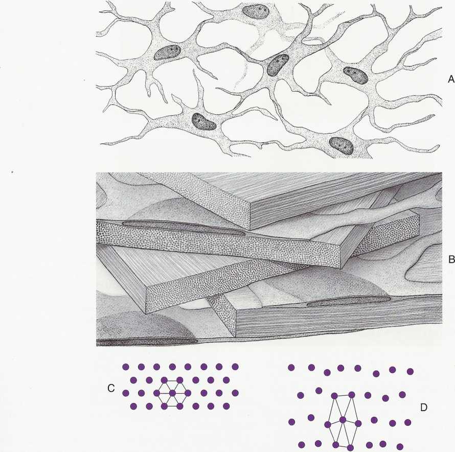
Summary diagram of corneal stroma. A, Fibroblasts. Diagram shows six fibrobiasts lying between stromal lamellae. Cells are thin and fiat with long processes that contact fibroblast processes of other cells lying in same piane. These cells once were believed to form a true syncytium, but electron microscopy has disproved this theory. There is almost always a 200-A-wide intercellular space that separates the cells. Unlike fibroblasts elsewhere, these cells occasionally join one another at a macula occludens. B, Lamellae. Cornea is composed of orderly, dense, fibrous connective tissue. Its collagen, which is a stable protein with an estimated half-life of 100 days, forms many lamellae. Collagen fibrils within a lamella are parallel to one another and run the fuli length of cornea. Successive lamellae run across cornea at an angle to one another. Three fibroblasts are seen between the lamellae. C, Theoretic orientation of corneal collagen fibrils. Each of the fibrils is separated from the others by an equal distance. Maurice46 has explained the transparency of cornea on the basis of this equidistant separation. As a result of this arrangement, stromal lamellae form a three-dimensional array of diffraction gratings. Scattered rays of light passing through such a system interact with one another in an organized way, resulting in elimination of scattered light by destructive interference. Mucoproteins, glycoproteins, and other components of ground substance are responsible for maintaining proper position of fibrils. D, Orientation of collagen fibrils in an opaque cornea. Diagram shows that orderly position of fibrils has been disturbed. Because of this disarrangement, scattered light is not eliminated by destructive interference, and cornea becomes hazy. Edematous fluid in ground substance also produces clouding of cornea by disturbing interfibrillar distance. (From Hogan M), Alvarado JA, Weddell JE: Histology of the human eye, Philadelphia, 1971, Saunders.)
Wyszukiwarka
Podobne podstrony:
SCAN0122 CHAPTER 2 ▼ Cornea and Selera 27FIGURĘ 2-17 Limbus. Limbal conjunctiva (A) is formed by an
SCAN0132 CHAPTER 6 ▼ Aqueous and Yitreous Chambers 111VIT R E O U S CHAMBER The vitreous chamber is
SCAN0144 CHAPTER 9 ▼ Ocular Adnexa and Lacrimal System 169 Supraorbital artery Lacrimal artery Super
84842 SCAN0124 CHAPTER 3 ▼ Uvea 43 The ciliary body can be divided into two parts: the pars plicata
SCAN0042 crop FIGURĘ 2-1 Corneal dimensions. A, Radius of curvature of cornea and selera. B, View fr
SCAN0127 CHAPTER 4 ▼ Retina 75 Temporal on retina Nasal Perimetric angles in degre
48206 SCAN0133 CHAPTER 9 T Ocular Adnexa and Lacrimal System 161 CHAPTER 9 T Ocular Adnexa and Lacri
więcej podobnych podstron