SCAN0127
CHAPTER 4 ▼ Retina 75
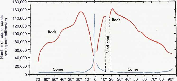
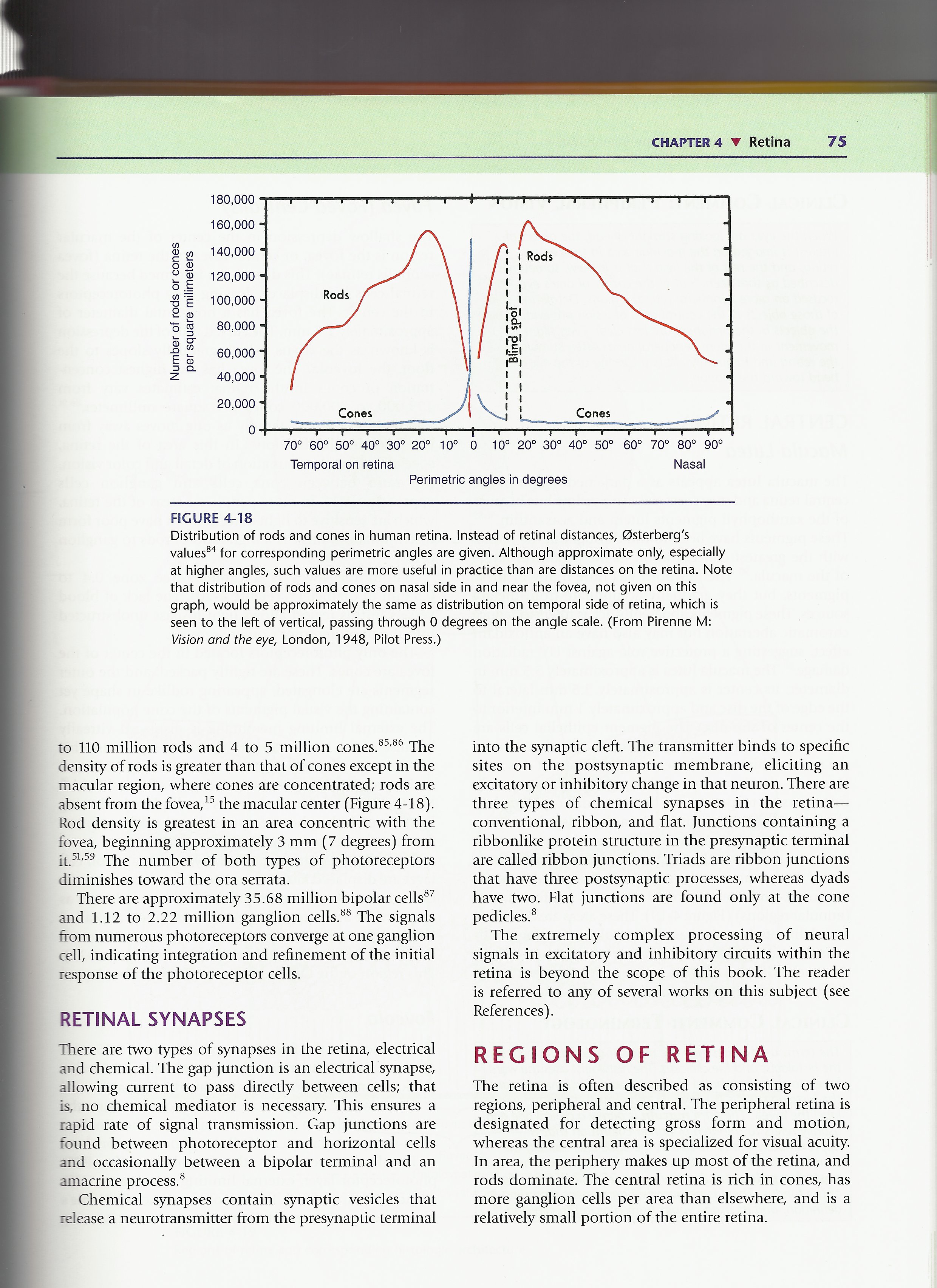
Temporal on retina Nasal
Perimetric angles in degrees
FIGURĘ 4-18
Distribution of rods and cones in human retina. Instead of retinal distances, 0sterberg's values84 for corresponding perimetric angles are given. Although approximate only, especially at higher angles, such values are morę useful in practice than are distances on the retina. Notę that distribution of rods and cones on nasal side in and near the fovea, not given on this graph, would be approximately the same as distribution on temporal side of retina, which is seen to the left of vertical, passing through 0 degrees on the angle scalę. (From Pirenne M: Vision and the eye, London, 1948, Pilot Press.)
to 110 million rods and 4 to 5 million cones.85,86 The density of rods is greater than that of cones except in the macular region, where cones are concentrated; rods are absent from the fovea,15 the macular center (Figurę 4-18). Rod density is greatest in an area concentric with the fovea, beginning approximately 3 mm (7 degrees) from it.51,59 The number of both types of photoreceptors diminishes toward the ora serrata.
There are approximately 35.68 million bipolar cells87 and 1.12 to 2.22 million ganglion cells.88 The signals from numerous photoreceptors converge at one ganglion celi, indicating integration and refinement of the initial response of the photoreceptor cells.
RETINAL SYNAPSES
There are two types of synapses in the retina, electrical and Chemical. The gap junction is an electrical synapsę, allowing current to pass directly between cells; that is, no Chemical mediator is necessary. This ensures a rapid ratę of signal transmission. Gap junctions are found between photoreceptor and horizontal cells and occasionally between a bipolar terminal and an amacrine process.8
Chemical synapses contain synaptic vesicles that release a neurotransmitter from the presynaptic terminal into the synaptic cleft. The transmitter binds to specific sites on the postsynaptic membranę, eliciting an excitatory or inhibitory change in that neuron. There are three types of Chemical synapses in the retina— conventional, ribbon, and fiat. Junctions containing a ribbonlike protein structure in the presynaptic terminal are called ribbon junctions. Triads are ribbon junctions that have three postsynaptic processes, whereas dyads have two. Fiat junctions are found only at the cone pedicles.8
The extremely complex processing of neural signals in excitatory and inhibitory circuits within the retina is beyond the scope of this book. The reader is referred to any of several works on this subject (see References).
REGSONS OF RETINA
The retina is often described as consisting of two regions, peripheral and central. The peripheral retina is designated for detecting gross form and motion, whereas the central area is specialized for visual acuity. In area, the periphery makes up most of the retina, and rods dominate. The central retina is rich in cones, has morę ganglion cells per area than elsewhere, and is a relatively smali portion of the entire retina.
Wyszukiwarka
Podobne podstrony:
59608 SCAN0047 CHAPTER 4 ▼ Retina 67 Internal limiting membranę FIGURĘ 4-11 Retinal cells and synaps
SCAN0122 CHAPTER 2 ▼ Cornea and Selera 27FIGURĘ 2-17 Limbus. Limbal conjunctiva (A) is formed by an
SCAN0132 CHAPTER 6 ▼ Aqueous and Yitreous Chambers 111VIT R E O U S CHAMBER The vitreous chamber is
84842 SCAN0124 CHAPTER 3 ▼ Uvea 43 The ciliary body can be divided into two parts: the pars plicata
SCAN0120 CHAPTER 2 ▼ Cornea and Selera 17FIGURĘ 2-10 Summary diagram of corneal stroma. A, Fibroblas
SCAN0144 CHAPTER 9 ▼ Ocular Adnexa and Lacrimal System 169 Supraorbital artery Lacrimal artery Super
SCAN0148 CHAPTER 11 ▼ Orbital Blood Supply 201 Retina ——^ Choroid - -< Circle of Zinn (Haller) Sh
więcej podobnych podstron