84842 SCAN0124
CHAPTER 3 ▼ Uvea 43
The ciliary body can be divided into two parts: the pars plicata (corona dliaris) and the pars piana (orbicularis ciliaris). The pars plicata is the thicker, anterior portion containing the ciliary processes (see Figurę 3-10). Approximately 70 to 80 ciliary processes extend into the posterior chamber, and the regions between them are called valleys (of Kuhnt). A ciliary process measures approximately 2 mm in length, 0.5 mm in width, and 1 mm in height, but there are significant variations in all measurements.1
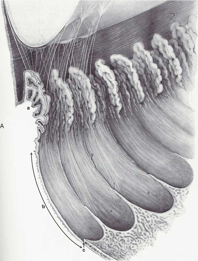
The pars piana is the flatter region of the ciliary body. It extends from the posterior of the pars plicata to the ora serrata, which is the transition between ciliary body and choroid. The ora serrata has a serrated pattern, the forward-pointing apices of which are called teeth or dentate processes. The rounded portions that lie between the teeth are called orał bays (Figurę 3-11, A).
The dentate processes are elongations of retinal tissue into the region of the pars piana.
The zonule fibers course from the ciliary body to the lens. Some of these fibers insert into the intemal limiting membranę of the pars piana region and travel forward through the valleys between the ciliary processes. Some attach to the internal limiting membranę of the valleys of the pars plicata (Figurę 3-11, B). The attachment to the vitreous, the vitreous base, extends forward approxi-mately 2 mm over the posterior pars piana.1
SUPRACSLIARIS (SUPRACSLIARY LAMINA)
The supraciliaris is the outermost layer of the ciliary body adjacent to the selera. Its loose connective tissue is arranged in ribbonlike layers containing pig-

FIGURE 3-11
A, Inner aspect of the ciliary body shows pars plicata (a) and pars piana (b). Ora serrata is at c, and posterior to it, retina exhibits cystoid degeneration (d). Bays (e) and dentate processes (f) of ora are shown; linear ridges or striae (g) project forward from dentate processes across pars piana to enter valleys between ciliary processes. Zonular fibers arise from pars piana beginning 1.5 mm from ora serrata. These fibers curve forward from sides of dentate ridges into ciliary valleys, then from valleys to lens capsule. Zonules coming from the valleys on either side of a ciliary process have a common point of attachment on lens. Zonules attach up to 1 mm from the equator posteriorly and up to 1.5 mm from the equator anteriorly. At equatorial border, attaching zonules give a crenated appearance to lens. Ciliary processes vary in size and shape and often are separated from one another by lesser processes.
Radial furrows (h) and circular furrows (i) of peripheral iris are shown. Continued
Wyszukiwarka
Podobne podstrony:
47005 skanowanie0074 (3) 12.1.1.1. Sub-skills of listening Listening can be divide
skanowanie0074 (3) 12.1.1.1. Sub-skills of listening Listening can be divided into
GSM Architecture The functions of the NMS can be divided into three categories: •
10 To realize its Intentions the Labour Market Agency runs programmes whlch can be divided into four
47005 skanowanie0074 (3) 12.1.1.1. Sub-skills of listening Listening can be divide
SCAN0126 45 CHAPTER 3 ▼ Uvea FIGURĘ 3-12 Light micrograph of transverse section of the ciliary body,
SCAN0132 CHAPTER 6 ▼ Aqueous and Yitreous Chambers 111VIT R E O U S CHAMBER The vitreous chamber is
SCAN0122 CHAPTER 2 ▼ Cornea and Selera 27FIGURĘ 2-17 Limbus. Limbal conjunctiva (A) is formed by an
SCAN0120 CHAPTER 2 ▼ Cornea and Selera 17FIGURĘ 2-10 Summary diagram of corneal stroma. A, Fibroblas
więcej podobnych podstron