23476 SCAN0121
CHAPTER 2 T Cornea and Selera 19
brane can be detached easily from posterior stroma.19 The adhesions between Descemet's membranę and the endothelium are not the typical hemidesmosomes but some variation.50
Endothelium
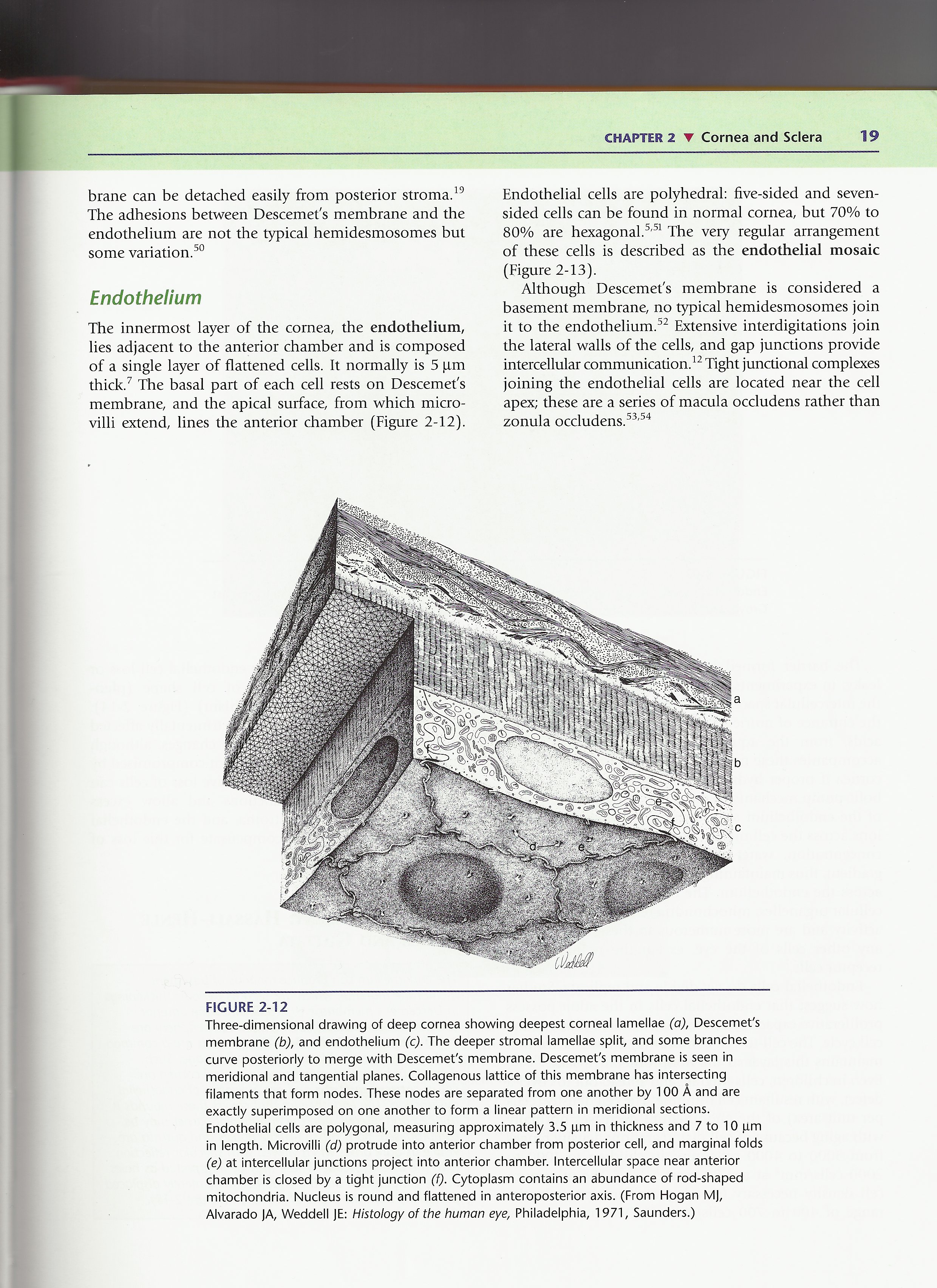
The innermost layer of the cornea, the endothelium, lies adjacent to the anterior chamber and is composed of a single layer of flattened cells. It normally is 5 pm thick.7 The basal part of each celi rests on Descemet's membranę, and the apical surface, from which micro-villi extend, lines the anterior chamber (Figurę 2-12).
Endothelial cells are polyhedral: five-sided and seven-sided cells can be found in normal cornea, but 70% to 80% are hexagonal.5,51 The very regular arrangement of these cells is described as the endothelial mosaic (Figurę 2-13).
Although Descemefs membranę is considered a basement membranę, no typical hemidesmosomes join it to the endothelium.52 Extensive interdigitations join the lateral walls of the cells, and gap junctions provide intercellular communication.12 Tight junctional complexes joining the endothelial cells are located near the celi apex; these are a series of macula occludens rather than zonula occludens.53,54
FIGURĘ 2-12
Three-dimensional drawing of deep cornea showing deepest corneal lamellae (a), Descemefs membranę (b), and endothelium (c). The deeper stromal lamellae split, and some branches curve posteriorly to merge with Descemefs membranę. Descemefs membranę is seen in meridional and tangential planes. Collagenous lattice of this membranę has intersecting filaments that form nodes. These nodes are separated from one another by 100 A and are exactly superimposed on one another to form a linear pattern in meridional sections. Endothelial cells are polygonal, measuring approximately 3.5 pm in thickness and 7 to 10 pm in length. Microvilli (d) protrude into anterior chamber from posterior celi, and marginal folds (e) at intercellular junctions project into anterior chamber. Intercellular space near anterior chamber is closed by a tight junction (f). Cytoplasm contains an abundance of rod-shaped mitochondria. Nucleus is round and flattened in anteroposterior axis. (From Hogan MJ, Alvarado JA, Weddell JE: Histology of the human eye, Philadelphia, 1971, Saunders.)
Wyszukiwarka
Podobne podstrony:
SCAN0122 CHAPTER 2 ▼ Cornea and Selera 27FIGURĘ 2-17 Limbus. Limbal conjunctiva (A) is formed by an
SCAN0120 CHAPTER 2 ▼ Cornea and Selera 17FIGURĘ 2-10 Summary diagram of corneal stroma. A, Fibroblas
SCAN0042 crop FIGURĘ 2-1 Corneal dimensions. A, Radius of curvature of cornea and selera. B, View fr
sa 07 Exercise 5.—Biceps and Forearms I lłis excrcise can be tlone either iu a sitting or a stamling
image001 .MWCttOMMPersephone and Hades T«cbool!»ov can be io kOI w pt»t«rvo kto 8ut Mft it (HMC
how to ąuillOuilling, sirnple and easy to learn. can be mastered by young and obi alike in a mat ter
calibre cover 6ESTSŚIIIS5 AUT HO# Of DOWN TO A SUNLESS S£A AND LOUISIANA BLUEDAVID "THERE CAN B
Program Option Modifiers Some options are “boolean” and control behavior that can be turned on or of
how to ąuillOuilling, sirnple and easy to learn. can be mastered by young and obi alike in a mat ter
c) Tłiey usually appear and disappear seasonalły. d) It can be s
więcej podobnych podstron