SCAN0130

94 Clinical Anatomy of the Visual System
ANTĘRIOR CAPSULE
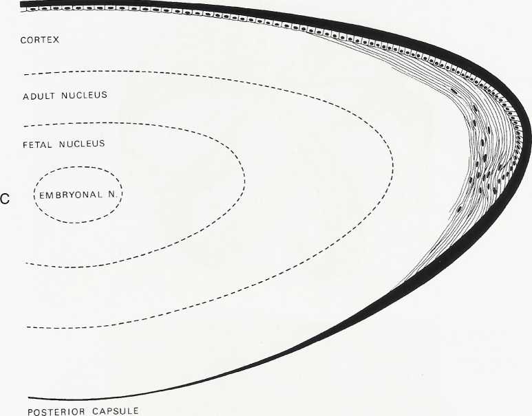
FIGURĘ 5-8 cont'd
C, Adult lens, showing nuclear zones, epithelium, and capsule. Thickness of lens capsule in various zones is shown. (From Hogan M), Alvarado JA, Weddell JE: Histology of the human eye, Philadelphia, 1971, Saunders.)
LENS SUTURES
As the lens fibers reach the poles they meet with the other fibers in their layer, forming a junction known as a suture. The anterior suture is formed by overlapping apical aspects of the fibers, and the posterior suture is formed by the overlapping basal aspects.3,10 The fibers formed during embryologic development meet in three branches, forming Y sutures. The anterior suture is an upright-Y shape and the posterior suture an inverted-Y shape (see Figurę 5-8, A).14 As growth continues and the lens becomes larger, the sutures become asymmetric and dissimilar. The limbs of the anterior and posterior sutures are offset, and the complexity of the sutures contributes to lens transparency.24 The sutures formed after birth are morę stellate shaped; sutures formed through early adulthood have 6 to 9 branches; and 9 to 15 complex branched stars are formed in middle to old age24 (see Figurę 5-8, B).
Clinical Comment: Slit-Lamp Appearance of Lens
An optic section through the lens demonstrates the biconvexity of the structure (Figurę 5-9). The first bright linę is convex forward and is the anterior lens capsule; posterior to this is a dark linę, the anterior epithelium; the next bright linę is the cortex; then various gray zones (zones of discontinuity) are seen. The anterior Y suture of the fetaI nucleus may be evident. The vertical center, the embryonic nucleus, has no anterior or posterior curvature. The posterior inverted-Y suture may be seen in the posterior aspect of the fetaI nucleus. Posterior to this, the zones of discontinuity are concave forward, with the finał zonę being the posterior capsule.25 A diffuse view of the anterior lens surface illustrates lens shagreen, with the lens surface resembling the surface of an orange, likely caused by the conformation of the capsule to the epithelial celi undulations.26 The zones of discontinuity are apparent because of changes in light-scattering properties (Table 5-1).
Wyszukiwarka
Podobne podstrony:
SCAN0131 96 Clinical Anatomy of the Visual System 96 Clinical Anatomy of the Visual System FIGURĘ 5-
SCAN0145 178 Clinical Anatomy of the Visual System Epimysium Perimysium EndomysiumFIGURĘ 10-1 Connec
SCAN0125 44 Clinical Anatomy of the Visual System mented melanocytes, fibroblasts, and collagen band
SCAN0131 96 Clinical Anatomy of the Visual System 96 Clinical Anatomy of the Visual System FIGURĘ 5-
SCAN0151 214 Clinical Anatomy of the Visual System Parotoid lymph nodeFIGURĘ 11-13 Lymphatic drainag
SCAN0152 218 Clinical Anatomy of the Visual SystemFIGURĘ 12-1 Orbit viewed from above showing branch
75024 SCAN0131 96 Clinical Anatomy of the Visual System 96 Clinical Anatomy of the Visual System FIG
więcej podobnych podstron