SCAN0134
168 Clinical Anatomy of the Visual System
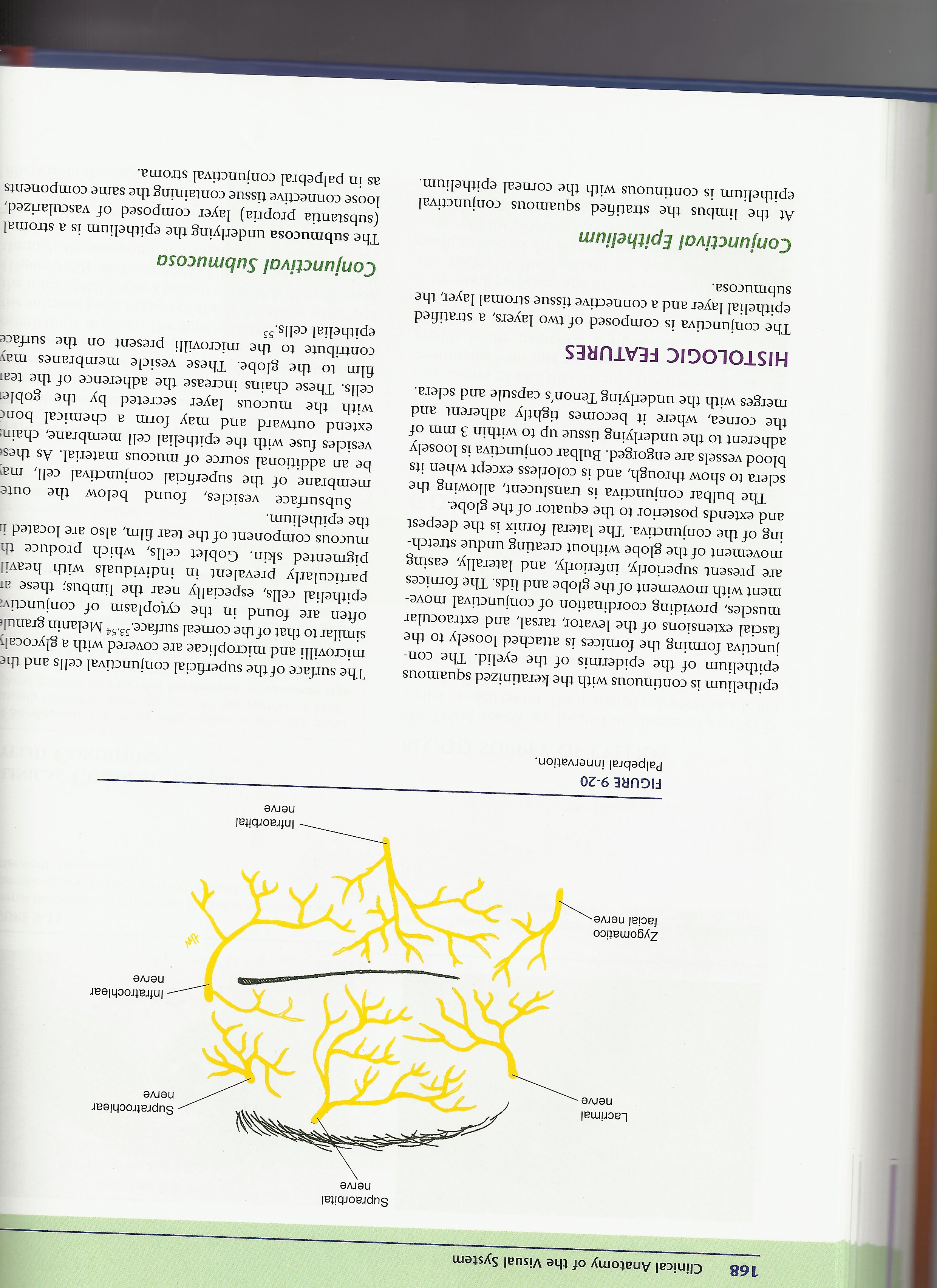
Lacrimal nerve -
Supraorbital
nerve
Supratrochlear
nerve
• Infratrochlear nerve
Zygomatico facial nerve-
Infraorbital
nerve
FIGURĘ 9-20
Palpebral innervation.
epithelium is continuous with the keratinized sąuamous epithelium of the epidermis of the eyelid. The con-junctiva forming the fornices is attached loosely to the fascial extensions of the levator, tarsal, and extraocular muscles, providing coordination of conjunctival move-ment with movement of the globe and lids. The fornices are present superiorly, inferiorly, and laterally, easing movement of the globe without creating undue stretch-ing of the conjunctiva. The lateral fornix is the deepest and extends posterior to the eąuator of the globe.
The bulbar conjunctiva is translucent, allowing the selera to show through, and is colorless except when its blood vessels are engorged. Bulbar conjunctiva is loosely adherent to the underlying tissue up to within 3 mm of the cornea, where it becomes tightly adherent and merges with the underlying Tenon's capsule and selera.
HISTOLOGIC FEATURES
The conjunctiva is composed of two layers, a stratified epithelial layer and a connective tissue stromal layer, the submucosa.
Conjunctival Epithelium
At the limbus the stratified sąuamous conjunctival epithelium is continuous with the corneal epithelium.
The surface of the superficial conjunctival cells and the microvilli and microplicae are covered with a glycocah similar to that of the comeal surface.53,54 Melanin granul* often are found in the cytoplasm of conjunctiv; epithelial cells, especially near the limbus; these ai particularly prevalent in individuals with heavil pigmented skin. Goblet cells, which produce th mucous component of the tear film, also are located ii the epithelium.
Subsurface vesicles, found below the oute membranę of the superficial conjunctival celi, ma1 be an additional source of mucous materiał. As thes< vesicles fuse with the epithelial celi membranę, chain: extend outward and may form a Chemical bonc with the mucous layer secreted by the goblei cells. These chains inerease the adherence of the teai film to the globe. These vesicle membranes may contribute to the microvilli present on the surface epithelial cells.55
Conjunctival Submucosa
The submucosa underlying the epithelium is a stromal (substantia propria) layer composed of vascularized, loose connective tissue containing the same components as in palpebral conjunctival stroma.
Wyszukiwarka
Podobne podstrony:
SCAN0131 96 Clinical Anatomy of the Visual System 96 Clinical Anatomy of the Visual System FIGURĘ 5-
SCAN0145 178 Clinical Anatomy of the Visual System Epimysium Perimysium EndomysiumFIGURĘ 10-1 Connec
SCAN0125 44 Clinical Anatomy of the Visual System mented melanocytes, fibroblasts, and collagen band
SCAN0131 96 Clinical Anatomy of the Visual System 96 Clinical Anatomy of the Visual System FIGURĘ 5-
SCAN0151 214 Clinical Anatomy of the Visual System Parotoid lymph nodeFIGURĘ 11-13 Lymphatic drainag
SCAN0152 218 Clinical Anatomy of the Visual SystemFIGURĘ 12-1 Orbit viewed from above showing branch
75024 SCAN0131 96 Clinical Anatomy of the Visual System 96 Clinical Anatomy of the Visual System FIG
więcej podobnych podstron