29514 skanuj0006 (456)

9J\
Sp\na\ Cord, Sogrrvervta\ Orga\\vzatVov\
Roof piąte
Roofpldte


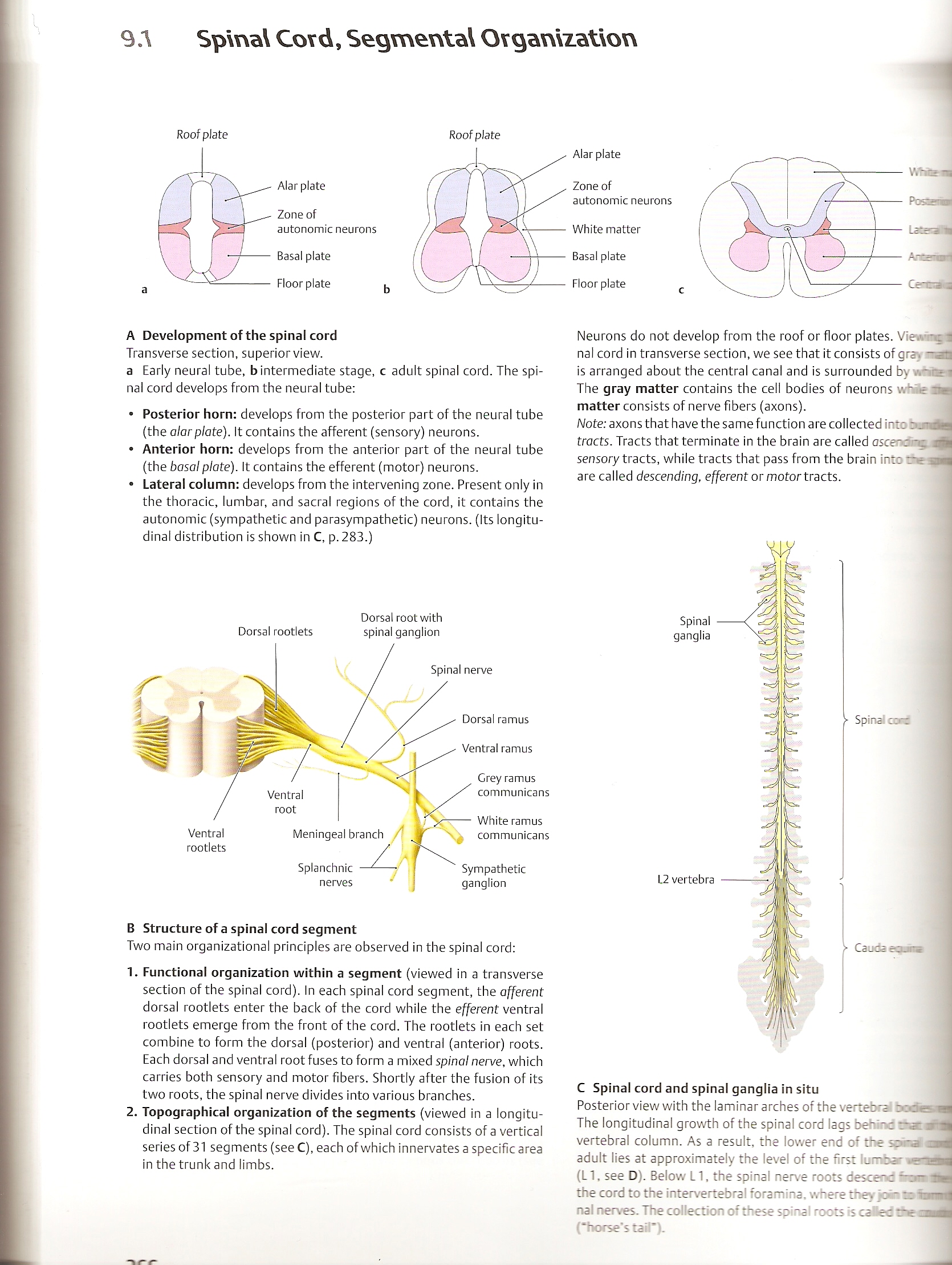
A Development of the spinał cord
Transverse section, superior view.
a Early neural tubę, b intermediate stage, c adult spinał cord. The spinał cord develops from the neural tubę:
• Posterior horn: develops from the posterior part of the neural tubę (the alarpiąte). It contains the afferent (sensory) neurons.
• Anterior horn: develops from the anterior part of the neural tubę (the basal piąte). It contains the efferent (motor) neurons.
• Laterai column: develops from the intervening zonę. Present only in the thoracic, lumbar, and sacral regions of the cord, it contains the autonomie (sympathetic and parasympathetic) neurons. (Its longitu-dinal distribution is shown in C, p. 283.)
Dorsal rootwith Dorsal rootlets spinał ganglion
Spinał nerve
Ventral
rootlets

Crey ramus communicans
White ramus communicans
Sympathetic
ganglion
Dorsal ramus
Ventral ramus
Splanchnic
nerves
Meningeal branch
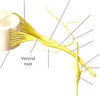
B Structure of a spinał cord segment
Two main organizational principles are obsen/ed in the spinał cord:
1. Functional organization within a segment (viewed in a transverse section of the spinał cord). In each spinał cord segment, the afferent dorsal rootlets enter the back of the cord while the efferent ventral rootlets emerge from the front of the cord. The rootlets in each set combine to form the dorsal (posterior) and ventral (anterior) roots. Each dorsal and ventral root fuses to form a mixed spinał nerve, which carries both sensory and motor fibers. Shortly after the fusion of its two roots, the spinał nerve divides into various branches.
2. Topographical organization of the segments (viewed in a longitu-dinal section of the spinał cord). The spinał cord consists of a vertical series of 31 segments (see C), each of which innervates a specific area in the trunk and limbs.
Neurons do not develop from the roof or floor plates. Vie.v’-ę 1 nal cord in transverse section, we see that it consists of grayadB is arranged about the central canal and is surrounded by The gray matter contains the celi bodies of neurons whSe na matter consists of nerve fibers (axons).
Notę: axons that have the same function are collected into ru uda traets. Tracts that terminate in the brain are called asce~z -ę am sensory tracts, while tracts that pass from the brain into re nil are called descending, efferent or motor tracts.
Spinał
ganglia
L2 vertebra

■ Spinał a*c
• Caudacojns
C Spinał cord and spinał ganglia in situ
Posterior view with the laminar arches of the vertebrai :>:de n
The longitudinal growth of the spinał cord lags behi-c rzdH
vertebral column. As a result, the lower end of the sona mm
adult lies at approximately the level of the nrst lumbar wonriH
(LI, see D). Below LI, the spinał nerve roots descecc tw
the cord to the intervertebral foramina. where thes
nal nerves. The collection of these spina I roots is ca Hec radli
(“horse*s taił*).
Wyszukiwarka
Podobne podstrony:
skanuj0059 (23) 180 MARTA DEREK objectives of the economic development of the commune. For example,
skanuj0026 Analiza firmy E Sp. z o.o. 1. Cel i zakres raportu: pozycja na rynku us
więcej podobnych podstron