1 Proteasom S


(a)
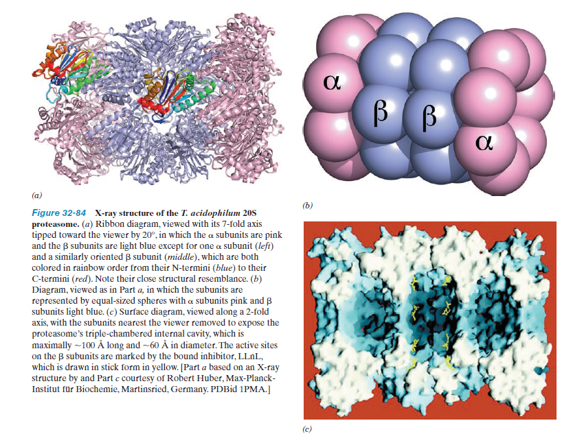
Figurę 32-84 X-ray structurc «f the i. acidophilum 20S protcasome. (a) Ribbon diagram. viewed with its 7-fold axis tipped toward thc viewer by 20°. in which thc a subunits arc pink and thc p subunits arc light blue exccpt for one a subunit (left) and a similarly orientcd (i subunit (middle). which arc both colorcd in rainbow order from their N-termini (blue) to their C-tcrmini (red). Notę their close structural resemblancc. (b) Diagram. viewed as in Part a, in which the subunits arc represented by cqual-sized spheres with « subunits pink and p subunits light blue. (c) Surfacc diagram. viewed along a 2-fo!d axis. with the subunits nearest thc vicwer removed to cxpose the protcasome's triple-chambered intcrnal cavity, which is maximally —100 A long and -60 A in diameter.The active sites on the p subunits arc marked by the bound inhibitor. LLnL, which is drawn in stick form in yellow. |Part a based on an X-ray structurc by and Part c courtesy of Robert Huber. Max-Planck-Institut fur Biochemie. Martinsricd. Germany. PDBid 1PMA.]

(c)
Wyszukiwarka
Podobne podstrony:
1 Proteasom S Figurę 32-84 X-ray struclure of the I. acidophilum 20S proteasomc. (a) Ribbon diagram
10 Ubikwitynacja oo II Ubiquitin —C—NH—Lys— Figurę 32-76 Kcactions involvcd in the
10 Ubikwitynacja oo II Ubiquitin —C—NH—Lys— Figurę 32-76 Kcactions involvcd in the
10 Ubikwitynacja oo II Ubiquitin —C—NH—Lys— Figurę 32-76 Kcactions involvcd in the
24 Kalmodulina Figurę 18-17 X-ray structure of rat testis calmodulin. *I*his monomcric 148-rcsidue
18 Chityna(1) Chitin Figurę 11-17 The priimiry structure «f chitin. ( hitin is a P(1 —► 4)-linkcd h
f32 4 FIGURĘ 32.4 The Fortune anplet
netter138 Smali Intestine Slructure: IGASTROINTESTINAL PHYSIOLOGY Figurę 7.16 Small Intfstine Struct
Michał PTAK Figurę 2. The structure of the support for renewable energy sources provided by regional
65 (166) T Figurę 32 Stave-church and tenth-century chamber grave beneath the medieval church at H^m
13 Peptydoglikan (a) jY-Acetylglucosamine Ar-Acetylmuramic acid Figurę 11-26 <lumk.il structuro
więcej podobnych podstron