33407 SCAN0128
90 Clinical Anatomy of the Visua! System
90 Clinical Anatomy of the Visua! System

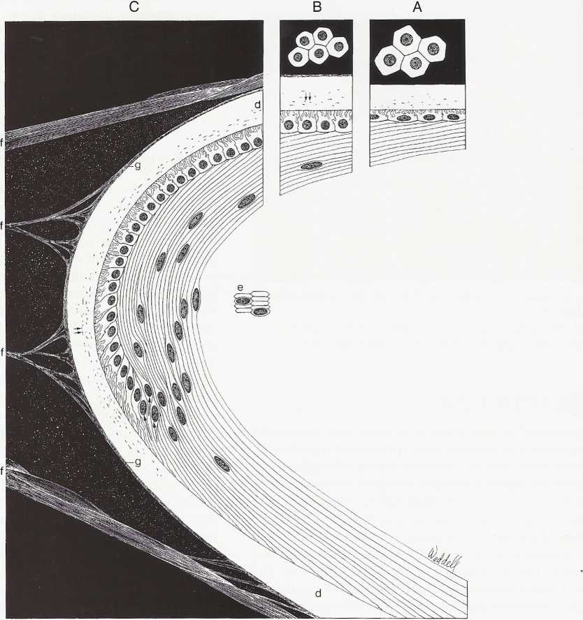
FIGURĘ 5-5
Composite drawing of crystalline lens, cortex, epithelium, capsule, and zonular attachments.
A, Anterior central lens epithelium, seen in fiat section and cross section. Size and shape of these cells can be compared with those of cells in B, intermediate zonę, and C, the equatorial zonę. Mitosis occurs in the preequatorial zonę and at the equator, the cells are elongating (arrows) to form lens cortical cells. As they elongate, cells send processes anteriorly and posteriorly toward sutures, and their nuclei migrate somewhat anterior to equator to form lens bow. At the same time, nuclei become morę and morę displaced into lens as new cells are formed at equator. Lens capsule (d) is thicker anterior and posterior to equator than at equator itself. Anterior and equatorial capsule contains fine filamentous inclusions (double arrows); these are not present posteriorly. Lens fibers elongate into flattened hexagons (e) in cross section. Zonular fibers (f) attach to anterior and posterior capsule and to equatorial capsule, forming pericapsular or zonular lamella of lens (g). (From Hogan MJ, Alvarado JA, Weddell JE: Histology of the human eye, Philadelphia, 1971, Saunders.)
Wyszukiwarka
Podobne podstrony:
SCAN0131 96 Clinical Anatomy of the Visual System 96 Clinical Anatomy of the Visual System FIGURĘ 5-
SCAN0145 178 Clinical Anatomy of the Visual System Epimysium Perimysium EndomysiumFIGURĘ 10-1 Connec
SCAN0125 44 Clinical Anatomy of the Visual System mented melanocytes, fibroblasts, and collagen band
SCAN0131 96 Clinical Anatomy of the Visual System 96 Clinical Anatomy of the Visual System FIGURĘ 5-
SCAN0151 214 Clinical Anatomy of the Visual System Parotoid lymph nodeFIGURĘ 11-13 Lymphatic drainag
SCAN0152 218 Clinical Anatomy of the Visual SystemFIGURĘ 12-1 Orbit viewed from above showing branch
75024 SCAN0131 96 Clinical Anatomy of the Visual System 96 Clinical Anatomy of the Visual System FIG
więcej podobnych podstron