86306 skanuj0001 (525)

i asacr- Bieeding from the bridg-^Kartfidalty opens the subdural ■i" ii the meningeal layer of the c upper layer of the arach-Hbfcane (subdural hematoma, see ■ cełts of the uppermost layer of
the arachnoid (neurothelium) are intero nected by a dense network of tight ju tions, creating a tissue barrier (blood-ce brospinal fluid barrier).
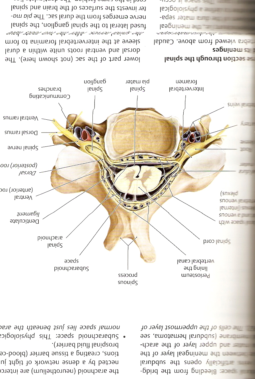
• Subarachnoid space: This physiologic; norma! space lies just beneath the arai
pai«eins
Spinous
process
Periosteum liningthe vertebral canal
Spinał cord
Subarachnoid
space
Spinał
arachnoid
ace with Btfaicnous rtemal .enous j*exus)

Ventral (anterior) roc
Dorsal
(pos tenor) roo
Denticulate
ligament
Spinał nerve Dorsal ramus Yentral ramus
Spinał pia mater
lntervertebral
foramen
Spinał
ganglion
Communicating
branches
sć section through the spinał fc meninges
tora viewed from above. Caudal
— a1, - | Jrrr-
—eningeal
■Nhrs —ater sepa-■>< piiysiological
ic nrr"
lower part of the sac (not shown here). The dorsal and ventral roots unitę within a dural sleeve at the intervertebral foramina to form
fused lateral to the spinał ganglion, the spinał nerve emerges from the dural sac. The pia mater invests the surfaces of the brain and spinał
^ —TL ! • ’ !—i ^-
Wyszukiwarka
Podobne podstrony:
skanuj0026 (162) L3 L4 L5 S1 Anterior side of thigh, passing obliquely downward from the approx
skanuj0019 4 Jhe Bubblęj- South Sea Company, trading slaves company, in 1720 got from the parliament
skanuj0044(1) Ej Repiace the underlined words to make these sentences true, Use the words from
f14 5 Bi Add Data Source Select which ODBC driver you want to use from the list, then choose OK. OK
Fox, Norman ?ctus?valier B ‘ Down from the hills in the early dawn light King Ćonover had co
google video 23.1 Solve the clues and complete the puzzle with words from the opposite page. 1
gramatyka0004 Choose from the list below for each sentence. Add an article or put the noun in the pl
helicoptermaze Directions: Tiy to find the path from the start to the finish in the maże below.
więcej podobnych podstron