4613347639
192 V. Bourdon et al. / Ann. Genet. 44 (2001) 191-194
2. Materials and methods
2.1. Patients
We studied 25 female patients with classical spo-radic RTT without any mutations identified in the MECP2 coding region after complete seąuencing and Southern biot analysis [6]. They were referred through the French Rett Syndrome Association and werc diagnosed according to the Rett Syndrome Diagnostic Criteria Work Group [22]. Blood sampies were ob-taincd after informed consent.
2.2. Fluorescence in situ hybridization
Metaphase spreads from contro! females and RTT patient lymphoblastoid celi lines were prepared according to standard procedures. A slides pretreatment by pepsin digest during 10 min (10 pg/mL) was madę to climinate cytoplasm. Chromosomal DNA denatur-ation was obtained in 70% formamide, 2X SSC, plJ 7 at 73°C during 2 minutes. The probe used was the PAC clone 671D9 containing only the MECP2 gene [accession number: AF030876, 20]. 1 lig was labcllcd with biotin-I6-dUTP by mek translation using Bio-Nick kit (Gibco-BRL, Germany). For each experi-ment, the labelled PAC DNA was used together with a CEP X Spectrum Aqua probe (Vysis, USA) specific to the X chromosome a-satellite region. The probe mixture was denatured for 10 minutes at 73°C and directly added to the slide. Slides were ineubated ovemight at 37°C, and washed for 2 min with 0,4XSSC/0,3% Nonidet P40, pH 7, and for 15 s with 2XSSC/0,1% Nonidet P40, pH 7. Biotin-labelled probe was detected according to the protocol of the fluorescein detection kit (Oncor-Appligcne, France). Finał ly, slides were counterstained with DAPI I (4’6 diamino-2 phenylindole) (Vysis, USA) and analyscd on a Zeiss cpifluorcscence microscope (Jena, Germany) with a Sensys CCD camera (Photometrics. USA) and IPLab Spectrum Imaging Software (Vvsis, USA).
3. Rcsults and discussion
Preliminary studies of the MECP2 coding region using a mutation screening strategy based on conformation-scnsitivc gcl electrophoresis, sequence analysis and Southern biot analysis did not revcal any mutation in 25 classic RTT patients. Thercfore, further analysis by FISH was performed in order to exclude large deletions of the RTT region in Xq28 in this

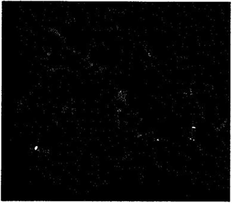
Figorc 1. Metaphase chromosome spread shows the 2 control aqua spots on the X centromcrcs. and the 2 green spots due to the PAC 671 d9 probe hybridisation on the 2 MECP2 alleles in Xq28 region. No deletion was detected in any of the 25 RTT patients studied.
cohort of 25 patients. AJ1 patients havc a normal female karyotypc. The 671 d9 PAC clone which con-tains the entire MECP2 gene, gave signals on the distal Xq-region on both X chromosomes in morę than 80% of the metaphases from both normal control females, and the 25 MECP2 mutation free patients (figurę 1). In addition, no duplication of the MECP2 loeus wras identified in any of the 25 RTT patients analyscd. Recently, Nielsen et al. [18] did no find any deletion of the entire MECP2 gene in 3 classical RTT patients, supporting our findings.
Repcated sequcnccs are vcry common in deletion prone regions [27]. As an illustration the steroid sulfatase gene (STS) is completely deleted in up to 90% of patients w'ith X-Linked-ichtyosis [13], or conversely, the phenylalanine hydroxylasc gene, poor in Iong highly homologous segments, is deleted in only 5% of patients with phenylalanine hydroxylase deficiency [10]. Based on the data presented herc, the MECP2 gene seems to belong to the second gene category; indeed complete loss (or duplication) of the MECP2 gene seems to be a rare event in typical RTT cases. Howcvcr, morę severc phenotype associated with congcnital malformations could be caused by contigous gene syndrome due to microdeletion in Xq28 region cncompassing the MECP2 gene.
in conclusion. gross rearrangements seem not to be a MECP2 gene mutational mechanism in RTF; an-olhcr way to illustrate a possiblc haplo-insufTiciency
Wyszukiwarka
Podobne podstrony:
18 H. Pridalova et al. Table 3. Average values for goat cheeses - physical and Chemical properties T
7. Opublikowane badania własne 66 J. Onet et al / Talanta I3S (2015) 64-70 (492 nm
ill coagulation (Koparal et Ogutveren, 2002; Mollah et al.y 2001; Barkley et al.y 1993; Vik et al.y
192 R.A.C.F. 32. 1993. 1979; BARBIEUX 1992; BONIN et VANGELE 1989; CHASTEL et al. 1988; FEMOLANT 198
56. Matematyka 2001 : poradnik dla nauczyciela : klasa 6 / [aut. Anna Bazyluk
44 KONU&PAYEYA ET AL for quantitativc analyscs and differcnt unit*, and some-times Ihc analytica
46 K0NU6PAYEVA ET AL Butik* rn hi m, N , M Mekkami!, N Rumiani, muł K. IlidaiH . 2
PRZETWARZANIE SŁÓW WIDOCZNYCH I MASKOWANYCH (BADANIE FMRI: DEHAENE ET AL, 2001) Czołowo-ciemien
Porównanie NOSów Alderton et al. Biochem J. 2001
Hartmann. Sweden - 23 - FASTH E. (1985) HAGBERG ET AL. (1986) HART M ANN J. (1977)
44 E. Cadier et al. sedimentaire) aux ressources en eau des grands bassins. L’insta!lation de nouvel
174 51. Dubois CM, Blanchctte F, Laprise MH, Leduc R, Grondin F, et al. (2001) Evi
więcej podobnych podstron