869568930
216
Tomasz Poboży, Maciej Kielar

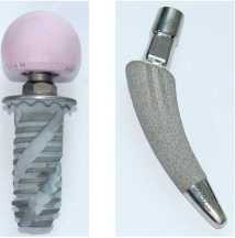
Figurę 1. Spiron® prosthesis Figurę 2. MiniHip™ prosthe-sis
The use of a short stem ensures a morę natural prox-imal stress distribution at the implant-bone interface.
The greatest advantage of this implant type results from the fact that a short stem preserves native bonę of the proximal end of the femur (bonę resection is max-imally limited). One example of a short-stem prosthesis is MiniHip™, also classified as a "partial collum” stem [26]. The composition of short-stem implants is uniąue, comprising a titanium alloy with a brushite coating. Brushite (BONIT®) is an amorphous calcium phosphate characterised by a morę rapid resorption, which accelerates implant osseointegration.
The stem surface texture increases the area of the implant-bone interface and directly increases prima-ry implant stability (Figurę 2).

Conventional, long-stem prostheses
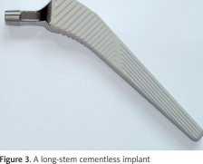
The last type of prostheses consists of conven-tional long-stem implants. Following resection of the femoral head and neck, the long-stem prosthesis is an-chored in the proximal and/or distal part of the femoral canal. Femur preparation involves cancellous bonę removal or, morę commonly, compaction with the use of special broaches shaped like the implant stem. Subseąuently the prosthesis stem is implanted into the prepared femoral canal. In the case of a cement-ed prosthesis the femoral canal must be thoroughly cleared of cancellous bonę. The implant stem is then anchored with the use of bonę cement. The surgeon must closely monitor the thickness of the cement layer surrounding the stem and seal the femoral canal distal to the stem to prevent cement from spreading too far into the bonę. Proper sealing is typically achieved with a special polyethylene stopper. Subseąuently, the cement is vacuum-mixed to eliminate any air being trapped within. In the case of a cementless anchoring techniąue, the stem must be integrated with the na-tive bonę to the extent ensuring maximum stability. During the procedurę, care must be taken not to crack the bonę (Figurę 3).
The bali of the prosthesis
While discussing the technical aspects of hip implant structure we should not omit the structure of the femoral head component, because it is mainly its structure and function that ultimately determine the everyday functioning of patients.
One important aspect is the size of the prosthetic head (bali). It is generally believed that larger prosthesis balls used during the procedurę ensure a greater hip joint stability, lower risk of dislocation, and re-duced long-term wear. One interesting solution is a bi-polar (double-bearing, "bali within a bali”) prosthesis, where a smaller head is placed inside a larger (typically polyethylene) head, which in turn articulates with the acetabulum.
Implant heads are typically composed of metal al-loys, but they can also be ceramic. Ceramic surfaces - in the case of both acetabular inserts and prosthesis heads - would seem to be optimal as they undergo minimal wear. However, the use of ceramic materiał has its disadvantages; one of which is that, although tough and generally durable, the materiał is not flex-ible, which increases the risk of cracks. Moreover, the use of a ceramic head reąuires the acetabular component to be absolutely perfectly set within the pelvic bonę (Figurę 4).
The acetabular component of the prosthesis
The head of the prosthesis is in direct contact with the cup, or acetabulum. The acetabular structure may vary depending on the head and prosthesis type.
Medical Studies/Studia Medyczne 2015; 31/3
Wyszukiwarka
Podobne podstrony:
218 Tomasz Poboży, Maciej Kielar C Figurę 5. Acetabular cups: A - uncemented press-fit ace-tabular
220 Tomasz Poboży, Maciej Kielar Figurę 6. Double-bearing, or “bali within a bali” bipolar
222 Tomasz Poboży, Maciej Kielar 34. Mohaddes M, Rolfson O, Karrholm J. Short-term
214 Tomasz Poboży, Maciej Kielar hip arthroplasty should be considered in situations where pain does
2 Figurę I. Walter Koste at the occasion of his 91st birthday at July 19th, 2003. highly productive
dr inż. KOROL TOMASZ 1. PREDICTING bankruptcy with the use of macroeconomic variab
Figurę 3: SCTP and M3UA the middle of this, the other nodes ąueries the HLR for information about wh
215 The use of yarious types of hip prostheses depending on the patienfs age and level of everyday a
217 The use of various types of hip prostheses depending on the patienfs age and level of everyday a
219 The use of yąrious types of hip prostheses depending on the patienfs age and level of everyday
221 The use of yarious types of hip prostheses depending on the patienfs age and level of everyday a
więcej podobnych podstron