
37
3
The Cremation Process
3.1 INTRODUCTION
The layman is clearly under the mistaken impression that a body can be easily
reduced down to ashes and thus not be recovered from a fire scene. On occasion,
forensic anthropologists are asked to recover and analyze human remains from a
fire scene. This concept of completely eliminating a body by fire has crept into
everything from religious doctrine that refers to decomposition (i.e., “…ashes to
ashes and dust to dust.”) to popular culture as evidenced by countless movie scripts
and books where fire is used by a perpetrator to destroy evidence. Yet, it is clearly
understood by any of us in the field that this is not the case.
As with any area of forensic recovery, if you do not know what you are looking
for, you will not recognize it when you encounter it. Therefore, it is crucial that, in
an endeavor to recover human remains from any fire scene, a team include someone
with training in human cremains recovery and analysis. In addition to expertise in
fragmentary bone recovery and documentation, the forensic anthropologist must
have an explicit understanding of how a body burns, and the effects this will have
on the various tissues of the body.
The burning of a body is not simply the combustion of the tissues resulting in a
pile of ash to be lamented over. The burning of a body proceeds in a regular fashion
under a particular set of circumstances. The tissues will pass through the various
degrees of burning, as seen in clinical contexts on living individuals. These differ-
ent degrees have well-defined diagnostic parameters. However, as a body does not
burn evenly, it is possible to see remains with all four degrees present. This fact may
aid an analyst in evaluating the temperature, location, and duration of a fire.
The position of the body in the context of the fire is also an important consider-
ation. Again, if the recovery team is not able to recognize charred bone fragments
and the skeletal elements to which they belong, it will not be possible to say any-
thing regarding the body position.
Finally, fire can have a profound effect on the area where there is suspected
trauma. The heat of a fire may mimic trauma on the remains, creating a pseudo
pathology (false pathology), or the pathology may be obscured by the action of the
fire.
The burning of human tissues will clearly cause macroscopic and microscopic
changes. Yet, these changes may affect our ability to conduct any kind of analysis.
Fortunately, there is a literature base to help resolve these problems; however, one’s
ability to do so is predicated on a firm understanding of the cremation process.
As indicated in
, the severity of the burn is dependent upon the inten-
sity of the heat and the time of exposure (Bohnert et al., 1998). Intuitively, most of
us know from day-to-day experience that meat in an oven may be burned if left for
too long. To speed up the cooking process, increasing the temperature may help in
© 2008 by Taylor & Francis Group, LLC

38
Forensic Cremation Recovery and Analysis
cooking meat, but if the temperature is too high the outer layers of the meat will
burn before the inside is cooked, depending on the thickness of the meat. Prolonged
exposure will eventually lead to charring over the entire surface of the meat. With
further exposure, the heat will affect the deeper portions of the meat and eventually
dry it out, and it may even reach a critical temperature to sustain its own flame. The
same can be said of bodies exposed to a heat source.
The temperature of a fire is linked to the type of material being burned. Differ-
ent types of wood are known to be hot and fast burning, while others provide a long,
steady burn at a consistent temperature. If we consider the potential fuel sources,
there is a wide array of temperatures possible in any given fire situation. However,
a fire in a house will not generate temperatures that we would see in some chemical
fires.
A confined space in which the heat of a fire may be concentrated will result in
an increased temperature. The most extreme example of this is a crematorium using
natural gas as its fuel source. The body of an adult can be reduced to bone and ash
at about 1500ºF in 1–1.5 hours (Spitz, 1993). The body of a child (5 years of age or
less) will be reduced faster due to the size difference, and lower level of mineraliza-
tion of the bones. Spitz (1993), in fact, relates that a body of a newborn infant can
be incinerated in an ordinary oven in less than 2 hours. There are, of course, other
factors that can influence how a body will burn (see below). However, these factors
cause minor variations that will be seen on the body as differential preservation.
The effects of fire on human tissue will vary according to the proximity of
the body to the fire, the temperature reached by the fire (which is a function of the
type of fuel), and the duration of exposure to the fire. Regardless of these factors,
a body will undergo heat-induced damage in a regular and predictable fashion. A
full appreciation of this process is a necessary first step in the analysis of cremated
human remains.
3.2 DEGREE OF BURNS
The clinically defined degrees of tissue burns are based on the intensity and dura-
tion of exposure to the heat source. Therefore, the degrees listed below also act as
an indicator of the process of burning.
3.2.1 F
IRST
-D
EGREE
B
URNS
Burns of the first degree are the most superficial. The outer layer of skin is damaged
in such a way that peeling of the upper layer of the epidermis of the skin may follow
the incident. It is usually characterized by a redness and swelling of the site of the
burn. Redness is due to the increase in blood to this area as a result of vasodilata-
tion. The accompanying swelling is due to an edema also found in the area. A mild
sunburn is a good example of a first-degree burn.
3.2.2 S
ECOND
-D
EGREE
B
URNS
The next step in the burn process is the destruction of the superficial layers of the
skin. In these cases, the hallmark of second-degree burns is the formation of blisters
© 2008 by Taylor & Francis Group, LLC

The Cremation Process
39
over the affected area. The base of the blistering does not go deeper than the epider-
mal layer of the skin. This category can be further subdivided into two other levels,
namely, 2a and 2b. The first of these sublevels deals with burns in the upper layers
of the epidermis, whereas the second deals with necrosis of the entire thickness of
the epidermis down to, but not including, the dermis itself.
3.2.3 T
HIRD
-D
EGREE
B
URNS
Once the heat passes beyond the superficial layers, the heat would then act directly
on the entire thickness of the dermis and proceed into the hypodermis. The depth of
these third-degree burns will result in the destruction of nerve endings, so the pain
that was experienced with the first two degrees would not be present in these cases.
It is in cases such as this that skin grafts are required for a patient’s recovery.
3.2.4 F
OURTH
-D
EGREE
B
URNS
Finally, fourth-degree burns are characterized by the destruction of all the layers of
the skin and the underlying tissues, including muscle, tendon, and ultimately, bone
(Spitz, 1993). Some authors, such as DeHaan (2002) list a fifth-degree categoriza-
tion of burns that include underlying muscle and bone. However, it is certainly clear
that fourth-degree burns involve the charring of underlying (i.e., beneath the skin)
tissue. It is equally clear that once charring of bone occurs there will not be any
subsequent viability of that tissue. That having been said, the burning of bone is a
different process than the burning of soft tissues, due to its hard inorganic matrix.
As with all other tissues, bone does not burn easily. Bone has a tough inorganic
matrix of salts that provides bone with its rigid structure, but there is also a softer
organic portion, largely composed of collagen and noncollagenous proteins that
provide structural flexibility. Among the organic portions of bone is bone marrow,
found in a marrow cavity running the length of the long bone, and also in a network
of spongy bone found in flat, short, and irregular bones. Once the soft tissue around
bone is eliminated, the fire then acts on the bone and all its components. However,
there are many other tissue effects to consider before the direct exposure to bone can
be discussed. The burning of soft tissues, such as muscle, can affect the position of
the body as well as the differential states of preservation of the remains.
3.3 THE CROW–GLASSMAN SCALE (CGS)
OF BURNED REMAINS
The Crow–Glassman Scale for describing the extent of burns to the remains of a fire
victim follows the premise that bodies decompose generally following a systematic
pattern based on increased exposure to fire temperature and duration (Glassman
and Crow, 1996).
The inspiration to develop such a system arose from the degree scale described
above for fire survivors. Further, Glassman and Crow (1996) note that by having such
a scale in place, first responders are able to describe the conditions of remains to the
medical examiner/coroner to better assess the types of consultants that should be at
© 2008 by Taylor & Francis Group, LLC

40
Forensic Cremation Recovery and Analysis
the scene. The other intent of this scale (as described in
3.1) is to standardize
the description of these remains for reporting purposes by professionals associated
with recovery and analysis of these scenes, and their associated remains. Glassman
and Crow (1996) also include in their levels recommendations on personnel that
should be considered in the recovery and identification process (
).
Traditionally, reference to the cremation of human remains in the forensic lit-
erature has separated the burning of soft tissue from hard tissue (Thompson, 2005).
This seemingly logical separation is due to the inherent differences in these matri-
ces. However, Thompson (2005) argues that these two tissue types are intrinsically
connected as a unified system. To that end, the nature of the burning of these tissues
should be considered jointly. As the literature base does tend to treat the combustion
3.1
The Crow–Glassman Scale for Burn Injury to Human Remains (compiled
from Glassman and Crow, 1996)
CGS Level
Description
1
Burn injuries characteristic of typical smoke death. The body may exhibit blistering of the
epidermis and singeing of the head and facial hair. Recovery of the body is similar to
that for other victims not involving burn injury. The body is recognizable for
identification at this level.
2
The body may be recognizable, but most often it exhibits varying degrees of charring.
Further destruction of the body is limited to the absence of elements of the hands and/or
feet, and possibly, the genitalia and ears. Additional searching near the body is warranted
for recovery of disarticulated elements. Identification is made, most often, by the
collaboration of the medical examiner and a forensic odontologist.
3
Further destruction of the body is demonstrated by missing major portions of the arms
and/or legs. The head is present at this level although identity is not evident. The search
area for associated disarticulated remains should be widened. A forensic anthropologist
should be included to facilitate successful search and recovery procedures at the death
scene. Identification is coordinated by a medical examiner who may require the aid of a
forensic odontologist. If needed, a forensic anthropologist may be called on to determine
sex, age, race, etc. from the skeleton.
4
The skull has fragmented and is absent from the body. Some portions of the arms and/or
legs may still remain articulated to the charred body. Search and recovery should be
aided by a forensic anthropologist using systematic bioarcheological methods including
screening procedures to locate small body fragments and dental elements. Identification
is coordinated by a medical examiner using a forensic anthropologist and an
odontologist as consultants as needed.
5
The body has been cremated and little or no tissue is present. The remains are highly
fragmentary, scattered, and incomplete. A forensic anthropologist should be an on-site
consultant for the identification and recovery of cremains. Personal identification is most
difficult at this level and a forensic anthropologist may be best trained to interpret
cremains for identifying physical attributes of the deceased. Recovery of dental elements
will require the expertise of a forensic odontologist. As with all fire deaths, a medical
examiner is, most likely, designated to coordinate consultant activities.
© 2008 by Taylor & Francis Group, LLC
The Cremation Process
41
of these two tissue types separately, the subsequent sections of this chapter will deal
with the effects of fire on these tissues in a similar format. Regardless of the argu-
ments posed by Thompson (2005), the fact remains that the burning of these tissues
is going to commence with the most superficial layers first and continue, with time,
to the deeper tissues.
3.4 SOFT TISSUE
Initially, soft tissue burns proceed as indicated in the descriptions found in Section
3.2. As outlined in the CGS above, postmortem burns do have a characteristically
different appearance than burns found on a live victim. Postmortem burns are never
reddened by the natural inflammation reaction exhibited in living tissue. Postmor-
tem burns exhibit a characteristically hard consistency with a yellowish appearance
(Spitz, 1993).
For the purposes of this book, the alterations of tissues due to smoke inhalation
will not be dealt with here. Although it is important for investigators to consider that
in cases of bodies found with intact soft tissue, CO levels in hemoglobin are key to
the investigation of the vitality of the person at the time of the fire. The assessment
of CO levels in hemoglobin, and the deposition of soot in the trachea are undertaken
by the pathologist and the toxicologist.
Given the degrees of burns that have been documented on living fire victims,
and those levels used in the CGS assessment of burned bodies found at the scene, the
broad spectrum of morphological manifestations of burns on any one set of remains
should be anticipated.
Soft tissue heat-induced damage can range from small foci of superficial burns
to areas demonstrating calcination of bone tissue.
summarizes the related
external and internal findings on bodies exposed to heat (Bohnert et al., 1998).
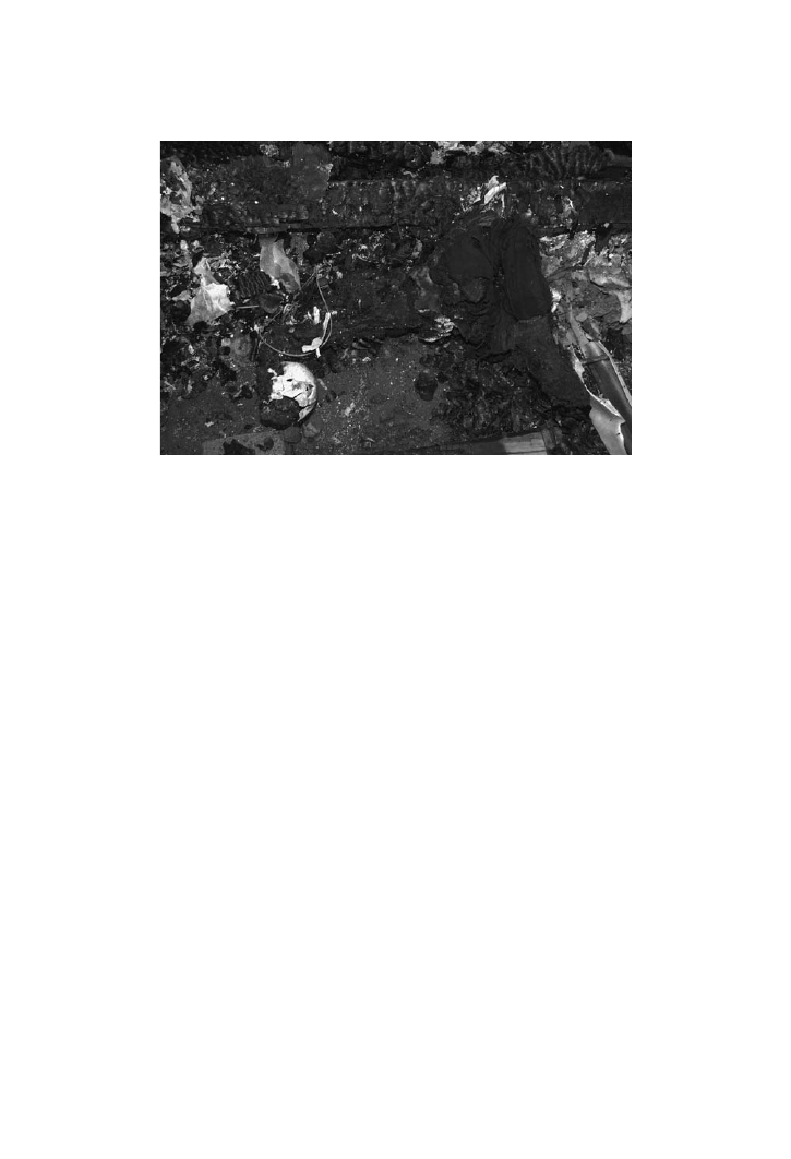
FIGURE 3.1
The Crow–Glassman Scale level #5 is exhibited by these remains. (By per-
mission, Regional Supervising Coroner, Northern Ontario.)
© 2008 by Taylor & Francis Group, LLC

42
Forensic Cremation Recovery and Analysis
The kind of heat a body is exposed to in a fire scene will have an influence on the
burning of the body. For example, the loss of body mass is more pronounced from a
direct fire than radiant heat (Bohnert et al., 1998). This difference is a result of the
body itself acting as a fuel source for the fire, whereas radiant heat acts to reduce
body mass by eliminating tissue fluids.
In general, as heat progresses, the epidermis and its appendages (i.e., hair and
nails) are also profoundly affected. The process of burning tissue is very much a pro-
cess of the tissues acting as either a direct fuel source or undergoing dehydration.
The initial reaction of skin to heat is a dilating of the dermal and epidermal
blood vessels. As heat exposure and/or an increase in temperature continues, the
circulation to this area ceases. As indicated by the description of degrees of fire
injury, burns then tend to proceed to blistering of the skin, which can include the
slippage and gloving of the epidermis from the dermis.
At the same time, hair is undergoing heat-dependent alterations. When a tem-
perature in excess of 300ºC is attained, the hair is charred. The keratin of hair
begins to melt at 240ºC. Finally, as the heat of the fire increases, the hair is con-
sumed in the fire.
A commonly encountered reaction to fire exposure is a heat rupture. Heat rup-
tures can occur before or after death. These ruptures, also referred to as splitting of
3.2
The Effects of Heat on the Body and Related External and Internal Findings
(drawn from Bohnert et al., 1998)
Effects of Heat
External Findings
Internal Findings
Burns
Burns of skin
Singing of hair
Consumption by fire
Burns and consumption of internal
organs and bone
Edema, mucosal bleeding, and
detachment of the mucosa of airways
Changes of content
and distribution of
tissue fluid
Skin blisters
Vaporization of body fluids
Rupture of abdominal wall with
prolapse of intestinal loops
Leakage of fluid from mouth and nose
Heat hematoma
Accumulation of fat in body cavities,
vessels, or heart
Heat fixation
Leather-like brownish fixation of
skin
Induration of internal organs and
muscles
Fragmentation of erythrocytes
Shrinking of tissue
Tightening of skin
Splitting of skin
Protrusion of tongue
Petechial hemorrhages of neck and
head
Pugilistic attitude
Shrinking of organs
“Puppet organs”
© 2008 by Taylor & Francis Group, LLC

The Cremation Process
43
soft tissue, superficially resemble lacerations or incised wounds. However, unlike
lacerations, there is no bleeding due to the coagulation of blood vessels by the heat.
In fact, blood vessels and nerves in the deepest portion of the rupture are intact. The
margins of ruptures are irregular and lack any signs of vital reaction. There is also a
lack of bruising around the ruptures. These ruptures can be found anywhere on the
body, including the scalp (Spitz, 1993; DeHaan, 2002).
As the burning of soft tissue proceeds, the contraction of the epidermis and the
underlying dermis, due to the fire’s dehydrating effects, exposes the hypodermis
and the underlying subcutaneous fat. In this situation, the fat can act as a source of
fuel for the fire (DeHaan and Nurbakhsh, 2001). The presence of clothing, assuming
it has survived the fire up to this point, can act as a wick for any adjacent fat (Spitz,
1993; DeHaan et al., 1999). The net effect is an increase in the rate of cremation as
well as the completeness of the burn. It should also be noted that tight clothing, such
as shoes, socks, clothing with an elastic, and even belts, may act to exclude air, and
hence hinder the progress of the fire on a victim.
Cremated bodies are most commonly encountered in house fires. It has been
purported that a body exposed to temperatures between 670º and 810ºC will show
the “pugilistic attitude” or “pose” after approximately 10 minutes (Bohnert et al.,
1998). After 20 minutes of exposure, the vault of the skull would be free of soft
tissue and even the outer table would exhibit fissures. If the body continues to be
exposed for another 10 minutes, the body cavities (i.e., internal organs) are visible.
At the 40 minute mark the internal organs have shrunk and demonstrate a “net-like
or sponge-like structure” (Bohnert et al., 1998). Fifty minutes into this process the
extremities “are destroyed” (meaning no longer in a complete state), leaving the
torso. From 1–1.5 hours the torso is broken down. In total, the incineration of the
body, in this temperature range, takes about 2–3 hours.
Bohnert et al. (1998) have not been the only ones to qualitatively assess the
effects of fire on human remains. Günther and Schmidt (1953) and Richards (1977)
have all examined the destruction of the body as a unit. These studies are very much
complements of one another. To that end,
, adapted from
Bohnert et al. (1998), deal with the findings of these three studies on the skull and
the remainder of the body, respectively. If we compare the condition of a body to the
surrounding structure, in the same amount of time, a timber would be charred to a
depth of half an inch or so (DeHaan, 2002).
Differential charring of tissues is a fact in cremation contexts. One study sug-
gests that exposures of the bones of the arms, rib cage, and face would occur at
1200ºF in about 20 minutes. This presumably means those areas that are closest
to the surface of the skin, as opposed to those areas that are deeper to associated
muscle. The anterior tibial margin is also reported to be exposed after 25 minutes at
the same temperature, with the femur and the rest of the tibia and fibula (i.e., lateral
side of the lower leg) not being exposed before 35 minutes (Spitz, 1993).
All of these studies presuppose a uniform temperature and an even exposure
to that temperature. Yet, the reality is that heat tends not to be uniform, especially
if the fire is not in a confined area, or there are various types of fuel present. In
fact, the flame temperatures and durations reached in a frame house fire will not be
sufficient to destroy skeletal remains of an adult or even all of the soft tissues of the
© 2008 by Taylor & Francis Group, LLC

44
Forensic Cremation Recovery and Analysis
torso because of the significant percentage of water found in those tissues (DeHaan,
2002). Yet, there will be significant alterations to those bones due to their exposure
to the fire.
Accelerants are materials that literally accelerate or enhance the burning pro-
cess. They tend to be highly inflammable substances that have been designed to act
as fuels in internal combustion engines of all sorts. In the forensic context, kerosene
and gasoline are the most readily available, and hence most commonly encountered.
Burns as a result of kerosene or gasoline result in a patchy distribution of charring
of variable degrees of burning. Readily recognizable aspects of the face and anterior
dentition may be severely damaged, while the chest and abdomen are not nearly as
severely burned (Spitz, 1993). Any ignited adipose tissue may undergo prolonged
smoldering in a defined area. As accelerants tend not to combust completely, resid-
ual accelerants may be detected in clothing and even underlying soil.
As the skin chars and heat ruptures appear, the heat will also have an effect
on muscle tissue. All of the muscles of the body contract due to the heat. Heat
contractures proceed in such a way that the dominant muscles, the major flexor
muscles of the body, overpower the contractions of the extensor muscles, producing
the characteristic “boxer’s” or “pugilistic” attitude or pose. The actual rigidity of the
3.3
The Effects of Fire on the Skull (drawn from Bohnert et al., 1998)
Time
Günther and Schmidt (1953)
1000º–1100ºC
Richards
(1977) 680ºC
Bohnert et al.
(1998) 670º–810ºC
8–10 min
Soft tissues of the face charred
Skull-cap free of soft tissue, soft
tissue of the face charred
13–16 min
Forehead and vertex free of soft
tissue, protruding facial bones
calcined
Bones of the
face showing
20 min
Skull showing
Sparse soft tissue remains in the
face, heat fractures of the
skull-cap
20–25 min
Severe shrinkage of soft tissues
at the skull, calvaria breaks,
brain superficially charred,
destruction of prominent parts
of the facial skull
30 min
Tabula externa of the calvaria
crumbling
40 min
Brain showing, bones of face begin
to disintegrate
50 min
Bones of face largely destroyed,
base of skull showing
45–75 min
Base of skull still intact, head
sometimes severed from trunk
© 2008 by Taylor & Francis Group, LLC

The Cremation Process
45
body, which develops during the cooling process, is due to the denaturation of the
muscle proteins (Spitz, 1993). In this context, the arms tend to be raised above the
shoulders, with the elbows flexed, and the fingers curled almost into a fist. The head
is often extended (looking superiorly) as a result of the contraction of the massive
muscle mass at the back of the neck. The masticatory muscles will contract, acting
to close the mouth unless the tongue was previously protruding out of the mouth.
The back may exhibit hyperextension. The thighs will likely be flexed relative to the
torso, with a marked flexion of the knees. The feet will be plantar flexed with the
toes curled. This position is not to be confused with postmortem rigidity or rigor
mortis. If a body is burned with fixed rigor mortis or has already passed through that
stage, a pugilistic pose will not be in evidence. This reaction of muscles to the heat
of a fire would account for the observation in outdoor funerary cremation pyres,
such as in the Hindu culture in India, of the body appearing to move, and even sit up
during the cremation process.
The pugilistic position of a body must be considered in the search for remains.
Different areas of the body, such as the arms and legs, will fall away from the rest of
3.4
Effects of the Fire on the Trunk and Extremities (drawn from Bohnert et al.,
1998)
Body Region
Time
Richards (1977)
680ºC
Bohnert et al. (1998)
670º–810ºC
Thorax/abdomen
20 min
Ribs showing
Thorax muscles charred, ribs and sternum
showing
30 min
Thoracic and abdominal cavity exposed, organs
blackened and shrunken
40 min
Shrunken, charred organs with bumpy surface
50 min
Organs largely consumed by fire
Arms
10 min
Arms badly charred
Pugilistic attitude
15 min
Arm bones showing
20 min
Hands are largely destroyed, ulna and radius
partially showing
30 min
Hands and distal forearms burned away
40 min
Forearms completely consumed, upper arms
largely free of soft tissue
50 min
Arms burned away
Legs
14 min
Legs badly charred
20 min
Carbonization of muscles
25 min
Shin bones showing
30 min
Tibia and distal femur free of soft tissue
35 min
Thighs and shins
completely bone
50 min
Calcined stumps of the thighs
© 2008 by Taylor & Francis Group, LLC

46
Forensic Cremation Recovery and Analysis
the body as the cremation progresses. However, if the intensity of the heat is main-
tained or increases, a pugilistic pose may not occur due to the pose generally being
assumed as the fire, and the body, cools.
As the skin and muscles undergo direct charring, the increase in heat to deeper
portions of the body starts to affect the internal organs. With increased heat inside
the skull, the formation of a heat hematoma may occur (Polson and Gee, 1973). A
heat hematoma may be originally interpreted as an extra-dural hematoma. The clot
has a light chocolate color or even a slight pink appearance due to blood saturated
with carbon monoxide (CO). This soft, and friable clot is not solid throughout. It
has more of a honeycombed appearance due to bubble formation from the heat. This
is not to be confused with an intracerebral hemorrhage as the cause of death (e.g.,
Chiba et al., 2003).
With the destruction of the skin and the progressive burning of muscle tissue,
the area of highest concentration of soft tissue, the trunk of the body (thoracic,
abdomen, and pelvic regions) will tend to be the last to go. Although fire may have
ignited subcutaneous fat, the heat will have the effect of cooking the internal organs
from the surface. Again, uniform burning would be the exception rather than the
rule. As a result, even though the limbs and head may be significantly damaged, the
internal organs may be well-preserved. This preservational state will often yield the
recovery of tissue samples and other fluids that may be subjected to toxicological
analysis, and in some cases, histological analysis.
By now it should be clear that the elimination of soft tissue by fire is not a simple
task. Yet, there are many forensic cases in which the circumstances have resulted
in the complete elimination of soft tissues and significant damage to the underlying
hard tissue.
3.5 HARD TISSUE DAMAGE
3.5.1 I
NTRODUCTION
Hard tissues usually refer to bones and teeth. However, cartilage is a precursor to
the development of bone, so in some instances it may also be included in this cat-
egory. Yet, cartilage will burn in much the same fashion as any of the soft tissues.
As a tissue, bone is a vascularized, living, and constantly changing mineralized
connective tissue. Bone consists of cells and an intercellular matrix. This matrix
is composed of organic materials, primarily collagen (~20%), and inorganic salts
composed largely of calcium and phosphate in the form of hydroxyapatite.
Bone tissue occurs in two forms: dense, compact lamellar bone, and spongy or
cancellous bone. The location and proportionality of these two types of bone tissue
will dictate how bone tissue is altered in a fire.
Teeth also have a hard matrix of inorganic salts; however, enamel does not regen-
erate once formed. Cementum, the tissue covering the roots, can have appositional
layers of cementum added over time. Dentin, deep to both enamel and cementum,
does not so much regenerate as, like cementum, it adds a layer from inside the pulp
chamber of the tooth, called secondary dentin.
The process of burning bone is essentially a process of dehydration and recrys-
tallization. Once muscle is eliminated, the next tissue to undergo heat stress is the
© 2008 by Taylor & Francis Group, LLC

The Cremation Process
47
periosteum, a thin epithelial tissue covering the bone, providing it with a blood and
nervous tissue supply. However, the heat easily disperses within the periosteum by
charring it directly and flaking off easily.
The direct burning of the bone is going to occur in the areas that are closest to
the surface of the body. These areas include the knuckles, elbows, the acromion of
the scapula, the neurocranium, the chin, the bridge of the nose, the knees, ankles,
and the phalanges of the hands and toes. Areas with a greater density of soft tissue
will be charred later in the process. Areas that are insulated from the fire, such as
the back and buttocks (if the body is in a supine position), will be the last to burn. In
other words, points of contact the body may have with an object or the ground can
result in delayed exposure. This differential burning will continue with the direct
charring of bone.
With the elimination of macroscopic soft tissue (i.e., the epidermis, dermis,
hypodermis, adipose, muscle, and other soft connective tissues) the bone will
undergo heat-induced alterations. As in diseases that leave a pathological finding on
bone, hard tissues have a limited repertoire of response to heat. Bone will undergo
dehydration and be subjected to the corresponding changes. Specifically, the elimi-
nation of water, and the subsequent consumption of the organic portion of bone
and the microstructural alterations to the hard matrix of bone resulting in a color
change, as well as splitting and warping.
3.5.2 H
EAT
-I
NDUCED
C
OLOR
C
HANGES TO
B
ONE
The most persistent remains encountered at cremation scenes are the remnants of
bones and teeth. The macroscopic appearance of the varying colors encountered on
these specimens has been of great interest to cremation researchers (e.g., Lisowski,
1968; Bonucci and Graziani, 1975; Gejvall, 1969; Heglar, 1984; Shipman et al.,
1984; Mayne, 1990; Mayne Correia, 1997). It is considered as fact that the color of
bone can provide information pertaining to the physical condition of the bone and
the context of the burn.
provides a summary of the interpretations of the
various colors encountered on cremated bone.
Although the colors exhibited on cremated bone may be indicators of fire tem-
perature, duration, and combustion circumstances, these colors may all be present
on the same bone at the same time. Variation in fuel load, oxygen availability, and
even bone contact with metals may account for the range of colors encountered on
bones from a fire scene.
Some sense of this process is best considered by examining the process of bone
being exposed to heat in relation to its relative anatomical position. This factor alone
can explain much of the variation in color encountered on the same bone and/or
adjacent bones.
The direct charring of bone on the extremities proceeds first. The extremities,
namely the arms, legs, and head, will have bone directly charred in the regions pre-
viously mentioned. The evidence of exposure to fire is indicated by a characteristic
color change to the bone. The bone will first exhibit a surface that appears to be light
amber. At this stage the organic components of the bone have not been completely
eliminated. In fact, the periosteum may still be intact in these areas. However, as
© 2008 by Taylor & Francis Group, LLC

48
Forensic Cremation Recovery and Analysis
the process continues, the bone begins to blacken. At this stage the periosteum has
been eliminated and the inorganic components of the bone, along with any leaching
bone marrow, are combusted. The bone itself is now feeding the fire. Researchers
studying the microstructure of burnt bone have found that the lamellar bone struc-
ture is intact (Holden et al., 1995a). This is important, as histological aging may be
a means of contributing to an age at death estimation. The black color indicates that
the bone, in this case cortical bone, has attained a temperature of approximately
300ºC (Holden et al., 1995a). This is the temperature of the bone itself and not the
air surrounding it.
In the temperature range of 200–400ºC the ultrastructural orientation of col-
lagen fibers is well preserved in older individuals who have well-mineralized bone,
such as that found in older adults.
Bone that has turned gray has reached a temperature of at least 600ºC. This gray
color is further evidence that the organic portion of the bone has leached out even
further. In this case, the bone exhibits the development of small spherical-type crys-
tals. These spherical-type crystals, as described by Holden et al. (1995a), measure
approximately 0.06 ± 0.007 µm in diameter. As the heat intensifies, these crystals
will alter their shape and size. The lamellar bone pattern is less well organized. As
an age effect, the size of the crystals exhibits a decreasing trend with age. This is,
once again, due to the degree of mineralization of the bone.
As the heat rises, the bone can take on a blue–gray appearance that will even-
tually yield to become white. The white color of bone is found to occur in bone
that has attained a temperature of at least 800ºC. Of course, the bone could attain
temperatures in excess of 800ºC, but the white color is an endpoint color in this
process. At this point the crystals are now a hexagonal-type, measuring from 0.25 ±
0.07 µm to 0.41 ± 0.09 µm in size, and there is no discernable lamellar pattern to the
bone (Holden et al., 1995a). As such, histological examination would be a fruitless
exercise. In older individuals the overall quality of the hexagonal-like crystal mor-
3.5
Interpretation of Color Change to Cremated Bone
Color
Interpretation
References
Brown
Hemoglobin and/or soil
discoloration
Gejvall, 1969; Lisowski, 1968
Black
Carbonized bone due to
burning in O
2
starved context
Herrmann, 1970
Gray-blue, gray
Pyrolized organic components
Dokládal, 1969, 1970
White
Calcination; complete loss of
organic portion and fusion of
bone salts
Mayne Correia, 1997
Other Colors: green, yellow,
pink, and red
Burning in the presence of
metals, including copper,
bronze, or iron
Dunlop, 1978; Gejvall, 1969;
Lisowski, 1968
© 2008 by Taylor & Francis Group, LLC

The Cremation Process
49
phology improved with the age of the deceased. Again, an age effect on the crystal
morphology is once again demonstrated.
It has been postulated by Holden et al. (1995a) that based on an examination
of the color, the degree of fraying of individual collagen fibers, the lamellar bone
orientation, and the formation of crystals, a bone may be placed into an age group.
Holden et al. (1995a) admit that this would only serve as a rough estimate of the
age, but it may assist in the sorting of commingled cremains in a highly fragmented
condition (
3.6).
Recently, the relationship of cremated bone color to organic content and oxygen
availability has been explored by Walker and Miller (2005). In this study, black
bone and white (or calcined) bones were produced in the same temperature scenar-
ios. Yet, with an increase in the exposure time, the influence of oxygen availability
on bone color was found to gradually diminish. As it was also found that bone col-
lagen persisted in specimens exposed to temperatures as high as 600ºC, bone color
was found to actually be an indicator of collagen content. Indeed, in a pilot study by
Marsh and Klem (2002) it was found that the depletion of collagen yield from bone
did decrease with the increased temperature and time of exposure. However, their
study did not attempt to correlate the collagen yield with cremation color.
The color change is associated with the elimination of the organic constituents
of the bone. The progression of color change is roughly from black to gray to white.
Some studies have indicated that some other colors occur, such as browns, and even
shades of red (e.g., Shipman et al., 1984). However, Holden et al. (1995b) found that
when bone has reached 600ºC it has consistently turned a gray color. The elimina-
tion of collagen fibers, proteins, and fats in the bone tissue was found to be complete
at 600ºC. Beyond this temperature the aforementioned production of spherical-type
and then hexagonal-type crystals proceeds at the ultrastructural level. In addition
to this, the color eventually changes to white. This white stage is also known as the
complete, “calcined,” or “calcinated” stage. This is the extreme outcome of bone
recrystallization.
3.5.3 H
EAT
-I
NDUCED
M
ORPHOLOGICAL
C
HANGES TO
B
ONE
The ultrastructural changes are all associated with the dehydration process, or dry-
ing, of the bone as the cremation progresses. A change in color is not the only
3.6
Bone Temperature and Resulting Bone Color (compiled from
Holden et al., 1995a)
Temperature (ºC)
Color Effects
300
Black color of cortical bone
200–400
Ultrastructural orientation of collagen fibers is well preserved
600
Gray color indicates a leaching out of the organic portion
800
White color of bone
© 2008 by Taylor & Francis Group, LLC
50
Forensic Cremation Recovery and Analysis
macroscopic alteration associated with heat. A structural breakdown of bone, as
suggested by the changes noted above, is also going to occur.
The dehydration process involved with burning bone is actually the combustion
of organic materials; this process then continues with a recrystallization of the hard
matrix and will result in a contraction (shrinkage) in the bone’s normal dimensions.
Further, there is also a warping effect on the bone. At the same time, cooling of the
bone also results in the development of cracks or fractures. All of these changes will
impact the metrical analysis necessary for the osteobiographical characterization of
the cremains.
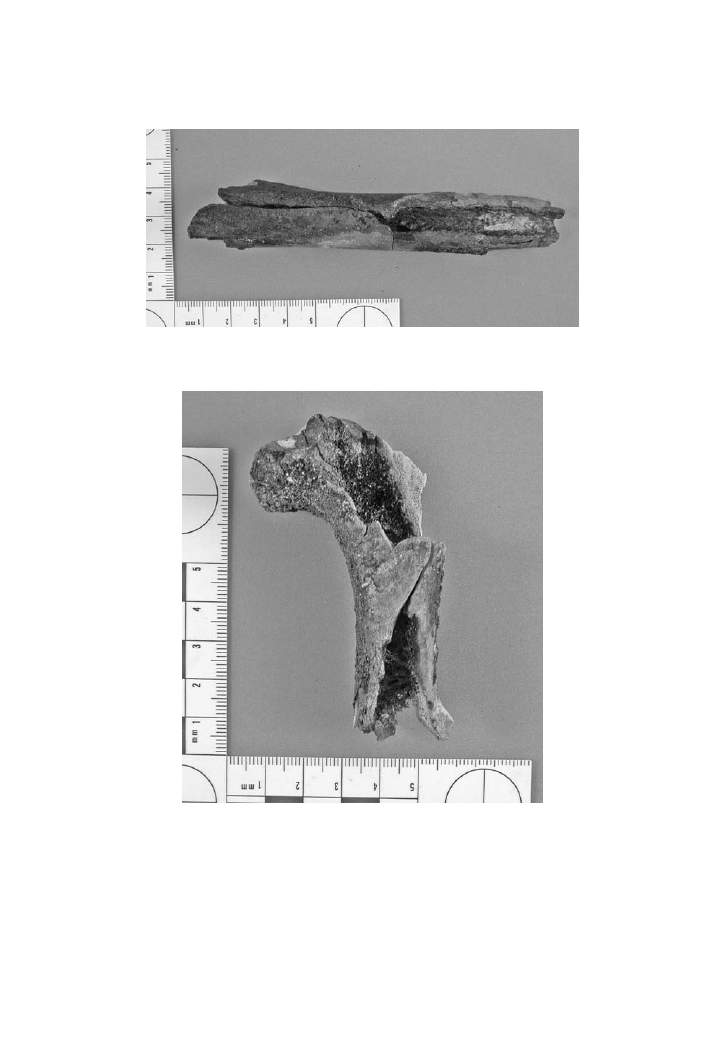
3.5.3.1 Heat-Induced Fractures
On long bones there are several different types of fractures (Stewart, 1979; Her-
mann and Bennett, 1999). Patina fractures are a type of fracture that is observed
on the surface of the bone, typically on flat bones, and even on the surfaces of
long bones (
3.2). These fine cracks do not penetrate through to the medul-
lary cavity of the bone. Longitudinal fractures follow the long axis of a long bone
and may penetrate to the marrow cavity of the bone (
). The longitudinal
direction follows the orientation of collagen fibers along the cylindrically oriented
osteons. Curvilinear (or curved transverse) fractures circumscribe the long bone
shaft proceeding around from one side of the bone to the other (
). They
may be extensions of longitudinal fractures and even exhibit an oblique orientation.
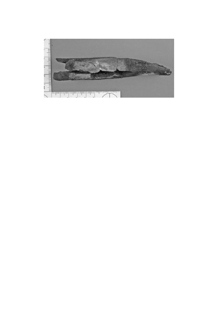
Transverse (or straight transverse) fractures are perpendicular to the longitudinal
axis of a long bone. Transverse fractures tend to penetrate through to the medul-
lary cavity and may even result in a complete transection of the bone (
).
Finally, delamination fractures appear as peeling or flaking of bone layers, particu-
FIGURE 3.2 The patina fractures on the surface of this bone are typically seen on the
surfaces of long bones or flat bones. (Photo by S. Fairgrieve.)
© 2008 by Taylor & Francis Group, LLC
The Cremation Process
51
larly with the separation of cortical from cancellous bone in the epiphyseal region
of a long bone.
Krogman (1943) claimed that a burned bone demonstrating sharp and “clear-
cut” heat fractures of the patina variety, as well as charring, calcinations, and splin-
tering is indicative of bone having a scant or thin covering of soft tissue. He further
FIGURE 3.3 The longitudinal fracture on this specimen has penetrated to the marrow cav-
ity in this case. (Photo by S. Fairgrieve.)
FIGURE 3.4 As the name describes, curvilinear fractures, as seen here, circumscribe the
long bone shaft. (Photo by S. Fairgrieve.)
© 2008 by Taylor & Francis Group, LLC
52
Forensic Cremation Recovery and Analysis
characterizes bone that is deeply embedded in the muscle as eventually undergo-
ing fusion due to the heat in a “molten condition.” Krogman’s comments clearly
indicate the need to be able to distinguish bones that have been recently defleshed
(referred to as “dry bone”) from those that have been burnt with soft tissue present
(referred to as “green bone”).
The problem with distinguishing dry bone from green bone has been examined
by several authors (Baby, 1954; Binford, 1963; Stewart, 1979; Buikstra and Swegle,
1989). Baby (1954) noted that dry bone does not demonstrate the warping found on
green bone. Additionally, dry bone is claimed to show “superficial checking” (likely
referring to patina fracturing), longitudinal fractures, and transverse splintering.
Binford (1963) reported dry bones as having straight transverse cracking. Green
bone, on the other hand, has curved, transverse cracking. Stewart (1979) summa-
rized his findings on defleshed and dried specimens as having a similar appearance
to that reported by Binford. Yet, Buikstra and Swegle (1989) do not support either
Baby or Binford based on their own study of bovid, human, and canine bone. They
report warping in both green and dry bones. Deep transverse cracks were not felt to
provide sufficient evidence of fleshed bone. However, they did conclude that it was
easier to interpret a bone as being either green or dry at the time of burning based
on the color rather than the fracture pattern.
In any event, the point to being able to identify heat-induced fractures is so
that they are not interpreted as being due to another origin, such as direct trauma.
Ultimately, the best way of chronicling fractures as either being due to the crema-
tion process or a mechanically induced trauma is to reassemble the broken portions.
Fractures due to trauma are generally found to exhibit characteristic patterns as
seen in unburned remains. This fact permits analysts to provide an explanation of
the origin of all fractures. Mayne (1990) conducted a study on precremation trauma
and the identification of trauma on cremated faunal bones. She was able to dis-
tinguish heat-induced fractures from those caused by tension, compression, and
shear dynamic forces. However, she cautions that in order to do so, the analyst must
adhere to a six-step procedure (see
).
Fractures to the cranial bones will also occur in a similar pattern (Bohnert,
et al., 1997). However, fractures to the cranial bones are also caused by increased
FIGURE 3.5 This completely transected long bone demonstrates a transverse fracture.
(Photo by S. Fairgrieve.)
© 2008 by Taylor & Francis Group, LLC
The Cremation Process
53
intracranial pressure as a result of the heat. Fractures penetrating both the inner and
outer table are largely due to this pressure and the weakness that may be inherent in
the various regions of the neurocranium. The release of pressure along the cranial
sutures is reported by most researchers to be a rare occurrence due to the ossification
process and the interdigitating osteophytes that make up the sutures. The structure
of the cranial vault often results in fractures separating the inner and outer tables
of bone exposing the diploë (
3.6). The bones of the facial skeleton are not
as readily charred as those of the vault due to the greater thickness of soft tissue.
Nonetheless, once exposed, the facial bones, which are less dense than the bones of
the cranial vault, will undergo the same process of shrinkage and cracking as any
other area of the skull. Areas closer to the surface will, of course, be subjected to a
more prolonged period of heat stress.
The exception to the aforementioned pattern of cranial heat-induced fractures
is said to be in situations where the cranial vault has been breeched due to a cranial
trauma (Rhine, 1998). For example, a traumatic perimortem cranial fracture that
penetrates the neurocranium would allow a means of escape for any increased pres-
sure inside the skull as a result of the heat. Hence, the fractures will not be associ-
ated with a sudden release of pressure. As Rhine (1998) points out, in such a case,
the cranium and its components will be in much better condition as a result of the
pressure being released through the breech. In this instance, Rhine would be refer-
ring to temperatures akin to those found in a house fire. At this temperature range,
cremains are typically identifiable as to their location and position in the house.
However, the head is not usually intact for the above reasons. If the head were in a
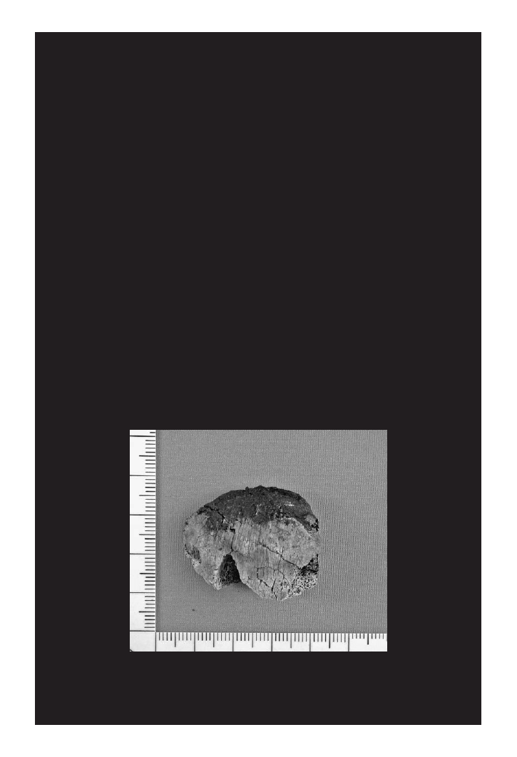
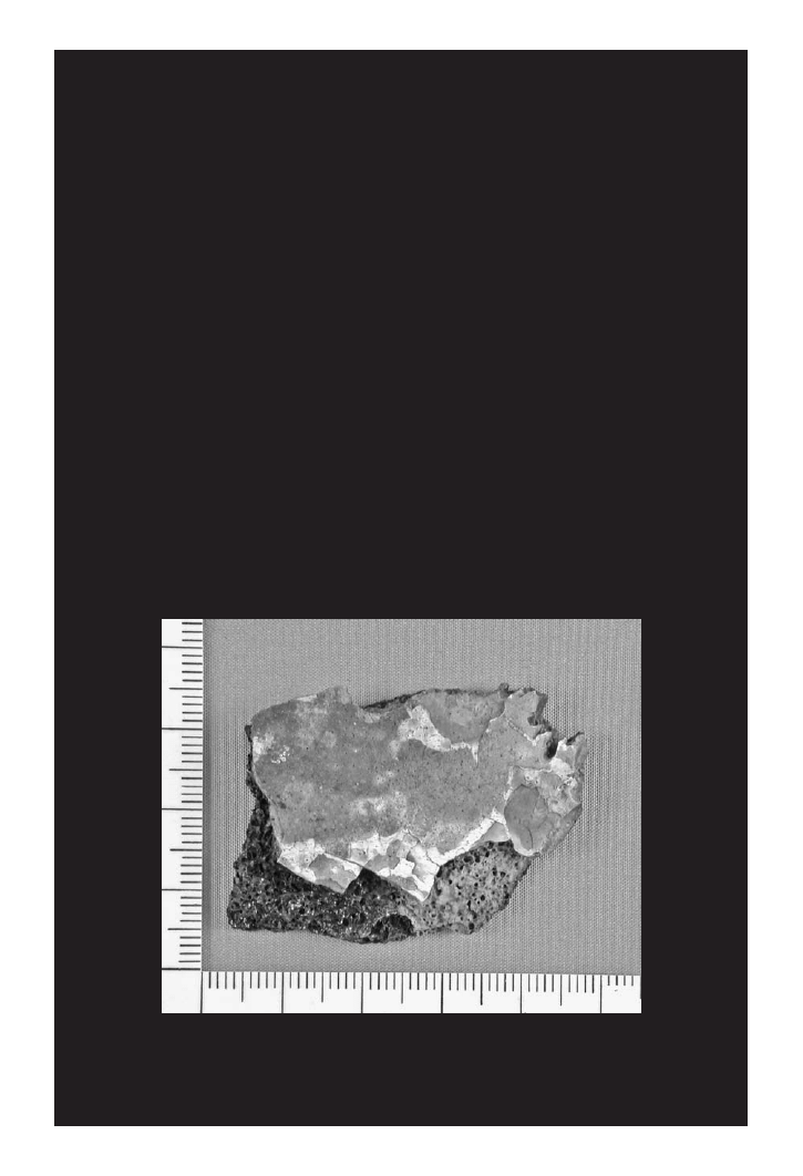
FIGURE 3.6 This cranial fragment from a victim of an aircraft crash demonstrates the
separation of inner and outer tables of bone. (Photo by S. Fairgrieve.)
© 2008 by Taylor & Francis Group, LLC
54
Forensic Cremation Recovery and Analysis
reasonable state of completeness one would certainly be suspicious of a perimortem
trauma. It is important to remember that, even if the skull were in minute pieces,
the skull should always be reconstructed to examine the fracture patterns for any
type of trauma.
In experiments using fresh pig carcasses in automobiles with accelerants, such
as gasoline, the head is typically rendered down to small fragments even with a
postmortem gunshot wound to the neurocranium (
3.7). As the temperature
of these fires exceeded 1200ºC, according to an infrared temperature monitoring
system, this well exceeds the temperatures reached in typical house fires. Therefore,
it would appear that in spite of a gunshot wound to the head, a high degree of frag-
mentation of the skull is certainly possible.
3.5.3.2 Heat-Induced Dimensional Changes
The burning of human tissues will often result in something being left to recover
and analyze. It is up to those individuals performing the recovery to recognize those
altered remains and document them for analysis and interpretation.
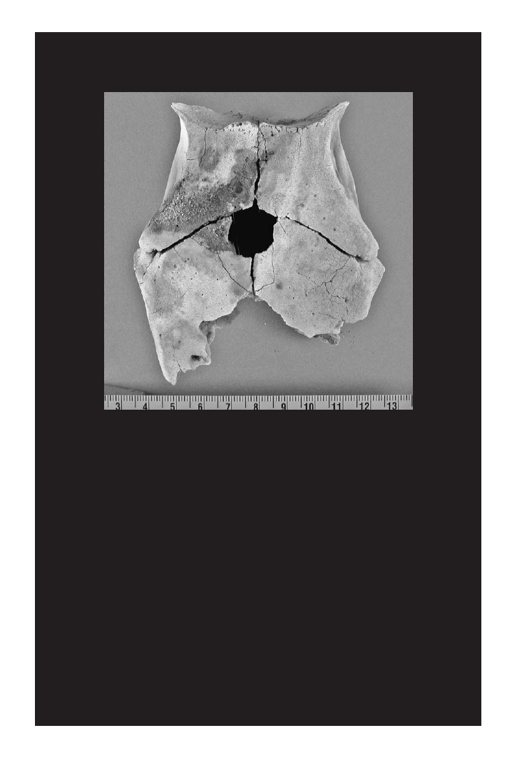
FIGURE 3.7 This cremated pig (Sus scrofa) skull from inside a car fire clearly demon-
strates the evidence of a gunshot wound. Note the sharp edges of the external aspect of the
wound. (Photo by S. Fairgrieve.)
© 2008 by Taylor & Francis Group, LLC

The Cremation Process
55
The investigation of skeletal remains, cremated or not, depends on the use of
multivariate statistical methods, including discriminatory analysis. This fact is par-
ticularly important in light of the additional standards required by U.S. and Canadian
courts pertaining to the admissibility of scientific evidence (i.e., Daubert vs. Mer-
rill–Dow; Regina vs. Mohan; and Regina vs. JLJ, respectively). This is a particu-
larly acute problem should the remains in question not yield a positive identification,
but a presumptive identification based on analytically derived information, such as
the sex, age at death estimation, stature estimate, and the location and types of docu-
mented pathologies (e.g., antemortem fractures). Van Vark (1974) concluded that the
two main causes of difficulties in the application of multivariate statistical analyses
are the changes in the size and shape of the bone in the cremation process, and the
fact that small and fragmentary remains are recovered. Although these factors are
a function of the temperature and duration of exposure to the fire, the fragmentary
nature of the cremains may be due to the action taken by a perpetrator to conceal
the cremation. The skill of these undertaking the recovery of the cremains may also
be a limiting factor in the completeness of recovery (see
). However, the
changes in size and shape should be ascertainable through experimentation.
So far, the discussion of heat-induced alterations to bone has treated these topics
as mutually exclusive events. The same cremation process that has changed the color
of the bone and produced fractures will also result in shrinking the bone and warp-
ing its dimensions.
3.7 summarizes the stage of heat-induced transformation
in bone based on some revised temperature ranges (Thompson, 2004). According to
this information, it is in the fusion stage, that is, the melting and coalescence of the
crystal matrix of bone, that dimensional changes are observed.
Thompson (2005) has conducted the most recent research into heat-induced
dimensional changes in bone. The two key and most widely accepted precepts that
explain why warping occurs, specifically the claim that warping is more appar-
ent in bone that is fleshed at the time of burning (Binford, 1963 and Kennedy,
1996), and that the burning process causes expansion of air in the medullary cavity
(Spennemann and Colley, 1989), are speculative and not substantiated by quantita-
tive data (Thompson, 2005). Further to this, nothing is mentioned about the actual
3.7
The Four Stages of Heat-Induced Transformation of Bone (Thompson, 2004)
Stage of Transformation
Evidence
Revised Temperature
Range (ºC)
Dehydration
Fracture patterns; weight loss
100–600
Decomposition
Color change; weight loss; reduction in
mechanical strength; changes in porosity
300–800
Inversion
Increase in crystal size
500–1100
Fusion
Increase in mechanical strength; reduction in
dimensions; increase in crystal size; changes
in porosity
700+
© 2008 by Taylor & Francis Group, LLC

56
Forensic Cremation Recovery and Analysis
structure of the bone contributing to the manner in which it predisposes the bone to
distort in a particular fashion.
Bone shrinkage in fire has been documented to affect both the length and width.
Malinowski and Porawski (1969) conducted a study of pre- and postcremation met-
rics of bone specimens. In their study, they found that the radial head diameter
decreased by 0.7 mm. Dokládal (1971) cremated one half of each of five cadavers
in a study to examine bone shrinkage. A comparison of the unburned side with the
cremated side yielded a 5 to 12% shrinkage. However, this study does not consider
the fact of asymmetry of intrapersonal dimensions. Herrmann, in a series of studies
(1976, 1977), based his experimentation on compact bone segments measuring 20
millimeters by 5 millimeters. This resulted in the formulation of three phases with
the following corresponding temperatures:
I. 150ºC–300ºC = 1–2% shrinkage
II. 750ºC–800ºC = 1–2% shrinkage
III. 1000ºC–1200ºC = 14–18% shrinkage
These studies by Herrmann led to the conclusion that there are four criteria for con-
sidering the shrinkage of bone in fire:
1. Distribution of bone types in the bone (i.e., compact, spongy, and
lamellar)
2. Temperature of exposure
3. Mineral content of bone
4. Aspects of the mineral content of bone tissue
It seems reasonable to assume that different types of bone tissue will respond dif-
ferently to heat. The mineral content is referring to the level of mineralization. This
can also refer to the relative amounts of the organic portions of bone (i.e., the col-
lagen), and the hard matrix. Finally, aspects of mineralization seem to relate to the
inherent variation of mineral content within the bone. Upon closer examination,
Herrmann found that the higher percentage of bone mineral results in a greater
amount of shrinkage. Grupe and Herrmann (1983) found that there is a 12% reduc-
tion in measurements for spongy bone. In the case of compact bone, Bradtmiller and
Buikstra (1984) found a 5% shrinkage when subjected to temperatures not exceed-
ing 600ºC. In a subsequent study, Buikstra and Swegle (1989) recommended the use
of a correction factor for measurements from 0 to 10%.
Changes in bone dimensions were not found to be consistent within the same
bone. Hummel and Schutkowski (1989) measured the length of compact bone and
found a 5% shrinkage up to a temperature of 1000ºC. However, they also found a
27% reduction in the cross-sectional diameter of the same bone. This indicates that
the orientation of the collagen fibrils has a significant influence on the manner of
shrinkage. Essentially, the fibrils arranged in a parallel and longitudinal axis relative
to the length of the long bone will undergo a smaller relative amount of shortening
© 2008 by Taylor & Francis Group, LLC

The Cremation Process
57
than will be observed in the transverse reduction. If this is borne out, then it would
be expected that other bones that have a less regular arrangement would also differ
in their amount of shrinkage. Holland’s (1989) anticipation of a 1–2.25% decrease
in the size of the cranial base exposed to fire would reflect the kind of orientation of
collagen in bone with an intramembranous ossification. With temperatures of up to
800ºC it was concluded that shrinkage in the cranial base was not significant.
Thompson (2005) recognized a contradiction in the literature as to whether
spongy bone or compact bone shrinks more readily with exposure to heat. Gejvall’s
(1969) work suggests that spongy bone shrinks only slightly and retains its original
shape. Gejvall (1969) and Gilchrist and Mytum (1986) suggest that compact bone
will shrink more than spongy bone. Conversely, McKinley (1994) and van Vark
(1974) argue that spongy bone shrinks the greater amount. Thompson’s analysis of
this split in the literature may be due to the interpretation of relative versus abso-
lute size. Additionally, Thompson (2005) also notes that Holden et al. (1995a) sug-
gest that older bone, with greater intermolecular cross-linkage of collagen, resists
shrinkage. But this is only to a point where the heat is so intense that collagen is
being destroyed.
Thompson’s (2005) study of heat-induced dimensional changes on 60 complete
sheep long bones is an attempt to address many of the questions raised above. The
strength of this study lies in its adherence to the methodology. However, it would
have been better for Thompson to utilize long bones of the same type and side (e.g.,
a left humerus) in an attempt to control as many variables as possible. The method-
ology involved the heating of these long bones for differing periods of time at par-
ticular temperatures. It is important to note that the methodology involved removing
soft tissue and then drying each bone on a rack. The unfortunate aspect of this is
that it does not simulate an actual fire situation with a fleshed victim. However, this
simple study is valuable as it is the actual property of heat-induced bone shrinkage
that is being studied.
Thompson (2005) reports that, with increasingly intense burns, there were
more long bones that fragmented and, hence could not be remeasured with cooling.
This indicates that a postcremation repair of the bones and measurement was not
attempted. This is a shame, as recording such measurements would have recorded
interesting data of importance to forensic anthropologists who are often faced with
fragmentary material from cremation scenes.
It is clear from Thompson’s study, and others, that the variation in the destruc-
tion of a bone by heat is dependent upon the architecture and constituents of the
bone itself.
One result that seemed to surprise Thompson was the fact that as the bone spec-
imen cooled, the dimension of the bone changed. In Thompson’s words, “...this tem-
poral influence means that heat-induced shrinkage is more dynamic than has been
previously realized.” This conclusion was reached by taking repeated measurements
at 5-, 15- and 25-minute intervals after removal from the oven. Although the amount
of difference between the recorded dimensions at these time intervals may have been
surprising to Thompson, the laws of thermodynamics would certainly lead one to
© 2008 by Taylor & Francis Group, LLC

58
Forensic Cremation Recovery and Analysis
expect that with the cooling, there would be a contraction of the bone’s dimensions
until it reaches an equilibrium with the room temperature at which the original pre-
cremation measures were taken. Of equal interest is the fact that, in some instances,
there was an increase in the dimensions over the original measurement with the
heating of the bone. A gradual contraction of the bone followed with cooling, how-
ever, in a few instances after 25 minutes of cooling, there was still a net increase in
the dimension. Again, this can be explained by the fact that 25 minutes may not have
been a sufficient amount of time to cool. However, given that this effect was seen to
occur in the epiphyseal width, the architecture of this area is certainly amenable to
dynamic forces on a regular basis, such as compression, shearing, and torsion. Con-
trary to Thompson’s statement that collagen in the epiphyseal region is “randomly
arranged” and thus has less structural support, collagen is actually arranged as the
structural proteinaceous basis for the intersecting bone spicules, or trabeculae, that
form a multiple arched support structure for the articular ends of long bones. It is
known that the arch is an extremely efficient architectural structure that provides
strength and stability with a minimal amount of mass. The intersecting arches of the
trabeculae that composes spongy (or trabecular) bone produce an extremely strong
and light framework. The trabeculae are surrounded in the intervening space by
marrow that can act to insulate the trabeculae. However, this type of structure does
expose a greater surface area of bone to the heat. Hence, trabecular bone is more
readily expanded by the heat, relative to the compact bone found in the diaphysis.
Therefore, a longer period of cooling will be required prior to taking measurements
to account for cooling of trabeculae at a greater depth. Likewise, if the temperature
and duration are increased, as done in Thompson’s study, the trabecular bone will
contract more than compact bone. This explanation, and Thompson’s findings, are
consistent with McKinley (1994) and van Vark (1974). The “nonwarping origin” of
increased dimension is nothing more than the expansion seen in a heated substance
according to the laws of thermodynamics. Thompson’s study certainly confirms the
finding of an increase in temperature producing an increase in the percentage the
bone tissue contracts.
The ultimate goal of studying the dynamics of bone contraction is to account for
the amount of contraction when undertaking an osteometric analysis. Thompson has
generated predictive equations utilizing step-wise regression for various recorded
dimensions. The recrystallization of the organic phase of bone is an important factor
in the changing dimension of the bone. Temperature was not found to be an accurate
predictor of the amount of contraction. Temperature, in combination with the dura-
tion of the exposure, will have a greater effect on bone contraction than temperature
alone. Logically, a bone may reach a specific temperature, but it is the duration at
that temperature that will directly influence the contraction of hard tissues. Further,
a principal components analysis indicates that the removal of the organic phase
from the bone has the greatest influence on the following variables: duration of
heating, weight loss, alterations in mechanical strength, changes in crystal size, and
microscopic porosity. A strong association between temperature, skeletal density
and microporosity (pores with diameters between 0.01–0.1 µ) implies the involve-
ment of the inorganic phase (Thompson, 2005).
© 2008 by Taylor & Francis Group, LLC

The Cremation Process
59
3.6 CREMATION SLAG AND “CLINKERS”
The appearance of a somewhat porous material directly associated with cremated
remains is often referred to as cremation slag or “clinkers.” A “clinker” is defined
by the Oxford English Dictionary (1989) as:
… a hard mass formed by the fusion of the earthy impurities of coal, limestone, iron
ore, or the like, in a furnace or forge; a mass of slag.
Wells (1960) described crystalline lumps of material found in direct association
with cremains. Wells theorized that these clinkers were formed from the keratin
contained in hair, and in combination with fat and tissue burned at the same time.
Wells’ conclusions were challenged almost 30 years later by Henderson et al. (1987).
In a search of the literature, they found a citation by Brandt (1960) referring to a
study by Von Stoker of “urn resin” or “urnenharz” from cremation burials from
northern German archaeological sites dating from the Stone Age to the Migration
Period. Von Stoker found that this resin was soluble in acetic acid and chloroform
and produced an aromatic smell. Von Stoker’s conclusion was that the resin was
derived from Scots pine (Henderson et al., 1987).
Analysis of cremation slag from Illington, in England, was found to be com-
posed of Si, Ca, Al, P, Mg, and Fe with smaller quantities of K, Mn, Ti, Zn, Na, B,
Zr, Cu, Ba, Sr, Ni, and Pb. There was a complete lack of organic material. Further,
when the slag was heated for two hours at 450ºC it lost only 2% of its original mass
(Henderson et al., 1987).
In 1962, the British Museum analyzed the same slag and found that it was chiefly
composed of sintered grains of silica (SiO
2
) mixed with small amounts of other
materials, such as a bead of iron/iron oxide, and fragments of bone (Henderson et
al., 1987). X-ray diffraction analysis of the quartz form of silica had been almost
completely converted in fused areas into the high temperature form of cristobalite.
The conversion of pure silica to cristobalite takes place at 1470ºC. In the presence of
impurities, the conversion temperature would be lower.
Henderson et al. (1987) conducted a study of cremation slag or “clinkers” in
order to investigate Wells’ claims and to provide macroscopic, microscopic, and
chemical analyses. Such an undertaking provided an opportunity to characterize
cremation slag and determine its likely origin. They determined that the raw mate-
rials needed to produce cremation slag include silica, alkali (such as K or Na) and
Ca from the cadaver or wood ash, with sufficient fuel to raise the temperature of
the pyre to a level that would result in fusing these components into the slag. The
sources for silica include the human body, wood, and plant ash.
The amount of energy it takes to burn a body has been previously discussed.
However, the body itself should also be considered a fuel. Consider that a body
weighing 140 pounds, placed inside an elm coffin weighing 90 pounds, will yield
over 800,000 BTUs as along as there is sufficient oxygen to complete the combus-
tion process (Polson et al., 1962 as cited by Henderson et al., 1987). If there is a lack
of a critical level of oxygen, the body will char rather than burn to ash. In this sce-
nario, the body and the coffin act as fuel and raise the temperature of the crematory
chamber higher than the flame generated by the supplied gas alone. Hence, achiev-
© 2008 by Taylor & Francis Group, LLC

60
Forensic Cremation Recovery and Analysis
ing the requisite levels of heat may not be a problem for this context, but in other
contexts, such as house fires, this may not be the case. Open cremation pyres of a
clandestine nature may reach such temperatures if the fire is tended by a perpetrator
through the addition of fuel.
Regardless of the mechanism by which cremation slag is produced, the slag is
derived from silica-bearing sandy soils fusing with material at a high-temperature
(Henderson et al., 1987). Its importance for forensic casework would serve to act as
a rough indicator of combustion temperature.
3.7 SUMMARY
This chapter has dealt with reviewing the combustion of human remains from a
forensic perspective. It is clear from the literature that it is impossible to completely
eliminate all evidence of a body through the act of burning it. Perpetrators obvi-
ously encounter this fact if they are actively tending to the cremation pyre. Hence,
their attempts to further render the remains to an “unrecognizable state.”
Understanding the mechanisms of body immolation will serve the investiga-
tor well at fire scenes. Of course, recognizing the fact that there are remains worth
recovering from fire scenes of various types is the first step to a successful recovery.
The next step is to assemble a team that will competently undertake the documenta-
tion and recovery of the cremains.
© 2008 by Taylor & Francis Group, LLC
Document Outline
- Table of Contents
- Chapter 3: The Cremation Process
Wyszukiwarka
Podobne podstrony:
9189ch5
9189ch8
9189ch7
9189ch1
9189ch4
9189ch6
9189ch2
9189ch5
9189ch8
więcej podobnych podstron