
141
7
Incineration of
Dental Tissues
7.1 INTRODUCTION
As most references on forensic identification of human remains will tell you, the
analysis of dental remains can provide information pertaining to the age of the indi-
vidual, as well as identity through comparison to antemortem records. Of course,
in order to do such a comparison, one must have an idea of whom the cremains rep-
resent. Any information with respect to the age at death, the sex, race (or ancestry)
will be of great assistance to establishing a positive identification (see
for
complete discussion on positive identification through other methods).
As with bone tissue, teeth are also made up of an inorganic matrix of hydroxy-
apatite. However, a tooth is actually composed of three related hard tissues. These
tissues—enamel, cementum, and dentine—will react differently from one another
when exposed to heat. The nature of any one tissue’s reaction is dependent upon its
chemical/structural composition as well as its location on the tooth. Once again, the
anatomical axiom of structure reflecting the function holds for the reaction of dental
tissues to elevated heat.
Teeth are amongst the most resilient structures of the human body. As such,
forensic odontologists are often called upon to undertake an examination of dental
structures in order to establish an identification of remains based on a comparison of
antemortem dental records. The dentition is commonly preserved, at least partially,
in a variety of instances where exposure of a body to a heat source may occur. How-
ever, in forensic cremations, the heat exposure can be of such an extent that the teeth
and associated alveolar bone may be highly fragmentary (
and b). Such
contexts necessitate careful recovery procedures, as fragmentary dental remains
may yield important evidence concerning antemortem dental work that may lead to
a positive identification (Fairgrieve, 1994).
This chapter provides a review of the structure of teeth and their associated tis-
sues. The reactions of these tissues to elevated heat in a cremation context are also
detailed. Specific attention is paid to the interpretation of heat-induced alterations
to these dental tissues.
7.2 BASIC DENTAL ANATOMY
A tooth, in general, consists of a crown, covered by enamel, and a root covered by
cementum. These two areas of the tooth meet at the neck or cementoenamel junc-
tion (CEJ). Beneath these tissues, the bulk of a tooth’s structure consists of dentine.
The dentine supports the enamel covering the crown and the cementum of the root.
© 2008 by Taylor & Francis Group, LLC
142
Forensic Cremation Recovery and Analysis
Within the dentine there is a central space known as the pulp cavity that is expanded
at the coronal end and tapers down the root (a root canal) and terminates at the tip
of the root as an apical foramen. In teeth with multiple roots, such as premolars and
molars, each root will have an apical foramen. The roots are surrounded by a tissue,
known as the periodontal ligament, that binds them to the alveolar bone that makes
up the socket in which the root sits.
Humans have a heterodontic dental arcade. That is to say, we exhibit a dentition
that has a variety of tooth forms rather than just one form as found in homodontic
species, such as sharks. We also have teeth that are permanent, or rather, adult, and
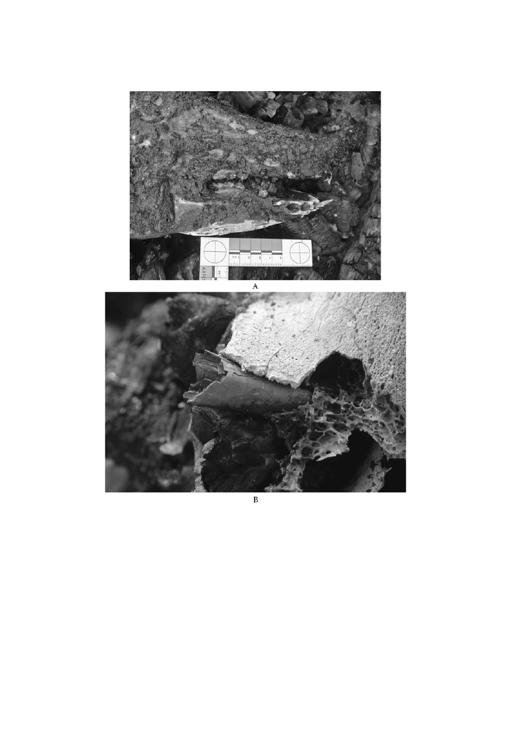
FIGURE 7.1 A) This photograph of the mandibular region of a pig carcass subjected to
a fire for 30 minutes demonstrates that the back teeth are more protected than the anterior
teeth. B) This detailed photograph of an alveolar remnant from a pig carcass subjected to 50
minutes in an automobile fire exhibits severe fragmentation. (Photos by S. Fairgrieve.)
© 2008 by Taylor & Francis Group, LLC

Incineration of Dental Tissues
143
teeth that are shed during childhood; hence, they are known as deciduous teeth. The
implication of this is that although teeth possess the same tissues, there is variation
in the actual structure and position of the tooth in the mouth. This in turn means that
teeth will not all react to heat in a uniform manner.
The human dentition consists of four morphologically functional forms: the
incisors, canines, premolars, and molars. The incisors and canines make up what
is known as the “anterior dentition.” This anterior position makes these teeth more
susceptible to heat stress, as they are directly adjacent to the opening of the mouth.
In contrast, the posterior dentition, also referred to as the “cheek teeth,” include the
premolars and molars. This position provides enhanced protection of the teeth due
to the insulative effect of the cheeks. The tongue also acts to insulate the lingual
surfaces of all teeth.
The teeth are organized into upper and lower dentitions. These opposing arches
are subsequently divided into halves, resulting in the four quadrants of the mouth.
The arrangement of teeth in these quadrants follows the order of placement along
that arch from the midline to the distal aspect. In each quadrant there are typically
two incisors, one canine, two premolars and three molars. This is known as the fol-
lowing dental formula: 2-1-2-3. As this is the same for upper and lower dentitions,
the more formal way to express this is 2-1-2-3/2-1-2-3.
The incisors are single-rooted teeth with a single incisal edge and somewhat
spatulate in shape. Their function is for shearing into items to be consumed.
Distal to the lateral incisor is the larger canine with a single cusp and a long
single root that acts to deeply anchor this tooth within the alveolus. The larger size
of the root results in a bulge in the maxillary alveolus known as the “canine emi-
nence.” The thinned alveolus in this area is subject to fracture in fire and exposes
the root of the upper canines to direct fire damage.
Distal to the canines are two premolars. These bicuspid teeth have one cusp
adjacent to the cheek (buccally), and the other lingually positioned (adjacent to the
tongue), separated by a mesiodistal fissure. The upper first premolars generally have
two roots, whereas the second upper premolar usually has one root.
At the distal end of each quadrant of the mouth are the three molars. Of all the
teeth so far described, these are the most well-protected, due to their overall mass
and deep position within the mouth.
In the case of a deciduous dentition, that is, children less than age six have five
teeth in each quadrant rather than eight; there are no deciduous premolars and there
is one less molar. These teeth are generally smaller than adult teeth and on a smaller
arch. Fire more readily affects these teeth due to their size and the smaller amount
of soft tissue protecting the back teeth. In children older than five the dentition is a
mix of deciduous and adult teeth. Those searching a cremation scene must be fully
cognizant of the possibility of victims with a mixed dentition and permanent teeth
developing within the alveolus.
It has been observed that with the heating of dental tissues they do not react in
an identical fashion to one another (e.g., Fairgrieve, 1994). This is largely due to
their respective microstructural anatomy. In order to understand how these tissues
react to heat, a brief description of the microstructure of enamel, cementum, and
dentine is outlined below.
© 2008 by Taylor & Francis Group, LLC

144
Forensic Cremation Recovery and Analysis
7.2.1 E
NAMEL
M
ICROSTRUCTURE
As demonstrated in the previous chapter, we are once again dealing with composites
of mineral and organic phases. This holds for enamel, cementum, and dentine. In the
case of enamel, the tissue covering the crown, its hardness, and rigidity have been
likened to a ceramic material. The justification of this comparison is due to 95–
96% of the weight being crystalline apatite, while less than 1% is from the organic
matrix, and the remainder being water (Williams et al., 1989). Once erupted, the
cells responsible for the formation of enamel (ameloblasts) are lost. This means that
once formed, enamel is not capable of further growth, or repair for that matter.
As a ceramic-like material, enamel is composed of tightly packed enamel prisms,
or rods, that are U-shaped in cross-section, and extend from the enamel–dentine
junction to within 6–12 µm of the surface. Each prism is packed with flattened
hexagonal hydroxyapatite crystallites. Between these prisms is an interprismatic
material that contains most of the organic material found in mature enamel.
The larger size of the crystals in enamel, relative to bone and dentine, will affect
its physical properties. This larger crystal size also means that the surface of the
organic phase is reduced and it is, therefore, less reactive (Cole and Eastoe, 1988).
During the growth of these crystals the amelogenins (proteins that predominate in
newly secreted enamel) are removed during the maturation process. It is at this stage
that the enamel becomes hard.
The orientation of enamel crystals will play an important role in the manner in
which it reacts to heat stress.
7.2.2 C
EMENTUM
M
ICROSTRUCTURE
Like enamel, cementum is also an outer covering, albeit of the tooth root rather than
the crown. The function of cementum is to encase the root of the tooth and receive
the uncalcified collagen fibers of the periodontal membrane (Sharpey’s fibers) in
order to anchor the tooth to the alveolus.
Cementum is approximately 50% by weight hydroxyapatite and amorphous cal-
cium phosphates. This tissue has a yellowish appearance. As one ages, this yellow
color becomes progressively deeper (Ten Cate et al., 1977).
Cementum is formed by specialized cells known as cementoblasts. The organic
matrix, or precement (or cementoid) is deposited first and is subsequently miner-
alized. In the active growth layer of cement, cementoblasts become entrapped in
developing tissue and they then become cementocytes.
Cementum deposition is a continuous process throughout the life of the indi-
vidual. The thickest areas of cementum deposition are the apices of the roots and the
point of bifurcation of the roots in multirooted teeth (Hillson, 1986). This is done to
maintain a small degree of eruption in order to compensate for occlusal wear.
Cementum is known to occur in layers. These layers can be observed as having
an incremental pattern that may be annually deposited (Hillson, 1986). This has
been the basis of an age-at-death estimation technique (e.g., Charles et al., 1989).
However, to my knowledge there has never been an attempt to use this technique on
cremated tooth remnants. This is likely due to the highly fragmented condition that
© 2008 by Taylor & Francis Group, LLC

Incineration of Dental Tissues
145
occurs when subjected to heat stress (see
below). It is of interest to note
that the deposition of cementum within the apical foramen (orifice) may cause a
strangulation of the vessels passing through it. Hence, this area should be examined
as a possible alternative for examining cementum annulations.
7.2.3 D
ENTINE
M
ICROSTRUCTURE
As with the previous two dental tissues, dentine is a mix of organic and mineral
components. An examination of this composition is listed in
7.1. This material
makes up the bulk of the tooth, as it is deep to, and in support of, both the enamel of
the crown and cementum of the root.
The major structural feature of dentine is the presence of densely packed den-
tinal tubules. The tubules are found to be passing perpendicularly through what
are described as felted mats of collagen fibrils that are piled one on top of another
(Hillson, 1986). The inorganic crystallites occur within this organic matrix. Fur-
ther, the tubules themselves enclose a single cytoplasmic process of an odontoblast,
containing microtubules, microfilaments, and mitochondria (Williams et al., 1989).
The cell body of this odontoblast is in the pseudostratified layer lining the pulpal
chamber encased within the dentine.
As dentine mineralizes it takes on the form of calospherites, microscopic spher-
ical aggregates of crystals. However, dentine is not subsequently remodeled, as is
the case with bone. Dentine does form what is known as secondary dentine inside
the pulpal chamber. This type of dentine is reparative in nature. It is the only mecha-
nism by which a tooth with a microfracture, due to some form of trauma, can repair
itself.
7.1
The Composition of Normal Human Dentine (drawn from
Cole and Eastoe, 1988)
% by Weight
Inorganic matter
75
Ash
72
Carbon dioxide
3
Organic matter
20
Collagen
18
Resistant protein
0.2
Citrate
0.89
Lactate
0.15
Chondroitin sulphate
0.4
Lipid
0.2
Unaccounted for (water retained at 100ºC, errors, etc.)
5
© 2008 by Taylor & Francis Group, LLC

146
Forensic Cremation Recovery and Analysis
7.2.4 D
ENTAL
P
ULP
The material contained within the confines of the pulpal chamber consists of a loose
connective tissue that is well-vascularized and known simply as the pulp. The pulp
communicates with the rest of the body via the apical foramen. There are several
arterioles and capillary loops along with myelinated and unmyelinated sensory
nerve fibers. These nerves respond to thermal, mechanical, or osmotic stimuli of
dentine by a pain response.
The pulp is of particular importance in cases involving fire, as it is a highly
protected environment that may yield nuclear or mitochondrial DNA that is suitable
for profiling (see
).
7.2.5 O
THER
T
ISSUES
The only other tissues of concern to the dentition are the gingiva and the oral mucosa
of the mouth. A healthy gingiva may be recognized by its pale pink color and lightly
stippled appearance. The gingiva is continuous with the oral mucosa via the muco-
gingival junction. The mucosa consists of stratified squamous epithelium that has a
smooth texture and red appearance due to vascularization.
The mucosa, along with the tissues that make up the cheeks, act to insulate the
teeth from heat stress during the initial stages of the cremation process. The only
gap in that protection is through the opening of the mouth.
7.3 INCINERATION OF DENTAL TISSUES
It has been recognized in the professional literature that teeth are indeed the most
reliant structure of the human body. In fact, enamel is often described as the hard-
est substance of the body. This statement is supported through the paleontological
recovery of teeth exceeding that of bone. As part of their inherent resiliency, teeth
are resistant to fire, desiccation, decomposition, and prolonged immersion in water
(Robinson et al., 1998). The experience of individuals involved with the recovery
of bodies from fires and blast events is that dental remains are often going to play
a key role in the identification process, and may help in answering other questions
relating to the scene.
This section outlines the effects that various temperatures have on dental tis-
sues. Each tissue is dealt with in turn.
7.3.1 I
NCINERATED
E
NAMEL
One of the most important papers in surveying the effects of heat on dental tissues
is by Harsányi (1975). In this study Harsányi exposed intact incisors and premolars
from fresh corpses from 35 to 45 years of age, representing both sexes, to a heat
source. An electric incinerator was used for these teeth to reach specific tempera-
tures. It took 5 minutes for the oven to reach each target temperature; once reached,
that temperature was maintained for 55 minutes. The teeth were then weighed and
examined.
is a compilation of Harsányi’s results for enamel.
© 2008 by Taylor & Francis Group, LLC

Incineration of Dental Tissues
147
Enamel appeared to go through a series of color changes ranging from a dark
grayish-brown at 300ºC to porcelain-white at 1000ºC. Structurally, the enamel
begins to crack and eventually break into fragments, separating from the underly-
ing dentine. Microscopically small crevices appear at 300ºC and increase in size
and number as the temperature increases. The formation of granules on the surface
of the enamel occurs at 700ºC, while a fusion of these granules appears at 900ºC. At
1000ºC the enamel microstructure is unrecognizable.
If a comparison is made to the results of a similar study by Shipman et al.
, many of the same types of physical alterations
are noted, albeit under heating regimes that were slightly different. Shipman et al.
refer to temperature ranges as opposed to a specific peak temperature as seen in
Harsányi’s study. However, the individual temperatures are within the range out-
lined in Shipman et al.’s study, save those in Harsányi’s study, that are in excess
of 900ºC. Both studies note that there is a structural change at temperatures below
200ºC. However, as 300ºC is approached, there are changes to the surface of the
enamel ranging from “dimple” development to the initiation of crevices. As tem-
peratures approach 500ºC, the presence of rounded particles is noted by Shipman
et al. At this same level, Harsányi reports a gray color and the establishment of a
microcrevice network. Shipman et al. also note the presence of fissures (crevices)
and enamel beginning to break into small fragments. Both studies agree that pre-
viously formed particles (granules) will coalesce into larger, smooth globules and
eventually fuse below 1000ºC. Once above that temperature, the enamel is “porce-
7.2
The Effects of One Hour of Exposure at Specific Target Temperatures on
Enamel at Macroscopic and Microscopic Levels (drawn from Harsányi, 1976)
Temperature
Macroscopic Observations
Microscopic Observations
200ºC
Color changes
None
300ºC
Dark grayish brown; small crevices; enamel
starts to peel off via small crevices; carries
are not narrowed
Small crevices; enamel intact from
crevice to crevice
500ºC
Gray enamel; longitudinal furrows
Divided by crevice network;
multiangular plates
700ºC
Light grayish-white; broken into fragments
Consists of fine grained granules;
original surface unrecognizable
900ºC
Almost white (equal in color to cementum
and dentine); enamel in smaller pieces
Enamel grains start to fuse-structure
unrecognizable
1000ºC
Enamel porcelain-white (equal in color to
cementum and dentine)
“Structureless” smooth plates
1100ºC
Small fragments, porcelain-white color
Same as at 1000ºC
1300ºC
Minute smooth porcelain-white fragments;
glass-like surface
Inorganic salts are fused into round
formations
© 2008 by Taylor & Francis Group, LLC

148
Forensic Cremation Recovery and Analysis
lain-white” and has fragmented. The effects of the heat on the hydroxyapatite are
the same as observed in bone. Ultimately, with temperature in excess of 1100ºC and
up to 1300ºC, the fusion of these salts results in the enamel developing a glass-like
consistency.
Although these studies examine the effects of varying temperatures on the
enamel, they do not indicate what will be observed in the field. The differing chemi-
cal composition of each of the three dental tissues will dictate how they will each
react at a given temperature. Of these three tissues, enamel has the smallest amount
of water. This is part of the reason that enamel, in relation to cementum and den-
tine, does not retract to the same degree. It is therefore quite common to encounter
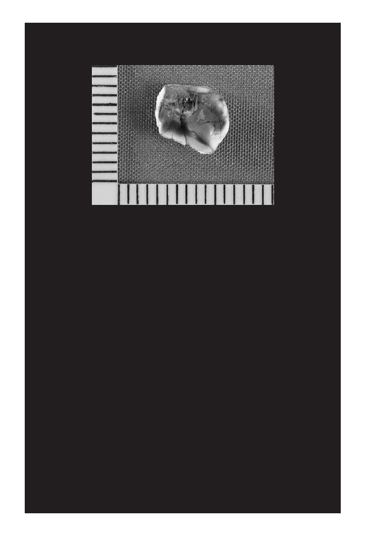
enamel crowns separated from underlying dentine (
7.2). In cases where the
enamel has some form of dental work, such as a filling composed of some sort of
amalgam, the separation of the enamel from the dentine will still occur, with the
FIGURE 7.2 This enamel crown has separated completely from its underlying dentine.
This is a common occurrence in forensic cremations. (Photo by S. Fairgrieve.)
7.3
Heating Stages of Enamel: Microscopic Morphology (drawn from Shipman et
al., 1984)
Stage
Temperature
Description
I
20–< 185ºC
Normal and unaltered
II
185–< 285ºC
Enamel develops dimples, but overall surface texture is smoother than in
Stage I
III
285–< 440ºC
Rounded particles appear, covering the surface
IV
440–< 800ºC
Appearance of vitrified or glassy particles separated by many pores and
fissures; enamel closer to CEJ, breaks into smaller fragments
V
800–940ºC
Particles coalesce into larger, smooth-surfaced globules and these fuse into
an irregularly shaped mass pierced by rounded holes
© 2008 by Taylor & Francis Group, LLC
Incineration of Dental Tissues
149
additional separation of the filling from the enamel itself (Fairgrieve, 1994; Bush et
al., 2006) (
7.3).
The alterations of enamel during incineration may appear to be dramatic on a
histological level; however, the enamel is nonetheless recoverable from cremation
scenes. Analysis of such enamel may provide indications of peak temperatures as
well as dental morphology that can be utilized as part of the identification process.
7.3.2 I
NCINERATED
C
EMENTUM
Cementum is protected by the gingiva, periodontal ligament, and the alveolus of the
mandible, or maxilla. As the soft tissue features of the face and parietal aspects of
the oral cavity are consumed by fire, the cementum is not directly affected until the
gingiva is consumed.
As mentioned above, the enamel of the crown is the first of the dental tissues
to be assaulted by the fire, starting with the anterior dentition. The cementum, the
second tissue to be direly affected by fire, would first be exposed at the CEJ and then
as the periodontal ligament and surrounding alveolus are eliminated; the remaining
portions of the cementum are subjected to the heat down to the apex of each root.
This explains the differentially shaded appearance of cremated tooth roots that have
not been completely incinerated to the porcelain-white stage.
summarizes the effects of an hour of exposure of cementum to fire.
Early on in the process, at 300ºC, the evaporation of water from the cementum
results in a lifting of this tissue layer from the underlying dentine. At this stage
the cementum has a “dark grayish-brown” color (Harsányi, 1975). At 500ºC the
root canal and apical foramen are preserved. The root appears to have large plates
with deep furrows in between (
). With a tooth having been liberated from
the alveolus, the root is now going to be directly accessible to the flame. With this
increased heat to 700ºC the cementum becomes a grayish-white color with a fine
FIGURE 7.3 This section of a crown exhibits the remnants of an occlusal filling that has
been eliminated during the cremation process. (Photo by S. Fairgrieve.)
© 2008 by Taylor & Francis Group, LLC
150
Forensic Cremation Recovery and Analysis
7.4
The Effects of One Hour of Exposure at Specific Target Temperatures on
Cementum at Macroscopic and Microscopic Levels (drawn from Harsányi,
1976)
Temperature
Macroscopic Observations
Microscopic Observations
200ºC
Color changes
None
300ºC
Dark grayish brown
Evaporating water lifts cementum from dentine,
“vesicles” with disrupted walls are formed;
denuded surface exhibits tubular orifices
500ºC
Light brownish-gray; root
canal is preserved
Aggregated into large plates, divided by deep
furrows; single plates are multiangular and are 30
to 60 µm in diameter
700ºC
Light grayish-white; root canal
narrowed but recognizable
Fnely granular surface; original structure no longer
visible
900ºC
Light, almost white color; root
broken into large pieces
Granular surface penetrated by deep and wide
crevices; original structure is decomposed
1000ºC
Porcelain-white color
Covers dentine as a homogeneous, melted
unconnected layer; some open orifices of dentine
tubules are visible
1100ºC
Porcelain-white color
No change
1300ºC
Minute smooth porcelain-
white fragments
Structure is decomposed; round formations of
various sizes
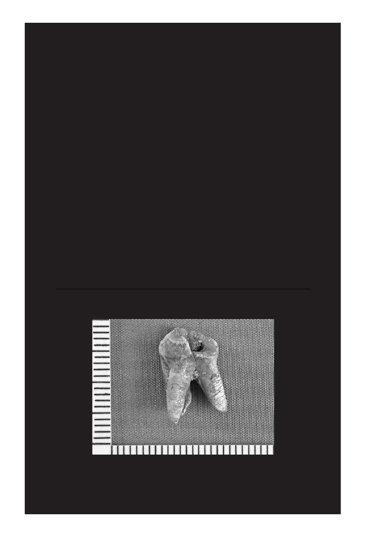
FIGURE 7.4 The roots of this upper first molar exhibit the platelike fragmentation of the
cementum. The graded shades from white to gray from the CEJ to root apex indicates the
differential exposure to heat due to its location in the alveolus. (Photo by S. Fairgrieve.)
© 2008 by Taylor & Francis Group, LLC

Incineration of Dental Tissues
151
granular surface at the microscopic level. In excess of this temperature, approxi-
mately 900ºC, the color of the cementum is white and the root has disintegrated
into large fragments. As the temperature increases the cementum becomes “porce-
lain-white” and appears to have undergone some form of fusion of surface particles
producing a melted layer over the dentine.
It is important to stress that the studies undertaken by Harsányi (1975) and
Shipman et al. (1984) used teeth that were already isolated from the alveolus. Any
conclusions drawn from those studies should be considered estimates in actual
casework where teeth are likely to be situated in the alveolus during cremation.
7.3.3 I
NCINERATED
D
ENTINE
As cementum is likely to persist over dentine, albeit in a fragile state, dentine will
typically be fully exposed at the crown once the enamel has separated. This is not
to say that the dentine is not subjected to any heat while the enamel is still present.
Heat will still reach the dentine via conduction through the enamel. As each of the
dental tissues has been treated separately, in order to facilitate an explanation of this
dynamic process, the tooth is a matrix of tissues, the majority of which consists of
dentine.
At temperatures up to 200ºC the dentine is unaffected by the heat (
).
Surface alterations of color are possible, but they are dependent upon the condition
of the superficial surfaces of enamel and cementum for the crown and root, respec-
tively. Such a tooth would be an excellent candidate from which to extract DNA
from the pulp.
As 300ºC is approached the dentine may become more of a light grayish-brown
color, due to the heating of material within dentine tubules. This coincides with an
opening of the tubules. However, the overall morphology is unaffected. The micro-
scopic examination of the peritubular matrix demonstrates a contraction in size and
separation from the intertubular matrix. The somewhat roughened appearance to
the dentinal surfaces are due to the openings (or asperities) of the tubules.
The pulp cavity remains intact up to 500ºC. The dentine has shifted to a dark
grayish-black color, indicative of the carbonization of organic components. The
asperities have melted and created a smooth surface. By this temperature Shipman
et al. found that tubule openings had elongated and the intertubercular matrix had
formed bars of fused material between these openings.
A further rise in temperature to approximately 700ºC seems to have eliminated
the majority of the organics present inside the dentine as demonstrated by the gray
color. The root canal and pulp chamber persist, and yet they are narrowed. The
tubules are narrowed inside, but with elongated and enlarged openings. The surface
has what is described by Shipman et al. as a “frothy” appearance due to the altera-
tion of the tubule openings. Some openings are irregularly shaped with a glassy
texture (
).
Upon reaching approximately 900ºC the dentine, as with the other dental tis-
sues, is white. Dentine tubules are narrowed and the frothy areas mentioned above
have coalesced into globules that fuse into nodular spikes. The spaces between
© 2008 by Taylor & Francis Group, LLC

152
Forensic Cremation Recovery and Analysis
these spikes are the remnants of the tubules and the spikes are the remnants of
intertubular bars.
Temperatures in excess of 1000ºC produce dentine that is porcelain-white with
a narrowed pulp chamber. The organic components have been eliminated at these
temperatures.
The temperatures noted above are those achieved by the tissue and should not
be used as an indicator of the amount of heat produced by a specific fuel, such as a
volatile ignitable liquid (VIL).
7.3.4 I
NCINERATION OF
D
ENTAL
R
ESTORATIONS
The level of damage to dental tissues from fire is a combination of the heat being
generated, the length of exposure, the type of tooth, the position of the tooth in the
7.5
The Effects of One Hour of Exposure at Specific Target Temperatures on
Dentine at Macroscopic and Microscopic Levels (drawn from Harsányi,
1976)
Temperature
Macroscopic Observations
Microscopic Observations
200ºC
Color changes
None
300ºC
Light grayish-brown
Structure preserved; tubules opened
horizontally or longitudinally; morphology
unaffected
500ºC
Dark grayish-black; pulp chamber and
root canal are preserved and not
narrow
Preserved-open dental canalicules, without
narrowing
700ºC
Pale gray color; parts of pulp chamber
and root canal recognizable but
narrow
Tubules are narrowed but visible;
peritubular zone is heat-resistant relative
to intertubular dentine, which contains
more organics and water
900ºC
Almost white; large pieces with root
Narrowed dentine tubules 1.5 to 1.7 µm in
diameter; anastomoses between tubules
cannot be seen
1000ºC
Porcelain-white; narrow pulp
chamber; root canal slightly
distinguishable
Tubular structure preserved; minute
“pearls” of material connected (0.1–0.2
µm) in a string formation
1100ºC
Root is porcelain-white; narrow pulp
chamber and root canal can be
observed
Tubular structure preserved; narrow
portions and anastomoses are not
observable; round plates and granules of
varying sizes are formed
1300ºC
Minute smooth porcelain-white
fragments; remains of narrowed pulp
chamber and root canal may be
observed
Structures have decomposed and fused into
granules of varying size
© 2008 by Taylor & Francis Group, LLC

Incineration of Dental Tissues
153
mouth, the physical condition of the tooth, and the presence of restorative materials
such as dental amalgam for fillings.
The presence of fillings, be they from an amalgam of metals or a composite of
resins and other materials, will yield fragments of teeth that differ in morphology
from those of unrestored teeth (e.g., Fairgrieve, 1994; Robinson et al., 1998; Bush
et al., 2006; Savio et al., 2006). The ability to recognize dental fragments and their
associated restorative materials will enhance the interpretation of the fire and, ulti-
mately, assist in establishing a positive identification (see
).
Teeth incinerated to the point of fragmentation and elimination or restorative
materials may be assessed for the type and position of a lost filling through the use
of scanning electron microscopy (Fairgrieve, 1994). Although a filling may detach
from the enamel to which it has been previously bonded, the striations left behind
by the dental drill used to prepare the carious lesion for filling may still be observed.
The usefulness of this technique is to enable analysts to identify teeth that have had
dental restorations, as well as the type and position of that restoration from inciner-
ated enamel fragments (
).
Initial examinations of dental restorative materials, such as amalgam and por-
celain, seemed to indicate that the former could resist temperatures of up to 870ºC,
and the latter would maintain stability up to 1100ºC (Robinson et al., 1998). Dental
resins have become more popular and have largely replaced dental amalgam. These
resins are as varied in their composition as they are in listed types and brand names.
Many of these brands produce X-ray spectra that assist in the identification of such
resins from incinerated remains (Bush et al., 2006). Structural and elemental com-
positions of these resins are capable of being identified. When high temperatures are
applied to these resins, the elemental composition is still capable of being identified
through energy dispersive X-ray spectroscopy (EDS).
7.6
Heating Stages of Dentine: Microscopic Morphology (drawn from Shipman
et al., 1984)
Stage
Temperature
Description
I
20–< 185ºC
Pulp cavity dentine surface is unaltered; calcospherites are visible and
pierced by smooth-edged dentine tubules
II
185–< 285ºC
Peritubular matrix is shrunken and separated from the intertubular matrix;
small asperities produce a roughened appearance
III
285–< 440ºC
Asperities have melted and smoothed out; tubule openings are elongated;
intertubular matrix forms a network of bars between openings
IV
440–< 800ºC
Surface has a frothy appearance due to particles and increasing elongation
and enlargement of tubule openings; some portions exhibit a glassy texture
and irregularly shaped openings
V
800–940ºC
Frothy areas have coalesced into globules that fuse nodular spikes; spaces
between spikes are remnants of tubules and the spikes are remnants of
intertubular bars
© 2008 by Taylor & Francis Group, LLC
154
Forensic Cremation Recovery and Analysis
Resins have been found to undergo color changes during heating that are brand-
specific (Bush et al., 2006). These colors include white, gray, and dark gray. If still
adhering to the tooth, distinguishing resin material from tooth matrix will be easily
done. However, if the resin is dislodged from the tooth, it will be more challenging
to locate this material amongst other debris. This is particularly true when a perpe-
trator is actively crushing dental structures.
The elemental composition seems to be the best hope of identifying the type of
resin used. The production of a list of resins and their respective spectra would seem
to be of value. Just such a listing was initiated by Bush et al. (2006) in their study
of ten commercially available resins (see
1 of their article). Yet, not all resins
lent themselves to EDS analysis. Those resins containing barium glass were not able
to be identified individually, due to changes in their elemental composition during
heating. Clearly, caution in undertaking an EDS analysis is called for when barium
glass may be indicated.
The basis for any comparison of dental samples with restorations must begin
with an understanding of heating regimes and their effects on unrestored teeth.
In a recent study that examined the radiographic appearance of teeth subjected to
high temperatures, a baseline study of unrestored teeth was done (see
). As
with the studies cited in the previous section, no changes were observed at 200ºC.
fissures between the crown and the underlying dentine formed at 400ºC. It was not
until 600ºC that fissures within the root dentine were observed. Fissures within
dentine of the crown were also observed at the same temperature. Fractures through
root dentine did not appear until 800ºC, while the crown still exhibited the same
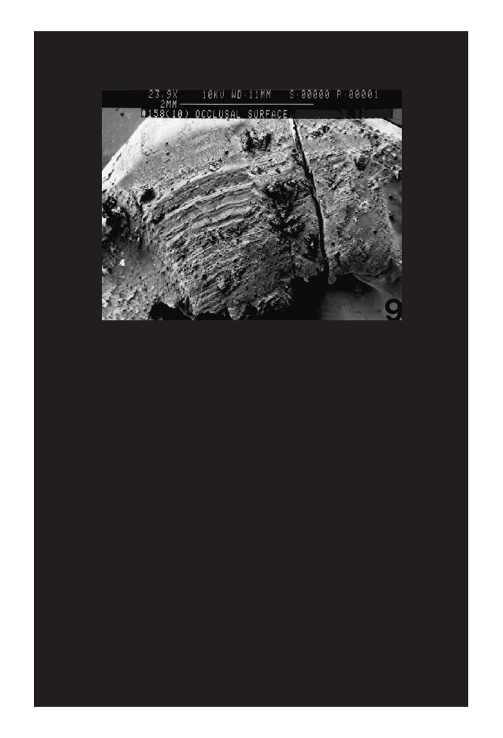
FIGURE 7.5 Scanning electron micrograph of a human and upper right second premolar
with visible drill striations. This evidence of an occlusal filling on this tooth was consistent
with the dental records of a victim of a cremation homicide. (Photo by S. Fairgrieve.)
© 2008 by Taylor & Francis Group, LLC

Incineration of Dental Tissues
155
effects as at 600ºC. The crown was not reduced to fragments until 1000ºC. Larger
fractures through root dentine were formed at 1000ºC and continued to 1100ºC.
Using an identical temperature regime, Savio et al. (2006) examined the effects
of heat on postmortem and antemortem restored teeth having amalgam fillings,
composite resin fillings, and endodontic treatments (treatments of the pulp cham-
ber) (
). They found that composite fillings were in place and maintained
their shape up to 600ºC while amalgam fillings maintained their shape and posi-
tion up to 1000ºC. Endodontic treatments, due to their protected context within the
tooth, were recognizable up to 1100ºC (the upper temperature limit of their study).
At this highest temperature the amalgam fillings demonstrated a partially altered
shape while the composite fillings remained in place despite being altered from a
solid to a fluidic state. The endodontic treatments were slightly altered in all samples
from 400ºC by having less regular radiopacity, radiotransparent areas, and their
shape and dimensions minimally altered. However, a “honeycomb” appearance to
the endodontic treatment became apparent at 600ºC. This is due to the change of the
material to a fluid and/or it boiling as the temperature increased.
It is clear from the foregoing that restorative materials, be they amalgams, com-
posite resins, or even endodontic restorations, will persist and serve as an aid to
both positive identification and an estimate of the temperature attained by the dental
structures in question. In the latter of these studies, Savio et al. (2006) heated their
test teeth in an oven at a rate of 30ºC per minute until the target temperature was
reached and then immediately removed them from the oven. Further experimenta-
tion with heating regimes to mimic different types of fire contents would be useful
given this baseline in the literature. Variation in size of restoration, and position, will
also be needed to fully elucidate the fate of restorative materials in fire contexts.
For now, estimates of temperature are possible. However, the fact that restor-
ative materials are hardier than some may imagine, detailed recovery of dental frag-
ments and their associated restorations is essential.
7.7
Differences Observed in the Radiographic Appearance of Postmortem vs.
Antemortem Unrestored Teeth after the Thermal Stress (drawn from Savio et
al., 2006)
Temperature
Crown
Root
200ºC
No changes
No changes
400ºC
Fissures between enamel and dentine
No changes
600ºC
Fissures between enamel/dentine and within
dentine
Fissures within dentine
800ºC
Fissures between enamel/dentine and within
dentine
Fractures through dentine
1000ºC
Reduced to fragments
Large fractures through the dentine
1100ºC
Reduced to fragments
Large fractures through the dentine
© 2008 by Taylor & Francis Group, LLC

156
Forensic Cremation Recovery and Analysis
7.4 RECOVERY OF DENTAL CREMAINS
As mentioned above, the recovery of dental fragments is an issue that needs to be
addressed. Most forensic identification officers do not have training in the recogni-
tion of bone, cremated or otherwise, let alone expertise in the recognition and recov-
ery of ashed teeth. Mincer et al. (1990) found that 66% of responders to a survey
7.8
Differences Observed in the Radiographic Appearance of the Postmortem vs.
Antemortem Restored Teeth after the Thermal Stress (drawn from Savio et
al., 2006)
Temperatures
Amalgam
Composite
Endodontic treatments
200ºC
No changes in shape and
dimension
No change in shape and
dimension
No changes
400ºC
No changes in shape and
dimension
No change in shape and
dimension
Radiopacity less regular,
presence of
radiotransparent areas,
shape, and dimension
slightly altered
600ºC
No changes in shape and
dimension (even in
samples with
detachment of crown
and fillings)
No change in shape and
dimension
Radiopacity less regular,
presence of many
radiotransparent areas,
“honeycomb” appearance,
shape and dimension
slightly altered
800ºC
Large fissures between
dental tissue and
fillings—no changes in
shape and dimension
(even in samples with
detachment of crown
and fillings)
In place in an altered
shape (state changing)
Radiopacity less regular,
presence of many
radiotransparent areas,
“honeycomb” appearance,
shape and dimension
slightly altered
1000ºC
Large fractures between
dental tissue and
fillings—no changes in
shape and dimension
(even in samples with
detachment of crown
and fillings)
In place in an altered
shape (state changing)
Radiopacity less regular,
presence of many
radiotransparent areas,
“honeycomb” appearance,
shape and dimension
slightly altered
1100ºC
Crowns reduced in
fragments—shape
partially maintained
In place in a remarkably
altered shape (state
changing)
Radiopacity less regular,
presence of many
radiotransparent areas,
“honeycomb” appearance,
shape and dimension
slightly altered
© 2008 by Taylor & Francis Group, LLC

Incineration of Dental Tissues
157
on incinerated teeth considered the fragility of such specimens to be a significant
forensic issue. Other respondents thought that difficulties may be overcome by care-
ful handling, in situ photography, and/or removal of the tongue through the floor
of the mouth, in order to facilitate radiography of the mandibular and maxillary
dentitions. Some of the responders may not have encountered any problem due to
the protection of the premolars and molars afforded by their position in the den-
tal arcade. With the persistence of roots in the alveolus, comparative radiography
would be possible.
The aforementioned comments do seem to indicate that cremains are more typi-
cally found with soft tissue, such as the tongue, demonstrating brief fire incidents.
However, in forensic cremations where the remains have reached Stage 5 of the
Crow–Glassman Scale, dental cremains will be in a highly fragmentary state and
may require some form of consolidation.
Consolidation was found by Mincer et al. (1990) to be beneficial in the recovery
of cremated teeth. Using a series of suggested materials (
Table
7.9) they found that
all were able to increase the physical stability of the teeth in order to preserve their
macroscopic characteristics and features. Overall, the most convenient stabilizer to
use was clear acrylic spray paint. The second of these was cyanoacrylate cement
(Mincer et al., 1990). If the setting time is not an issue, polyvinyl acetate (PVA, or
white school glue) is a good choice as it is also water soluble.
Perpetrators are not above manually destroying dental tissues using imple-
ments before, during, and after the cremation of a body. In fact, commingling of
elements and actively crushing structures are not atypical in these types of scenes.
7.9
Materials Tested for Stabilizing Incinerated Teeth
(drawn from Mincer et al., 1990)
Material
Application
Cyanoacrylate (Superglue®, liquid)
tube/drop
Acrylic spray (Krylon®, clear No. 1303)
spray*
Hair spray (Style Superhold®, unscented)
spray*
Spray varnish (Illinois Bronze®, clear, satin)
spray*
Fingernail polish (Hard as Nails®, clear)
brush
Epoxy cement (Devcon 5-Minute®)
brush
Epoxy cement (Devcon 5-Minute®) 1:1 in acetone
brush
Household cement (Duco®)
brush
Household cement (Duco®) 1:1 in acetone
brush
PVA polymer (Union Carbide®) ~5% in acetone
brush
PVA polymer (Union Carbide®) ~1% in acetone
brush
Acrylic resin (Coe®, orthodontic, self-cure, clear)
brush
*Applied in 3 coats with 3-min. drying between coats.
© 2008 by Taylor & Francis Group, LLC
158
Forensic Cremation Recovery and Analysis
The implication of this is that the recovery of dental cremains need not be confined
to a single area of the scene itself. This poses many challenges in the recovery of
cremated teeth.
Cremated dental remains can be scattered through many layers of a scene, par-
ticularly if obfuscated by the perpetrator. Once the likely boundaries of the human
cremains have been determined, areas peripheral to this region should be cleared.
The region containing the cremains can then be approached systematically pro-
ceeding from the highest to the lowest levels. If the cremains are buried, forensic
archaeological procedures must be followed (see
).
The use of finer brushes to remove ash will be necessary to expose cremated
dental tissues. Alveolar tissue can be extremely fragile and may flake away on con-
tact. Consolidation should be considered at this time.
Individual padded containers for teeth will help to protect them from fragment-
ing during transportation to the laboratory. If one is not confident on how to pro-
ceed, it is best to consult a forensic odontologist or forensic anthropologist.
Once in the laboratory, the sorting and reconstruction of dental cremains can
begin.
7.5 RECONSTRUCTION OF DENTAL CREMAINS
Highly fragmented dental cremains may first be sorted according to the roots that
have been recovered. The conformation of roots may often be fitted with a portion
of alveolus that contains intact or partial sockets (
7.6). As roots are going to
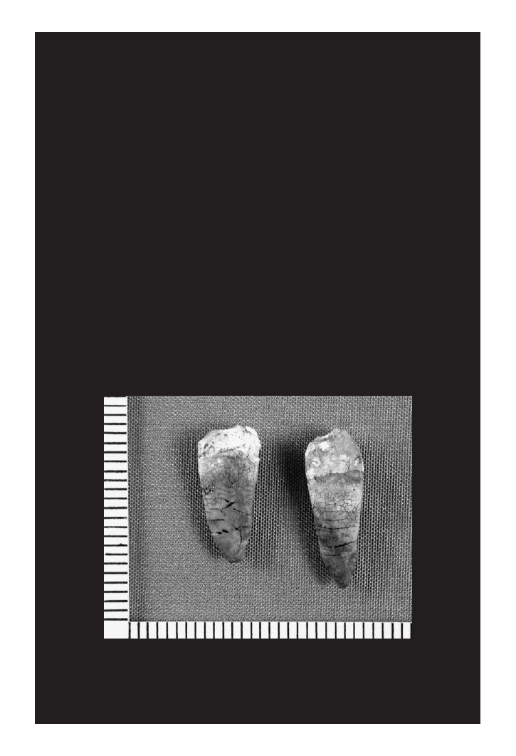
FIGURE 7.6 A pair of cremated tooth roots from the same individual. Roots often possess
dentine that supports the enamel of the crown that has previously separated. (Photo by S.
Fairgrieve.)
© 2008 by Taylor & Francis Group, LLC

Incineration of Dental Tissues
159
be the most complete dental structures to recover, they will likely serve as the best
means of achieving a positive identification (Smith, 1992). Root form and posi-
tion, along with preserved endodontic treatments, are readily visible in antemortem
radiographs, particularly when centered on the periapical area of a root (see
for aspects of positive identification from cremains).
Once the roots have been sorted as to possible tooth type, the crowns are then
sorted in a similar fashion. As the roots will often have the dentine that was deep
to a now separated enamel crown, it may be possible to simply match these to one
another. However, as stated above, the enamel and dentine contract at different rates
and extents when subjected to fire. It is for this reason that the enamel crown will not
be a perfect size match to the dentine. A consistency in fit may be achieved. If dental
restorations are present, or only a fragment of enamel with evidence of a previously
adherent restoration, they may be matched for consistency with antemortem dental
records once the position on the dental arch has been considered.
Reconstruction of enamel is possible with collected fragments. This is particu-
larly important to do for the anterior dentition as the form of these teeth may be
compared to antemortem photographs exhibiting the incisors and canines of the
victims. This reconstruction may be undertaken using any sort of adhesive. The use
of PVA glue will make it possible to separate fragments should the need arise.
7.6 SUMMARY
The incineration of dental tissues initially presents a daunting challenge for recovery
and analysis in those cases in which a body has been subjected to high heat for a pro-
longed period. Nonetheless, dental cremains are recoverable and may be utilized in
estimating temperature, and in the process of establishing a positive identification.
Ultimately, dental cremains will play an important role in the identification process,
as DNA analysis may not be possible in these cases.
With careful attention at the scene and thoughtful reconstruction, dental cre-
mains may also provide evidence of a thorough recovery strategy. This will lend
further credibility of the analysis in the minds of a judge and jury.
© 2008 by Taylor & Francis Group, LLC
Document Outline
- Table of Contents
- Chapter 7: Incineration of Dental Tissues
Wyszukiwarka
Podobne podstrony:
9189ch5
9189ch8
9189ch1
9189ch4
9189ch6
9189ch3
9189ch2
9189ch5
9189ch8
więcej podobnych podstron