
161
8
Positive Identification
of Cremains
8.1 INTRODUCTION
The establishment of identity is one of the most significant aspects of a forensic
investigation. The identity of remains, cremated or otherwise, permits authorities
to begin tracing a series of events that led to the demise of the identified victim. If
the remains are forensically relevant, the identity allows the authorities to contact
family, friends, co-workers, and even casual acquaintances and contacts about the
actions of a decedent, and who may have been associated with those actions. Hence,
identity is a pivotal aspect in the analysis of human remains.
The means of establishing a positive identification (i.e., an identification that
has been proven beyond any reasonable doubt and to the exclusion of all other pos-
sibilities) has changed significantly with advances in science and technology. In the
latter nineteenth century, Bertillon had introduced a system of measurements (or
anthropometry) of the body with full-length and profile photographs accompanied
by a detailed description, in order to confirm an individual’s identity if they were
subsequently arrested or their remains were recovered in some context (Saferstein,
2007). The premise of using anthropometry rested on the assumption that mea-
surements of the skeletal structure of an individual would remain the same from
postadolescence to death. Although this statement may have a degree of validity to
it, Bertillon’s system was somewhat inconvenient due to the act of measuring and
storing the various types of data collected. This system was only in use for 20 years
or so when it was replaced by a new system of identification based on the friction
ridge patterns found on digits (a.k.a. fingerprints).
Fingerprints revolutionized the field of human identification like no other sys-
tem. Fingerprints are relatively easy to record and store. They also provide a degree
of uniqueness that has been accepted by the courts for nearly a century.
More recently, the second revolution in human identification has been in the sci-
ence of genetics. The direct analysis of DNA and its bases, the fundamental source
of variation in all living things, is now the gold standard of forensic identification.
The use of fingerprints, DNA, and even Bertillon’s system are all dependent
upon finding and utilizing antemortem data from individuals who are candidates for
identification. The advantage of DNA is that known blood relatives may also act as a
source of known DNA to compare to the DNA from an unidentified individual. The
other obvious drawback is that the source of data for comparison is derived from
soft tissue. When it comes to positive identification from hard tissues alone, it can
become somewhat more challenging.
© 2008 by Taylor & Francis Group, LLC

162
Forensic Cremation Recovery and Analysis
It is true that forensic scientists do not have the luxury of choosing the quality
of the specimens that they will use in their analysis. Information and specimens
from the scene usually have limitations in completeness or preservational quality.
fire is a notoriously efficient mechanism of destroying biological specimens that
are depended upon for traditional means of establishing a positive identification.
The one advantage of hard tissues is that they do persist in fire, although to varying
degrees. In victims who have undergone some form of decomposition to the extent
that facial features are not sufficient to allow a visual identification, the use of sur-
viving dental work or DNA is considered.
Of course, modern methods of positive identification are not restricted solely
to those methods mentioned above. Additional methods under research or used to
assist in the identification process include stable isotope ratios, ear prints, soft and
hard tissue pathology, surgical procedures and implants, radiographic comparisons,
facial imagery analysis, iris patterns (in living individuals), footwear patterns, body
modifications, and biometrics, including retinal comparisons (for a review, see
, 2007). However, it is fair to say that those methods that are
based on soft tissue structures will not be applicable to cremated remains, especially
those that have reached Stage V of the Crow–Glassman Scale.
This chapter presents the issues of establishing a positive identification from
charred and cremated remains. The ability to apply these various methods is entirely
dependent upon the condition of the remains and the availability/suitability of ante-
mortem records.
The most common of these methods is based on dental comparisons. How-
ever, as discussed in the previous chapter, dental cremains present their own set of
difficulties. Other comparisons use osteological structures, as seen in antemortem
radiographs as a basis for identification. One of the most commonly applied of these
is the use of frontal sinuses. Other structures such as palatal rugae morphology
recorded through orthodontic casts may be of use as well.
The reconstruction of cremains from fragments (see
) has been the
basis of establishing morphological characteristics of remains in support of de facto
identifications. Some of these characteristics may include evidence of antemortem
trauma.
Finally, the application of genetic analysis to DNA recovered from cremains has
become increasingly important in the identification of charred bodies. There have
also been attempts to obtain analyzable DNA from dental remains.
Ultimately, attempts to identify human cremains can take an analyst down many
analytical pathways. It is the discretion of the analyst and their own experience with
cremains that will dictate how this process is undertaken.
8.2 RADIOGRAPHIC COMPARISONS
8.2.1 I
NTRODUCTION
The utilization of antemortem radiographs necessitates that the cremains be care-
fully collected from the scene, cleaned ultrasonically, and then reconstructed to a
level permitted by the recovered fragments (Grévin et al., 1998). This may prove to
© 2008 by Taylor & Francis Group, LLC

Positive Identification of Cremains
163
be more difficult than it sounds due to the level of fragmentation that can occur (see
). However, reconstruction is the first step in the identification process of
cremains.
The comparison of antemortem and postmortem radiographs is done to exam-
ine, in detail, points of structural congruity between the images being compared.
A reconstruction of as much of the skeleton as possible is essential for this pro-
cess to have any validity. Assuming that an analysis of age, sex, and ancestry have
permitted the generation of a list of candidates for identification, reconstruction
of cremains may prove to be of sufficient quality to lend itself to a comparison of
radiographs.
The most common radiographic comparison used for identification would be
dental records. However, as dental identifications have many considerations that
need to be examined in greater detail, those details may be found in Section 8.3. The
next most common type of radiographic comparison is through structurally conser-
vative features. Such features must have a lasting quality with slow to negligible
alterations through time. In other words, these features need to last and not undergo
structural changes that will render them useless for comparison purposes.
The simple fact of the matter is that the skeletal structures of the body are
physiologically active. As such, bone has a rate of turnover, albeit an uneven one,
throughout the entire skeleton. The question is whether or not this turnover of bone
is going to significantly alter any of the structures that will be of interest to us for
making a comparison. This means that the age of the individual will have an effect
on our ability to identify cremains by this method. The most obvious individuals
who would not be good candidates for radiographic comparison would be preado-
lescent children. Children’s skeletal structures are not only less well ossified than
those of adults, but are also still very much in the process of developing the very
structures that we would evaluate in adults. For example, frontal sinuses (see below)
are not present in infants and young children. However, with growth, paranasal
sinuses are being formed into the adolescent stage and beyond (Williams et al.,
1989). Another area of consideration is the comparison of the trabecular structure
orientation found within vertebral centra. Again, postadolescent individuals who
have had radiographs of the spinal column may have these used as a basis of com-
parison if the corresponding area of the skeleton is recovered.
Regardless of the area, the comparison of skeletal structures may be used in the
identification process. However, the strength of such an evaluation must be consid-
ered in light of other studies of variation in the structures being examined, and the
extent to which those structures have been preserved.
8.2.2 F
RONTAL
S
INUS
C
OMPARISON
One of the earliest notations of the potential of frontal sinus radiographs as a means
of establishing an identification was by Schuller (1943). Recognizing the potential of
such a system, Ubelaker (1984) applied the comparison of frontal sinuses from found
human remains to antemortem radiographs of a suspected identification. A match
was noted in this case. Part of Ubelaker’s study was to examine the frontal sinus
patterns of skulls from the Terry Collection at the Smithsonian. This study failed
© 2008 by Taylor & Francis Group, LLC
164
Forensic Cremation Recovery and Analysis
to find any other frontal sinus patterns that matched that of the identified remains.
Other studies have examined the use of frontal sinuses as a means of identifying
remains (e.g., Kirk et al., 2002; Harris et al., 1987a; Marlin et al., 1991; Harris et al.,
1987b; Yoshino et al., 1987). More recently, the use of frontal sinuses as a means
of identification and, subsequently, testifying to the validity of this methodology in
court has come under scrutiny (Christensen, 2004). Under the current U.S. Federal
Rules of Evidence, there is concern that the methodology used by forensic anthro-
pologists, pathologists, and radiologists, in applying frontal sinus comparison, has
not sufficiently established the uniqueness of the patterns to establish identity. The
reason for this concern is that there is a lack of standardized methods. Without a
standardized methodology it is difficult for us to apply the results in a reproducible
manner such as found in other sciences as chemistry and toxicology. However, this
will only become an issue in the courts if identifications are challenged and the
forensic anthropologist must demonstrate how the identity was determined with this
methodology. In the meantime, the examination of frontal sinuses will continue to
provide evidence of identity in cases where it is unchallenged.
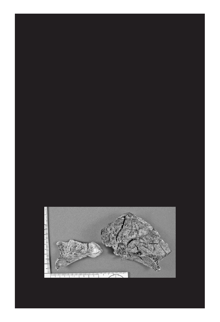
The frontal sinuses are often exposed in Stage V cremains (
8.1). In fact,
the recovery of the entire frontal bone may not be possible. Hence, only a portion
of the frontal sinuses may be present for comparison. The same may be said of the
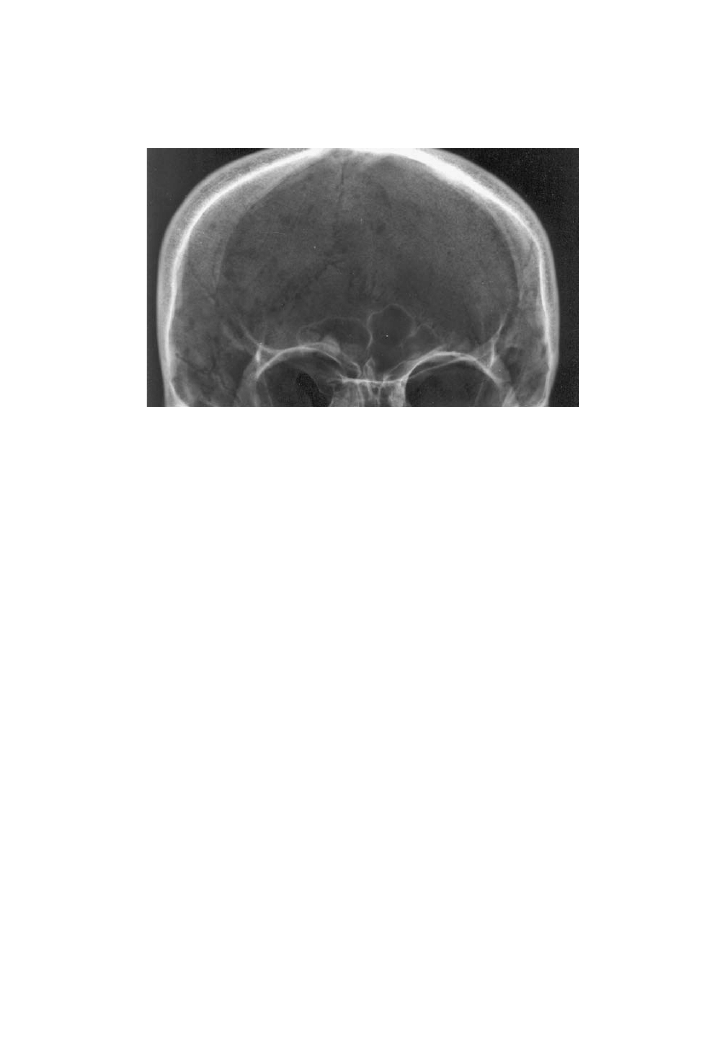
antemortem radiographs that are available for comparison. A complete P–A view
(posterior–anterior view) of the skull in which the skull was placed with the fore-
head placed directly onto the radiographic cassette will typically provide a suitable
image for comparison (
). However, in some cases, the antemortem radio-
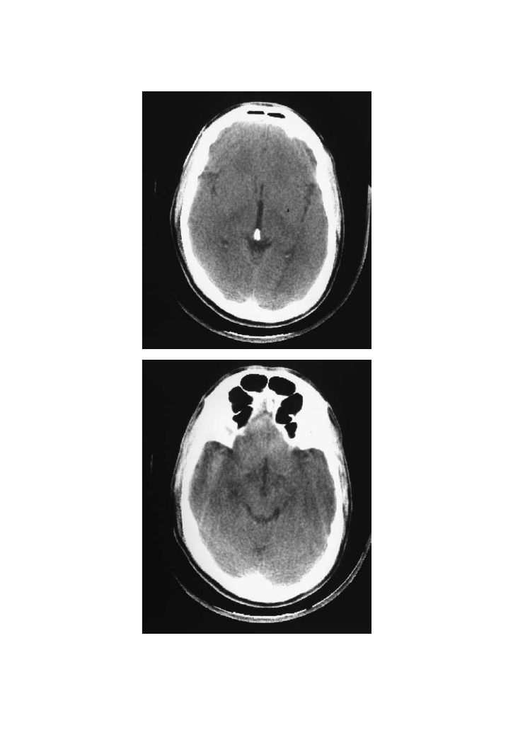
graphs are in the form of CT-scan images (
). If the scanned images are
available in electronic format, they may be compiled in order to enhance the area of
the frontal sinuses for comparison. Likewise, a CT scan of the reconstructed frontal
bone (or fragment) may yield images of the frontal sinuses from the unidentified cre-
mains. CT-scan images have nonetheless been used to confirm human identification
(Haglund and Fligner, 1993; Tatlisumak et al., 2007).
FIGURE 8.1 Partial frontal sinuses are evident in this fragmentary frontal bone from a
cremation homicide victim. (Photo by S. Fairgrieve.)
© 2008 by Taylor & Francis Group, LLC
Positive Identification of Cremains
165
Other complicating factors to the evaluation of frontal sinuses in complete skulls
would include the positioning of postmortem radiography of the skull to mimic the
antemortem position (Kirk et al., 2002). Likewise, the position of the head of the
living patient may not be at an optimal angle for the purposes of comparison, but
be of sufficient exposure for diagnostic purposes. One would then have to try to
mimic the same position and orientation if measured comparisons are to be made.
Therefore, measurements made on the antemortem and postmortem radiographs for
purposes of comparison may produce a degree of error. This makes inherent sense,
as a radiograph is a two-dimensional representation of a three-dimensional object.
Those areas deep to the glabellar portion of the frontal bone will tend to have the
clearest margins, as they are in a plane that is approximately parallel to the X-ray
cassette. The lateral margins of the frontal sinuses will tend to lack clarity if they
extend to the curvature of the frontal bone away from the cassette.
A caveat to keep in mind with cremated remains is that the frontal sinuses are
in a position that is extremely close to the surface of the skull and lacks the protec-
tion of a thick layer of tissue to protect that area from the heat of cremation. The
skull is one of the most vulnerable areas of the body and is often the first to undergo
heat-induced fractures (see
). It is for this reason that caution must be
exercised, as there are no studies of the postcremation form of the frontal sinuses
and their utility in identification.
The presence of unusual morphological features that may contribute to the
uniqueness of the individual should also be considered in an assessment. Owsley
found this to be the case in examining cremains with a fragmentary frontal bone
(Owsley, 1993). In this case, an asymmetrical frontal sinus and a well-defined sul-
cus were evident on the antemortem radiographs and the fragmented frontal bone.
However, if challenged, one must be prepared to provide other evidence that would
support the identification.
FIGURE 8.2 A P.A. radiograph of a human skull demonstrating a clear outline of the fron-
tal sinuses. (Photo by S. Fairgrieve.)
© 2008 by Taylor & Francis Group, LLC
166
Forensic Cremation Recovery and Analysis
FIGURE 8.3 These CT-scan images are the only antemortem record of the frontal sinuses
in
. Unfortunately, these images were not saved electronically with other views.
(Photos by S. Fairgrieve.)
© 2008 by Taylor & Francis Group, LLC

Positive Identification of Cremains
167
If there is sufficient material to reconstruct the frontal bone, and hence the fron-
tal sinuses, it is recommended that a comparison is made of ante- and postmortem
radiographs of this area. However, due to the lack of sufficient scientific examina-
tion of this technique on cremains, it is recommended that this type of examination
be used to assist in confirming, or at least failing to exclude, the individual in ques-
tion as the identity of the cremains.
8.2.3 O
THER
R
ADIOGRAPHIC
C
OMPARISONS
Certainly, other areas of the body that have been radiographed have been used as a
basis for identification. The overall form and radiographic features have served as
points of comparison to at least note structural similarities. Ubelaker (1990) uti-
lized the axillary (or lateral) border of a recovered human scapula and its distinctive
morphology as the basis for identification with the antemortem radiograph show-
ing the same characteristics. The argument justifying the use of these features was
that they were compared to 100 right scapulae from the Smithsonian’s Huntington
Collection and 100 right scapulae from skeletal collections of American Indians in
the Smithsonian. Because of the uniqueness of the axillary border of the recovered
skeleton, when compared to the 200 scapulae cited above, Ubelaker considered the
“well-developed extended notch” to be sufficient for identification in this case.
The above case was done on a set of human remains that were subjected to car-
nivore scavenging, but not cremation. The effects of cremation on skeletal features
have been documented in
and
. There is no way of knowing
if the act of cremation has sufficiently altered the morphology of bone such that a
comparison to antemortem radiographs is without justification.
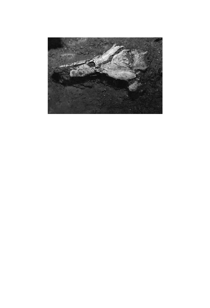
Anatomical features particular to a specific bone are most certainly preserved
, the distal end of a humerus from a homicide
cremation victim was found to have a preserved septal aperture. This feature, in
life, permits the person to hyperextend their elbow joint. If the decedent is known to
have been able to perform such an action, then this would be further congruence of
the presumed identity, but not sufficient for identity.
Further investigation of anatomical features and their recognition after crema-
tion is certainly recommended. A recent study by Muthusubramanian et al. (2005)
of the palatal rugae of burn victims was compared to cadavers. However, this study
did not extend to taking the cadavers to Stage V cremains. Superficial burning that
does not affect the bone directly is of interest to cases of identification in which the
charred remains are at Stage III and lower.
Identification from any area of the body for which there are any antemortem
radiographs should certainly be attempted. However, the question of a particular
feature’s or set of features’ validity for use as a means of individuating remains must
be approached through further study of documented collections. Again, without the
scientific studies, the analyst may, at best, fail to reject an identification rather than
state that a positive identification has been established.
Trabecular pattern recognition from antemortem radiographs has been recog-
nized as a means of identification. For example, the trabecular pattern of the distal
femur and the proximal tibia (the knee joint) has been found to be distinctive (Mann,
© 2008 by Taylor & Francis Group, LLC
168
Forensic Cremation Recovery and Analysis
1998). In fact, a minimum of four distinct “osseous features” were found by Mann
(1998) to be reliable indicators for identity from that area of the skeleton.
In a recent study to validate the use of antemortem and postmortem radiographs
of the hand as a basis for identification, the authors found that it is indeed a valid
method for identification (Koot et al., 2005). Further, the use of the hand has the
potential of employing features present in 27 bones. A significant feature of this
study is that it compared “fleshed” cadaver hands that were radiographed, and sub-
sequently removed and defleshed to simulate decomposition. This fact coupled with
a test of the reliability for identification by other forensic anthropologists satisfies
the Daubert guidelines. This is a further enhancement to the use of trabeculae in
identification. Certainly, this method holds much promise. However, given the level
of damage (shrinkage, warping, and cracking) that goes on in a cremation, care
would have to be exercised in applying such a methodology to cremains.
Trabecular densitometry has been undertaken using image analysis software as
a basis of comparison for antemortem and postmortem radiographs (Kahana and
Hiss, 1994) and radiographs of the wrist in living subjects taken years apart (Kah-
ana et al., 1998). As these methods and direct comparisons of trabecular patterns
have been successfully developed and used on noncremated remains, the question is
how differing levels of cremation will affect these methodologies.
Other anatomical features such as mastoid sinus and meningeal artery patterns
(Rhine and Sperry, 1991), vertebral body and spinous process form (Mundorff et al.,
2006), and even pathological conditions affecting the morphology of osteological
structures (Sudimack et al., 2002; Hulewicz and Wilcher, 2003) have been used to
confirm identification.
Features such as those listed above have been applied in cases that have yielded
human remains that have been extensively fragmented and been bent and warped
through taphonomic processes (Owsley et al., 1993). A case in which human
FIGURE 8.4 The cremated remains of a distal right humerus in situ. A septal aperture
joining the olecranon fossa with the coronoid fossa would have permitted hyperextension of
the elbow joint in life. (By permission, Regional Supervising Coroner, Northern Ontario.)
© 2008 by Taylor & Francis Group, LLC

Positive Identification of Cremains
169
remains were purposefully rendered down to such a condition by Jeffrey Dahmer,
demonstrated that a positive identification can be made even from remains that are
in a condition similar to that of cremains (Owsley et al., 1993).
In one extreme example, cremated and commingled human remains eventually
resulted in positive identifications based on a comparison of antemortem and post-
mortem radiographs in addition to medical, dental, and other background records
(Owsley et al., 1995). In this incident, members of the Branch Davidian sect had
secluded themselves in Mount Carmel, Texas, and were eventually cremated in a
fire on that property. It was demonstrated that there were many points of concor-
dance in the comparison of premortem and postmortem radiographs for individual
MC27. Hence, the process of cremating remains did preserve enough detail of osteo-
logical features for identification. However, it is worth noting that apparently not all
the regions of the remains from the Branch Davidian compound were reduced to
calcinated bone. Some soft tissue, albeit charred, did survive; however, crania and
other areas of the body susceptible to cremation did indeed reach Crow–Glassman
Stage V.
Finally, the use of cranial suture pattern matching from radiographs has been
examined from the perspective of Daubert criteria (U.S.A.) and the Mohan ruling
(Canada) (Rogers and Allard, 2004). It has yet to be explored in cases of cremated
cranial fragments. However, cranial fragments with sutures are typically salvaged
from cremations. As lambdoidal sutures are associated with the occipital bone it is
more likely to be preserved due to the thickness of that region of the vault.
8.3 DENTAL IDENTIFICATION
The use of dental tissues as a means of establishing a positive identification is a
common enough practice that there are full-time forensic odontologists. Although
forensic odontologists are involved with examining dental structures in deceased
individuals, they are also involved with assessing injuries to teeth, jaws, and oral
tissues in living victims (Hardy, 2007). In the case of cremation analysis, forensic
odontologists will work closely with forensic anthropologists, as the latter will have
been involved with the recovery and curation of dental cremains.
A forensic odontological examination of dental features used as a basis of com-
parison comprises a substantial list of features, many of which are based on the
assessment of soft tissues (for a review, see
, 2007). Yet, in the
context of forensic cremations, many of these features are either lost or significantly
altered. Those features that are more likely to be encountered in Stage V cremains
are summarized in
. Although this table would seem to indicate that there
is a plethora of details that may provide a basis for identification, in practice, many
of these features are obscured or eradicated in the cremation. It is nonetheless pos-
sible for cremated dental tissues to exhibit some of these features.
The actual analysis of dental cremains will follow a specific set of procedures
that will facilitate identification. The following section details those procedures and
the dental tissues to which they apply.
© 2008 by Taylor & Francis Group, LLC

170
Forensic Cremation Recovery and Analysis
8.1
Tooth and Periodontal Features Typically Encountered in Human Cremains
(drawn from
10.1 in Hardy, 2007)
Teeth
Teeth present
a. Erupted
b. Unerupted
c. Impacted
Missing teeth (based on sockets)
a. Congenitally
b. Lost antemortem
c. Lost postmortem
Tooth type
a. Permanent
b. Deciduous
c. Mixed
d. Retained primary
e. Supernumerary
Tooth position
a. Malposition
Crown morphology
a. Size and shape
b. Enamel thickness
c. Contact points
d. Racial variation
Crown pathology
a. Caries
b. Attrition, abrasion, erosion
c. Atypical variations, enamel pearls, peg lateral, etc.
Root morphology
a. Size
b. Shape
c. Number
d. Divergence of roots
Root pathology
a. Dilaceration
b. Root fracture
c. Hypercementosis
d. Root resorption
e. Root hemisections
(Continued)
© 2008 by Taylor & Francis Group, LLC

Positive Identification of Cremains
171
Pulp chamber/root canal morphology
a. Size, shape, and number
b. Secondary dentine
Pulp chamber/root canal pathology
a. Pulp stones, dystrophic calcification
b. Root canal therapy
c. Retrofills
d. Apicectomy
Periapical pathology
a. Abscess, granuloma, or cysts
b. Cementomas
c. Condensing osteitis
Dental restorations
1. Metallic
a. Nonfull coverage
b. Full coverage
2. Nonmetallic
a. Nonfull coverage
b. Laminates
c. Full Coverage
3. Dental implants
4. Bridges
5. Partial and full removable prosthesis
Periodontal Tissues
Alveolar process and lamina dura
a. Trabecular bone pattern and bone islands
b. Residual root fragments
Maxillary sinus
a. Size and shape
b. Relationship to teeth
Anterior nasal spine
a. Incisive canal (shape) (likely damaged)
b. Median palatal suture
Mandibular canal
a. Mental foramen
b. Diameter, anomalous
c. Relationship to adjacent structures
(Continued)
TABLE 8.1
(Continued)
© 2008 by Taylor & Francis Group, LLC

172
Forensic Cremation Recovery and Analysis
8.3.1 T
OOTH
R
OOT
A
NALYSIS
Dental crowns, as established in the previous chapter, are quite commonly frac-
tured. Although these crowns may be reconstructed, if recovered, they may not be
complete due to the absence of dental restorative materials.
Tooth roots are more commonly preserved intact due to the protected nature of
being situated in an alveolus. Roots may also be in poor condition due to the actions
of the fire context or the actions of a perpetrator. Root repairs may also be under-
taken if the roots have only been partially recovered. Single rooted teeth tend to have
well-preserved roots, as they will drop out of the alveolus more readily. Although
this would seem to make them more vulnerable, it has been my experience with
cremation homicides in which the perpetrator fragments the cremains, that single
tooth roots are reasonably intact. However, where a tooth has multiple roots, each
one of these roots tends to be broken off from the rest of the tooth. All roots must be
collected and an attempt be made to match these with broken root fragments.
Tooth roots may also be retained in the alveolus of the mandible or maxilla. In
this case, it will be desirable to radiograph such a fragment for direct comparison
to antemortem radiographs. Should the socket be intact and the root of the tooth be
missing, an alternative would be to use a method such as that suggested by Smith
(1992). This method requires the analyst to place inside the tooth socket a radi-
opaque substance, such as barium sulfate, in order for a radiograph to be taken,
simulating the presence of the tooth root.
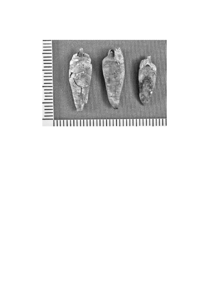
Tooth roots may also preserve evidence of antemortem dental work.
demonstrates some tooth roots from a cremation homicide in which a post has been
preserved. These may then be compared to dental records for concordance. Other
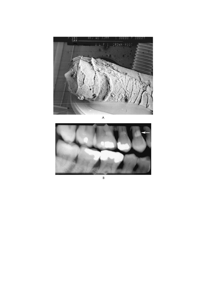
evidence of dental restoration that may be helpful would include discontinuities in
the surface adjacent to, or part of, the cemento-enamel junction (CEJ). Such areas
may be the remnants of fillings that are no longer present.
demonstrates
a tooth from a cremation homicide with a crescent-shaped concavity that conforms
to a resin-based composite filling at the CEJ. The location of that filling on an ante-
Coronoid and condylar process
a. Size and shape
b. Pathology
Temporomandibular joint
a. Size, shape
b. Hypertrophy/atrophy
c. Ankylosis, fracture
d. Arthritic changes
Other pathologies
a. Evidence of surgery
b. Trauma – perimortem or antemortem (wires, pins, and other implanted materials)
TABLE 8.1
(Continued)
© 2008 by Taylor & Francis Group, LLC
Positive Identification of Cremains
173
mortem radiograph is demonstrated in
. Confirmation of evidence of
dental work using a scanning electron micrograph (SEM) is recommended in these
situations (Fairgrieve, 1994).
8.3.2 C
ROWN
A
NALYSIS
Although the challenges of crown repair have been outlined above, the crown frag-
ments may be key indicators of antemortem dental restorations.
The most common form of dental restoration to the crown is a filling. As outlined
in the previous chapter, dental fillings may range from a metal amalgam of varying
composition to composite resins. Regardless of the type of filling material, the cari-
ous lesions to be treated must be prepared in order to receive the restorative agent.
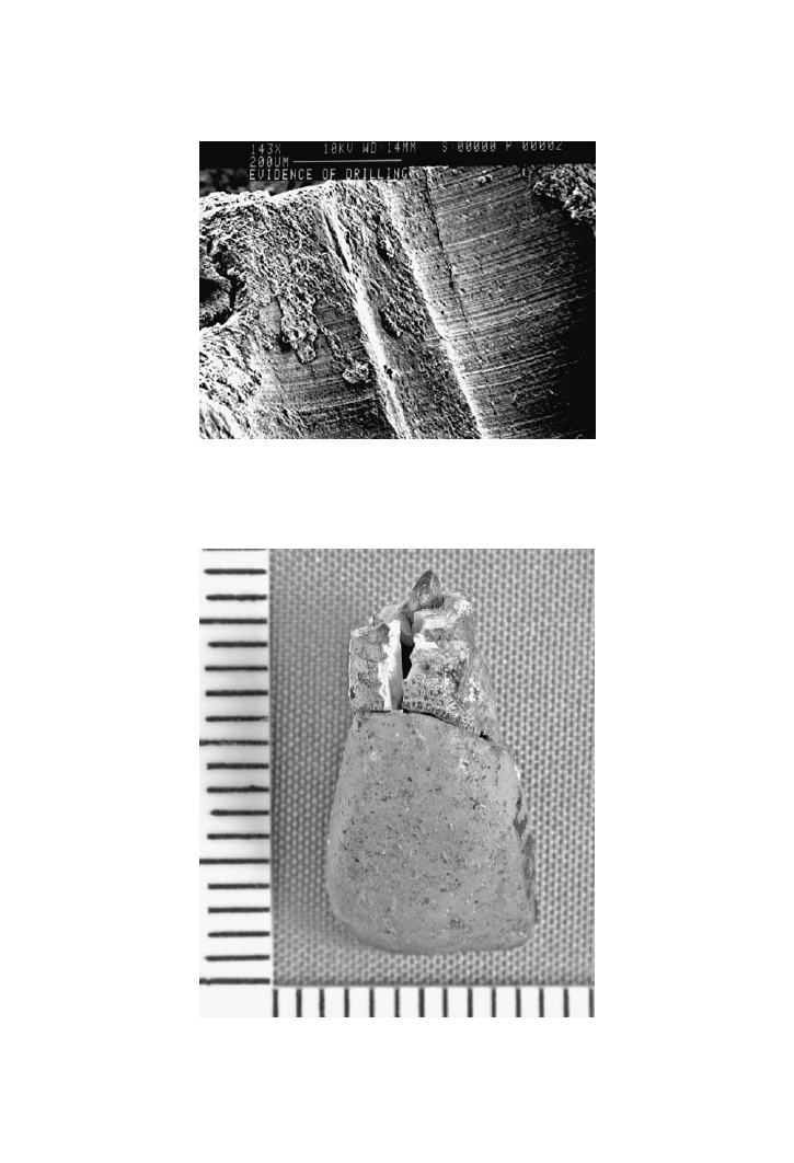
The preparation of a carious lesion is undertaken by drilling directly inside
the lesion and creating a chamber that will accommodate the restorative material.
The action of drilling directly inside the lesion and creating a chamber that will be
filled by the restorative material will leave behind a physical signature of this action
directly on the enamel. This signature is in the form of bore marks and striations.
The head of the drill, as it proceeds down into the enamel, will leave a well-defined
cylindrical mark. As several passes of the drill may be needed to clean out the lesion,
the bore marks will overlap. Examination of the inside of one of these bore marks
will yield evidence of horizontal, parallel striations from the rotational action of the
drill-head. These can be easily distinguished using an SEM or even a light micro-
scope at 40X magnification. The examination of a cremated tooth fragment will
show evidence of these indicators even under low magnification (see
).
In particularly hot fires, the dental crown will fragment into small pieces. It
is unusual to find a crown, not only in one piece, but sill attached to a tooth root.
FIGURE 8.5 Dental posts are often preserved in cremated tooth roots such as these from a
cremation homicide. (Photo by S. Fairgrieve.)
© 2008 by Taylor & Francis Group, LLC
174
Forensic Cremation Recovery and Analysis
is a lingual view of an upper incisor from a cremation homicide victim.
Note that the area of the crown adjacent to the CEJ has a metallic look to it and also
exhibits a gap between the crown and the root. This is an artificial crown that has
survived the cremation process and remained affixed to the underlying root post.
Although the surface of the crown has some persistent adherent material, the form
of the crown has been perfectly maintained.
The fragmentation of the crown, as described above, also means that resins will
be present amongst the detritus of the cremation. Bush and colleagues (2006), rec-
ognizing this fact, subjected various restorative resins to incineration and compared
the pre- and postincineration energy dispersive x-ray spectroscopy (EDS) elemental
analysis. They found that the elemental analysis is not only unique to the manufac-
turer of the resin, but, it was also “almost unchanged” after incineration. This is yet
another factor that may be used to assist in the identification process. It is their feel-
ing that the wear, alignment, and combination of missing, restored, and unrestored
FIGURE 8.6 A) The crescent shape concavity at the CEJ of this tooth is indicative of a
filling in this position. B) The corresponding location is demonstrated by the arrow as the
confirmed location of such a filling. (Photos by S. Fairgrieve)
© 2008 by Taylor & Francis Group, LLC
Positive Identification of Cremains
175
FIGURE 8.8 An upper first incisor with an artificial crown mounted on a post in the root.
FIGURE 8.7 An SEM of a first molar demonstrating vertical bore marks and striation in a
uncremated tooth. (Photo by S. Fairgrieve.)
© 2008 by Taylor & Francis Group, LLC

176
Forensic Cremation Recovery and Analysis
teeth are as unique as a fingerprint. The chemical signature of restorative materials
is surely another factor to add to the above suite of traits.
In the case of remains that have been charred, but not necessarily to Crow–
Glassman Stage V, the procedures followed are outlined in
8.2 (from Delattre,
2000). However, in the case of forensic cremains, removal of soft tissue and resec-
tion are not necessarily steps that will need to be taken.
8.4 DNA AND IDENTIFICATION
The goal of DNA analysis is to differentiate between individuals who have 99.5%
of their DNA in common. The fact that a differentiation can be made is testimony
to the amount of DNA a person has in a single cell. Hence, only a small portion of
DNA is going to exhibit any variability that forensic geneticists will exploit in dif-
ferentiating between individuals.
The variability of DNA is in the form of short tandem repeats (STRs). The STR
is typically characterized by having a short sequence of base pairs (four in most
cases) that repeat a variable number of times. A specific locus may have a variety
of possible alleles (segments that differ in the number of repeats). These segments
of DNA with repeating short sequences are bounded by nonrepeating sequences
known as flanking DNA.
As the method of analysis depends on analyzing the occurrence of various alleles
at specific loci that are relatively short, such a sample is not only easy to characterize
and interpret, but it is also ideal for profiling samples of poor quality, as is often the
case with forensic contexts. Amongst these contexts is the cremation scenario.
The source of DNA in unburned human remains usually comes from muscle
tissue, skin, hair, biological fluids, and even bones and teeth. In forensic cremations,
this will leave us with bones and teeth as a source of DNA. The extraction of DNA
from hard tissues has been studied in forensic and archaeological contexts (e.g.,
Alonso et al., 2001; Pääbo, 1989; Pääbo et al., 1989; Hagelberg et al., 1991; Jeffreys
et al., 1992; Hummel et al., 1999).
8.2
Description of the Steps Included in the Four Stages of Examination and
Documentation of the Charred Dentition (drawn from Delattre, 2000)
Stage I:
Noninvasive extra-oral visual examination
Preliminary charting of visible teeth
Extra-oral photography
Stage II:
Soft tissue removal for direct visualization of dentition
Continue documenting and photographing the dentition
Attempt to pry open the jaws for intra-oral access; if successful go to Stage III
Stage III:
Mandibular/maxillary resection
Radiographs and more photographs as needed
Loose, individual teeth should be identified and radiographed
Stage IV:
Place all loose dentition and resected items in a labeled container
© 2008 by Taylor & Francis Group, LLC

Positive Identification of Cremains
177
8.4.1 DNA
FROM
C
REMAINS
It has only been recently that the technology for isolating, detecting and quantifying
DNA from burned human remains has been pursued (for a review, see
et al., 2004).
The extent of the cremation is a major determining factor in the success of
isolating and amplifying DNA. In house fires, an examination of a scene can yield
cremated bone to the calcine stage as well as some charred soft tissue, as related
in some reports (e.g., Wickenheiser et al., 1999). If there is surviving fibrous mus-
cle tissue and cartilaginous material, then there is a strong possibility of obtaining
DNA. The use of surviving soft tissue from charred bodies is more commonly used
for DNA identification (Barbaro et al., 2003).
The elimination of all soft tissue during the cremation process or soft tissue
charred to the point of being of no value for DNA extraction will require us to con-
sider the remaining bones and teeth as a potential source of DNA. Because bone
houses the marrow, it may act as an insulator, for a time, from the severe heat of a
fire. Hence, attempts at extracting DNA from the innermost regions of a bone may
hold some promise (Staiti et al., 2004). As would be expected, larger bones found
deep inside soft tissues will be better candidates for such extractions. Therefore,
based on the model of cremation presented in
, the bones of the lower axial
skeleton, such as the lumbar vertebrae, are ideal candidates to test. Additionally,
parts of the ilium, ischium, and os pubic may also be worth considering. In general,
bones closer to the surface and those in the appendicular skeleton are less likely to
yield usable DNA.
In extreme cases of forensic cremations, the perpetrator has managed to elimi-
nate the body of all adherent soft tissue. The most extreme cremation of a body
takes place in commercial cremations. Once the body has been reduced to calcined
bone the fragments are then ground in a mill. In one test of such remains, the DNA
extracted prior to cremation was compared with that extracted from commercially
prepared cremains (von Wurmb-Schwark et al., 2004). It should not be too surpris-
ing that the postcremation DNA did not conform to that of the precremation DNA
that was profiled using STRs. The postcremation samples were likely contaminated
through processing and handling. To this point in time, DNA extraction from cre-
mains from commercial crematoria is unreliable at best.
This only leaves us with DNA from teeth as a possible means of identification.
It makes sense that the tooth pulp chamber of a tooth would be a highly protected
environment and, hence, a possible source of DNA (Duffy, 1989; Sweet and Sweet,
1995). Sweet and Sweet (1995) used the dental pulp from incinerated human remains
in order to generate a DNA profile. Although the soft tissue was degraded to the
point of being considered useless for DNA extraction, the dentition was preserved
and in the alveolus. Further, the teeth were not shattered and the roots appeared to be
intact. In particular, a third molar that was as yet unerupted was extracted for DNA
analysis. Teeth still situated within the alveolus and unerupted would be the most
ideal candidates. In general, teeth that are in the anterior dentition are more likely to
be affected by heat stress than those located distally on the dental arch. Molars are
certainly preferable, due to their overall size and thickness of dental tissues.
© 2008 by Taylor & Francis Group, LLC

178
Forensic Cremation Recovery and Analysis
The use of DNA from incinerated deciduous dentition as a means of sexing
cremains was pioneered by Williams et al. (2004). They found that they were able
to isolate and analyze DNA, specifically the amelogenin locus, to determine sex.
Deciduous teeth were subjected to temperatures from 100–500ºC for 15 minutes.
Although not always successful, some teeth that were heated to 400ºC were able to
have their DNA profiled.
The insulative properties of teeth were demonstrated by Duffy et al. (1991) in
their study of fleshed pigs’ heads subjected to an open fire. A fire temperature of
500–700ºC produced a temperature of only 75ºC in the pulp chamber of the pigs’
teeth. It was also found that intact nuclei were found in unextracted teeth in fleshed
jaws of pigs subjected to 300ºC for over an hour. As encouraging as this study
may be, forensic cremations with dental remains that are calcined, and exhibit heat-
induced fractures, are not candidates for DNA analysis of any type.
8.5 MASS DISASTERS AND IDENTIFICATION
OF CREMAINS
A mass disaster is usually recognized as any event in which there is a sudden occur-
rence of large numbers of deceased individuals. One usually thinks of plane and
train crashes; however, more recently, the events of 9/11 have added terrorist attacks
to our collective consciousness.
Large numbers of dead may also occur in industrial accidents and, of course,
conflicts. Natural disasters such as earthquakes, tornados, floods, forest and brush
fires, and even avalanches can produce a large number of dead in a short period of
time (Wagner and Froede, 1980).
One of the most challenging aspects of mass disaster scene processing is the
coordination of resources in an efficient manner. Although the process of mass
disaster scene processing is beyond the scope of this book, it is nonetheless impor-
tant for readers to familiarize themselves with these procedures (for a review, see
Wagner and Froede, 1980; and
, 2007).
The identification of human remains from mass fatalities is largely accom-
plished using dental remains. This task is made somewhat easier if there is some
record of the occupants of a building, or even a crashed aircraft. The implication
of having some form of manifest is that there is a list of potential candidates, and
hopefully, associated medical/dental records. Logistically, the task is now to sort
the remains and determine identity. Fragmentary human remains undergo an inven-
tory and cataloguing process in order to reconstitute individuals with the recovered
anatomical structures.
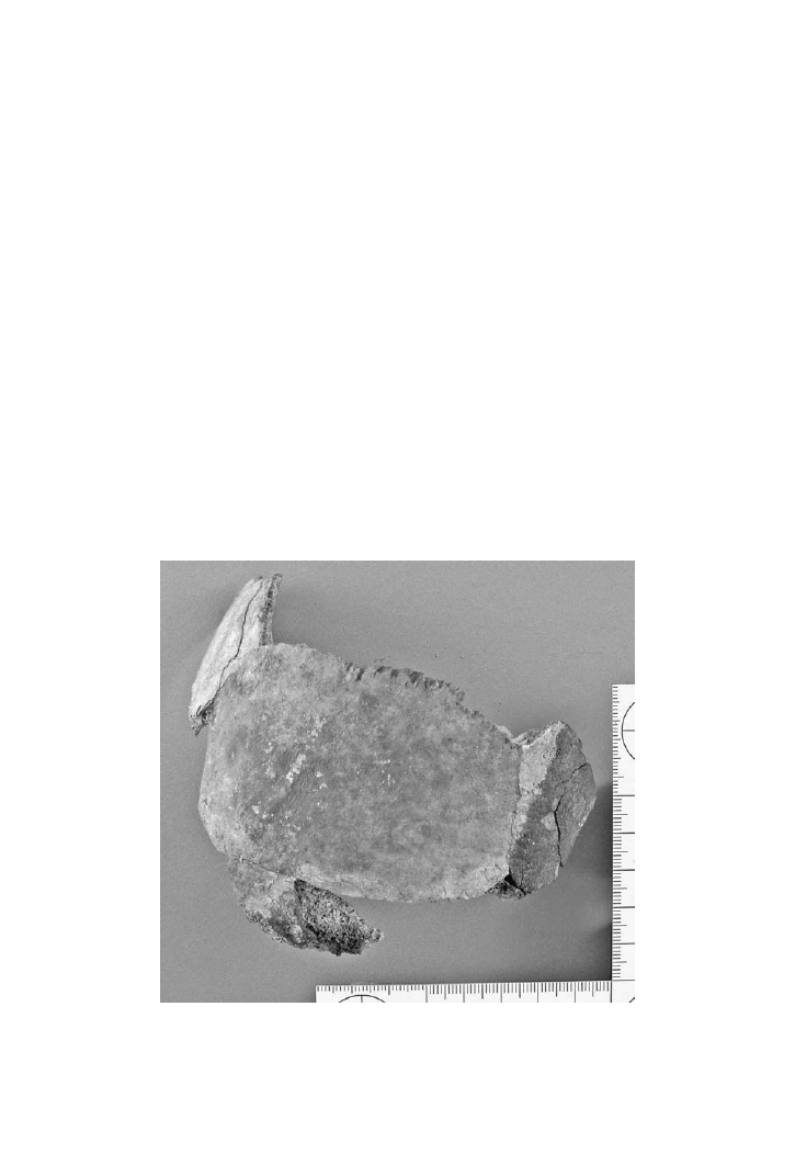
The charring of human remains in these events tends to be uneven. In cases
of aircraft crashes and fires, jet fuel will act as a very fast-burning and extremely
hot accelerant. Cremation of remains to the point of calcination may occur, but it is
more likely that the body will burn differentially. Of course, there are instances in
which a body will be engulfed by a fire and be subjected to explosive forces. How-
ever, the differential cremation of a body at a mass disaster scene is a real possibility
(
).
© 2008 by Taylor & Francis Group, LLC
Positive Identification of Cremains
179
Aircraft crash victims are usually identified through the use of recovered den-
tal structures (e.g., Barsley et al., 1985). More recently, the use of DNA on a wide
scale has been used to assist in establishing the identification of highly fragmented
remains (e.g., Mackinnon and Mundorff, 2007). DNA analysis in combination with
dental, anthropological, and pathological analysis will continue to be used in mass
fatality incidents.
As noted above, the challenge of any mass fatality incident is the documentation
and collection of remains. When those remains have been cremated, such as in the
Hinton, Alberta train crash in 1986, and commingled with wreckage, the challenge
is now separating and sorting the cremains from melted plastics and metal (Strat-
ton and Beattie, 1999). Although separation of cremains from other materials may
be time consuming, the process of identifying remains is a challenging task due to
their cremated state.
Nonmetric features have been suggested by Stratton and Beattie (1999) to be the
only means possible for the identification of cremains. Sex and age may be assigned
to cremains should the morphological features be present to do so. In addition to
these clinical x-ray comparisons, even associated personal effects can be of impor-
tance in the identification process.
It is clear from the foregoing that the condition of cremains in mass disaster sce-
narios is of paramount importance to the identification process. One must be open
FIGURE 8.9 A cranial vault fragment from a small plane crash. Note the charred and
uncharred areas present on the vault. (Photo by S. Fairgrieve.)
© 2008 by Taylor & Francis Group, LLC

180
Forensic Cremation Recovery and Analysis
to the use of many analytical avenues in order to pursue a positive identification. It
is equally clear that a team approach to the identification of disaster victims, par-
ticularly when cremated, will enhance the chances of arriving at an identification.
For cremains that have reached Crow–Glassman Stage V, the use of DNA will be a
foregone conclusion. However, morphological comparisons to antemortem records
may still be possible.
8.6 IMPLANTS AS A MEANS OF IDENTIFICATION
As with dental restorations, surgical procedures may also require the implantation
of materials in the body that may be of assistance in the identification of cremains.
The usefulness of orthopedic devices, due to manufacturer logo and a means of
tracking these devices to specific patients through unique serial and lot numbers,
has been documented as a likely source of information for positively identifying
associated human remains (Ubelaker and Jacobs, 1995). Pacemakers have been a
commonly traced implant in the identification of remains (Sathyavagiswaran et al.,
1992).
Although there are many forms of implants, one would have to consider the like-
lihood of survival of such implants in cremation contexts. Fixative devices, screws,
and surgical plates are ideal candidates for examination in cremains. However, even
more complicated devices, such as an osteostimulator, an implanted device that is
used to induce the regeneration of bone tissue by means of a stimulating electrical
current, have been of assistance in the identification of cremains (e.g., Bennett and
Benedix, 1999).
All materials that are directly associated with human cremains that can pos-
sibly be a component of an implanted device must be explained in the analysis of
recovered materials. The challenge is to recognize these cremated components
from amongst other fire-altered materials associated with, or around, the human
cremated material.
8.7 IDENTIFYING CREMAINS OF HISTORIC FIGURES
On occasion human cremains are recovered in a context that is consistent with an
expected or purported individual. The challenge is no less difficult in attempting
to identify these remains as with many others. To serve as examples of the chal-
lenges of such identifications, I have chosen to summarize the cases of attempting to
identify the charred remains once purported to be those of King Philip II of Mace-
donia, father of Alexander the Great (Andronicos, 1994; Bartsiokas, 2000), and
the attempts to identify the cremains of Adolf Hitler (Laurier et al., 1994; Kaleka,
1993).
The cremated remains of what was subsequently identified as belonging to an
adult male were discovered in “Royal Tomb II” at Vergina, Greece, in 1977 by
Andronicus (1994). The richness of the grave goods and the context (ca. 336 B.C.)
served to identify the tomb as that of King Philip II of Macedonia. As noted by Bart-
siokas (2000), this conclusion has been challenged due to another estimated date of
317 B.C. and, hence, belonging to King Philip III Arrhidaeus, a half-brother of Alex-
© 2008 by Taylor & Francis Group, LLC

Positive Identification of Cremains
181
ander the Great. As Philip II is said to have suffered an arrow wound to his right eye,
the detection of evidence of such an injury on the cremains would be thought to have
served as a strong indicator of the identity of these cremains. In a forensic context,
if a pathology is noted in a medical record with accompanying radiographs, one
would be more confident of utilizing a pathology as a basis for identification. The
strongest possible conclusion that could be made in this case, should the cremains
provide unequivocal evidence of an antemortem eye injury that is consistent with
that reported in history, would be that we failed to exclude Philip II as the identity
of the cremains. Examination of the margins of the supraorbital margin of the right
eye did not yield any evidence of a healed fracture or callus formation (Bartsiokas,
2000). However, is it possible for an arrow to enter the right orbit and result in the
loss of an eye without leaving any evidence on the bone? The obvious answer is yes.
Although it may not seem likely, documentation of the incident is not sufficient to
say one way or the other. Another point of consideration is the interval of time that
has passed from the incident to the time of death. A lack of evidence of a callus
formation on the bone may indicate that sufficient time has passed for resorption of
the bony callus. Although Bartsiokas (2000) concludes that there is no evidence to
support traumatic injuries to the face, and any asymmetries can be attributed to the
warping and shrinkage associated with the cremation process. It is true that cranio-
facial skeletal elements are highly susceptible to the aforementioned heat-induced
changes. However, given the above, it cannot be categorically stated one way or
another that the cremains are, or are not, those of Philip II of Macedonia. A rigorous
forensic approach would prevent us from rendering a conclusion one way or another.
Hence, the cremains should be considered as unidentified.
In the twentieth century, the most infamous person whose charred remains were
under severe scrutiny for identification were those of Adolf Hitler. In spite of such
interest, the purported cranial and dental fragments held by Russian authorities
still have questions related to identification issues (for a review, see
al., 2005). The issues surrounding Hilter’s remains lie in the fact that the remains
recovered were subjected to fire, using gasoline as an accelerant. According to
Bezymenski (1968), the charred remains recovered represented an adult male of
approximately 50–60 years of age with a stature of approximately 165 centimeters.
Enough soft tissue was present in order for there to be an examination of internal
organs, even to note the absence of the left testicle. The skull consisted of an occipi-
tal, left temporal, “lower cheek bones,” nasal bones, and the mandible and maxilla.
The dental remains are of significance due to the presence of bridgework, artificial
teeth, crowns, and fillings. Given the above condition of the remains, this would put
them into either level 2 or 3 of the Crow–Glassman Scale.
A reexamination of documents concerning the discovery of the grave contain-
ing the corpses of a man and a woman outside of Hitler’s Berlin bunker yielded a
reference to two fragments from a skull found at a depth of 50 to 60 centimeters
(Petrova and Watson, 1996). One fragment is noted as having a bullet hole. This
hole, interpreted as being an exit wound, is from a close contact gunshot through the
mouth or the chin. The original autopsy report, cited by Bezymenski (1968) makes
no mention of such fragments. However, mention was made of splinters from a glass
ampule in the mouth of the male body (presumably containing cyanide). Further,
© 2008 by Taylor & Francis Group, LLC

182
Forensic Cremation Recovery and Analysis
cranial vault fragments representing the “back of the parietal and part of the occipi-
tal” were discovered and examined in 1995 (Petrova and Watson, 1996).
The nature of the conflicting reports as to whether or not Hitler shot himself
or took cyanide (or both), is very much dependent upon an identification of the
mandibular and maxillary fragments as well as those of the skull. An identification
based on mtDNA from Hitler’s maternal cousins, as suggested by Marchetti et al.
(2005) would be an important step in resolving this problem. However, this case is a
classic example of poor controls at a scene and subsequent chain of custody. By dis-
sociating the remains, questions such as the above become issues of intense interest.
Modern forensic science would hopefully not let things get this far.
The two cases outlined above are merely examples of some of the problems that
are inherent to such work. Even remains that are charred and comparatively low on
the Crow–Glassman Scale (i.e., Hitler’s remains) may be more problematic due to
the methods of recovery and curation of materials.
8.8 SUMMATION
The identification of cremains is a very credible pursuit. Although it is a challenge
that faces any forensic analyst, it is indeed the case that success will depend upon
the suitability of the recovered specimens, and the availability and quality of medi-
cal and dental records. Care must be taken when examining morphological features
in light of the degree of shrinkage and warping that may occur during the cremation
process.
© 2008 by Taylor & Francis Group, LLC
Document Outline
- Table of Contents
- Chapter 8: Positive Identification of Cremains
Wyszukiwarka
Podobne podstrony:
9189ch5
9189ch7
9189ch1
9189ch4
9189ch6
9189ch3
9189ch2
9189ch5
więcej podobnych podstron