
221
Neuroprotection as a Treatment for Nerve Agent Survivors
Chapter 6
NeuroproteCtioN as a treatmeNt
for Nerve ageNt survivors
Gerald P.H. BallouGH, P
h
d*; JonatHan newmark, md
†
; eric S. levine, P
h
d
‡
;
and
marGaret G.
FilBert, P
h
d
§
iNtroDuCtioN
Neuropathology aND the meChaNism of Nerve-ageNt–iNDuCeD
Damage
speCifiC relevaNCe of NeuroproteCtioN to Nerve ageNt
survivors
NeuroproteCtaNts with proveN effiCaCy agaiNst Nerve-ageNt–
iNDuCeD seizure-relateD BraiN Damage
gangliosides
poly(aDp-ribose) polymerase inhibitors
ryanodine receptor antagonists
N-methyl-
d
-aspartate receptor antagonists
aDDitioNal NeuroproteCtive approaChes
free radical scavengers
mitochondrial permeability transition inhibitors
Neuroprotective hypothermia
summary
* Professor of Biology, La Salle University, 1900 West Olney Avenue, Philadelphia, Pennsylvania 19141-1199
†
Colonel, US Army, Deputy Joint Program Executive Officer, Joint Program Executive Office for Chemical/Biological Defense, Skyline #2, Suite 1609,
5203 Leesburg Pike, Falls Church, Virginia 22041-3203
‡
Assistant Program Manager, Science Applications International Corporation, 3465 Boxhill Corporate Center Drive, MS 23, Abingdon, Maryland
21009
§
Special Assistant to the Commander, US Army Medical Research Institute of Chemical Defense, 3100 Ricketts Point Road, Aberdeen Proving Ground,
Maryland 21010-5400

222
Medical Aspects of Chemical Warfare
Portions of this chapter appeared as: Filbert m, levine e, Ballough G. neuroprotection for nerve agent-
induced brain damage by blocking delayed calcium overload: a review. Journal of Medical, Chemical, Biologi-
cal, and Radiological Defense. 2005;3:1–21. available at: http://jmedcbr.org/issue_0301/Filbert/Filbert_1105.
pdf. accessed march 2007.

223
Neuroprotection as a Treatment for Nerve Agent Survivors
iNtroDuCtioN
early use of an anticonvulsant does not guarantee
that seizures, once stopped, will not return. the recur-
rence of seizures is often observed in animal studies in
several species and is of concern in human exposures.
although neuropathology is reduced in diazepam-
treated animals, the incidence and degree of protection
afforded by diazepam is not complete.
9,20–23
moreover,
switching the fielded anticonvulsant to another benzo-
diazepine, such as midazolam or lorazepam, does not
entirely solve the problem of refractory Se.
Seizures and Se are key causes of brain damage re-
sulting from nerve agent poisoning, and their preven-
tion or alleviation should be the primary objective.
24–26
However, because of the refractory nature of seizures
and especially Se, prevention and alleviation become
increasingly difficult as more time elapses before
therapy begins. also, there is high probability that
seizures will return when anticonvulsants wear off.
therefore, it is reasonable to anticipate a high incidence
of brain damage connected to the increased survival
rate of nerve agent victims.
casualties exhibiting seizures and Se can be an-
ticipated not only from terrorist attacks but also from
battlefield scenarios involving troops who were not
in full protective ensemble at the time of the attack.
27
in the confusion following a terrorist attack or on the
battlefield, prompt treatment of nerve agent casualties
can be expected to be problematic, and some victims
undergoing seizures may not receive anticonvulsants
inside the antiseizure therapeutic window. it is also
possible that some victims may undergo noncon-
vulsive Se, a state of continuous seizures without
observable clinical movement.
28
For these victims,
treatment might be inadvertently delayed beyond the
therapeutic window. under the Small Business innova-
tive research Program, the uS army funds efforts to
field a far-forward, simple seizure detector to identify
these casualties.
this chapter presents a detailed overview of nerve-
agent–induced neuropathology and explains the mech-
anisms of action of candidate neuroprotectants that have
shown promise in various animal and human studies,
especially those that have received uS Food and drug
administration (Fda) approval for other indications.
organophosphorus nerve agents are the principal
chemical warfare agents known to produce brain
injury. they block hydrolysis of the neurotransmitter
acetylcholine by inhibiting the enzyme acetylcholin-
esterase, resulting in greatly increased postsynaptic
acetylcholine levels. this causes a spectrum of effects,
including miosis, excess secretions, nausea, vomiting,
and muscle fasciculations. at moderate to high doses,
nerve agents also cause seizures and associated con-
vulsions. if left untreated, seizures rapidly progress to
status epilepticus (Se) and cause irreversible seizure-
related brain damage (SrBd).
1,2
the international
classification of epileptic Seizures defines Se as any
seizure lasting at least 30 minutes or intermittent sei-
zures lasting longer than 30 minutes between which
the patient does not regain consciousness.
3,4
For over a decade acute therapy has effectively saved
those poisoned by nerve agents on the battlefield,
5
after
accidental exposures,
6
and in terrorist attacks, as in the
Japan subway attacks in 1994 and 1995. one lesson
learned from the 1995 tokyo attack was that, lacking
acute antidotal treatment, many survivors arrived at
hospitals in convulsive Se. the tokyo experience illus-
trates the necessity of acute antidotal therapy, such as
the regimen adopted by the uS military. this regimen
is aimed primarily at treating cholinergic crisis with
a postexposure anticholinergic (atropine sulfate) and
an oxime reactivator (2-pralidoxime [2-Pam cl]). in
specific intelligence-driven situations, pyridostigmine
bromide (PB) pretreatment is added. although these
medications greatly reduce morbidity and mortality,
they do not always prevent seizures and brain damage
in nerve agent casualties; therefore, the regimen now
includes the anticonvulsant diazepam.
2
even with diazepam, however, the treatment regi-
men has limitations. the decision to include diazepam
was based on animal data showing that it could
terminate nerve-agent–induced seizures and convul-
sions and enhance survival when given in conjunction
with the acute therapy described above.
7–11
However,
the therapeutic window for arresting seizures and
Se with diazepam is less than an hour following on-
set; after that, both are refractory to anticonvulsant
therapy.
7,8,10–19
Neuropathology aND the meChaNism of Nerve-ageNt–iNDuCeD Damage
although there is little neuropathological data
from patients who have survived nerve agent attacks,
abundant evidence is available from animal models,
many of which involve persistent Se. the profound
brain damage produced by nerve agents was first
described by Petras
29
; lemercier et al
30
; and mcleod
et al.
31
Since then, numerous studies have greatly en-
hanced the understanding of neuropathology resulting
from nerve agent intoxication.
23,32–38
these studies have
established that prolonged seizures and Se resulting

224
Medical Aspects of Chemical Warfare
from nerve agent exposure are directly responsible
for the vast majority, if not all, of the neuropathology
produced by these agents. the associated damage is
typically bilaterally symmetrical and most severe in
temporal lobe structures (ie, piriform and entorhinal
cortices, hippocampus, and amygdala) as well as in
the thalamus.
Brain damage resulting from agent-induced sei-
zures is the result of the complex, multiphasic response
of individual neurons to numerous extracelluar and
intracellular events. Following inhibition of acetyl-
cholinesterase and accumulation of acetylcholine at
cholinergic synapses, the hyperstimulation of cholin-
ergic receptors on postsynaptic membranes triggers
seizures.
10,39,40
Subsequently, recruitment and excessive
activation of the glutamatergic neurotransmitter sys-
tem occurs. Glutamate, the most abundant excitatory
neurotransmitter in the brain, is responsible for sus-
taining soman-induced seizures and promoting the
development of Se.
1,24,41–44
large pathological eleva-
tions in the concentration of intracellular sodium and
(especially) calcium are caused by excessive stimula-
tion of ionotropic glutamate receptors, as is prolonged
depolarization of postsynaptic membranes. this
initiates a harmful cascade of pathological processes,
most of which center around a prolonged increase in
intracellular free calcium or delayed calcium overload,
leading to excitotoxic cell death.
1,24,45–47
transient elevation in intracellular free calcium is a
ubiquitous signaling mechanism and regulator of in-
tracellular processes, from cell growth and metabolism
to cell death.
48–50
cytosolic free calcium is also a critical
neuronal mediator of learning and memory.
51
How-
ever, when normal homeostatic control of intracellular
calcium is lost and a sustained elevation occurs, the
delayed calcium overload triggers neuronal cell death
by necrosis or apoptosis (a form of programmed cell
death).
52–56
in neurons, the majority of calcium influx
occurs through N-methyl
d
-aspartate (nmda) iono-
tropic glutamate receptors as well as voltage-gated cal-
cium channels (eg, l-type). calcium influx also occurs,
though to a lesser extent, through the other two classes
of ionotropic glutamate receptors (alpha-amino-3-
hydroxy-5-methylisoxazole-4-proprionic acid and
kainate receptors).
57
excessive stimulation of nmda
receptors is the first step in glutamate excitotoxicity.
24,45
the release of intracellular stores is also responsible
for increased cytosolic free calcium. the endoplasmic
reticulum (er) releases calcium following binding
of the second messenger, inositol triphosphate, to
ionotropic receptors located on the er membrane.
calcium is released from the er via ryanodine recep-
tors. these ionotropic receptors are also located on the
er membrane and open following binding of cytosolic
calcium; thus, cytosolic free calcium augments its own
concentration by stimulating calcium release from the
er.
49
the er plays a critical role in normal calcium
homeostasis. excessive release or impaired uptake of
calcium has been implicated in pathology resulting
from calcium overload.
49,52
Brain mitochondria are
important for calcium buffering as cytosolic concentra-
tions rise, and their ability to sequester calcium is de-
pendent on adenosine triphosphate (atP).
58
However,
when calcium overload occurs, mitochondria undergo
a permeability transition characterized by loss of
mitochondrial transmembrane potential, curtailment
of atP synthesis, mitochondrial swelling, release of
stored calcium, and neuronal death by necrosis.
59–62
the majority of soman-induced SrBd results from
glutamate excitotoxicity and the delayed calcium over-
load that follows.
1,24,42,43
delayed calcium overload in
neurons initiates a pathological sequence characterized
by activation of several potentially damaging enzymes.
these include oxygenases, phospholipases, and nitric
oxide synthase, which produce reactive oxygen spe-
cies such as superoxide radical, hydrogen peroxide,
hydroxyl radical, nitric oxide, and peroxynitrite.
neuronal injury induced by reactive oxygen species
stems from direct damage to cell membranes, dna,
and intracellular proteins, and also induction of cyto-
chrome c from mitochondria with subsequent caspase
activation.
62
release of cytochrome c, caspase activa-
tion, and dna fragmentation are molecular hallmarks
of apoptosis (Figure 6-1).
56,62,63
cysteine proteases called calpains are also activated
by sustained elevations in intracellular free calcium.
calpains degrade various intracellular proteins, in-
cluding those of the cytoskeleton, membrane channels,
and metabolic enzymes, and cause neuronal death by
necrosis.
56,62,63
(necrosis produces localized inflamma-
tion, which exacerbates damage, while apoptosis is
not associated with inflammation.) the culmination
of these events may result in cell death hours or days
after the initial insult.
53–55
necrosis and apoptosis are not an either/or phe-
nomena, that is, they are not completely distinct forms
of cell death with no overlap; a necrosis versus apop-
tosis dichotomy is a misleading over-simplification.
64,65
martin and colleagues proposed an “apoptosis-necrosis
continuum,” reporting that dying neurons can exhibit
intermediate forms between apoptosis and necrosis.
66
recently, Baille and colleagues confirmed that neuronal
injury, resulting from soman-induced seizures, exhibits
a large variety of hybrid forms between necrosis and
apoptosis, but that the majority show more necrotic
features.
67
whether soman-induced neuropathology is
mostly necrotic, as it is in the piriform cortex of rats,
38
or contains elements of apoptosis as first proposed
225
Neuroprotection as a Treatment for Nerve Agent Survivors
by Ballough et al in 1997 and definitively assessed
by Baille et al is less important than the fact that both
forms of neuronal cell death are triggered by nerve-
agent–induced seizures.
38,67,68
candidate drugs may alter the relative propor-
tions of neurons undergoing death by necrosis versus
apoptosis. Studies have reported that insufficient atP
availability is an important determinant of whether
a cell that has been triggered to undergo apoptosis
is instead forced to die by necrosis.
55,69,70
therefore, it
is conceivable that a neuroprotectant candidate that
enhances atP availability (for example, poly(adP-
ribose) polymerase [ParP] inhibitors) could suppress
necrosis while facilitating apoptosis. neither possibil-
ity should be excluded during pathological evaluations
of neuroprotectant candidates.
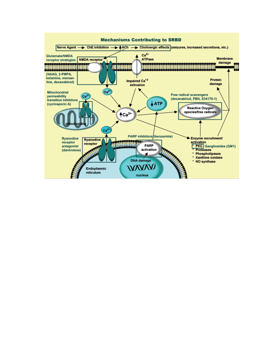
fig. 6-1. mechanisms contributing to nerve agent-induced SrBd. calcium plays a pivotal role in glutamate excitotoxicity. a
number of pharmacological approaches to neuroprotection have been investigated. various sites in this pathway have been
targeted. nmda receptor antagonists block calcium entry through this glutamate ionotropic receptor. Gangliosides promote
calcium extrusion indirectly by blocking Pkc translocation (not indicated). ParP inhibitors enhance functionality of ion
pumps and calcium extrusion by increasing atP availability. dantrolene blocks calcium release from intracellular stores.
Free radical scavengers include free radical “traps” and endogenous free radical scavenging enzymes and small molecules
prevent oxidative damage.
speCifiC relevaNCe of NeuroproteCtioN to Nerve ageNt survivors
the term “neuroprotection” is defined as “pharma-
cological intervention that produces enduring benefits
by favorably influencing underlying etiology or patho-
genesis and thereby forestalling the onset of disease or
clinical decline.”
71,72
within this broad definition, neu-
roprotection has acquired many different connotations.
as a result, a search of the term “neuroprotection” on
the national library of medicine’s Pubmed search
page produces several thousand studies, mostly on
disease states in which subsets of neurons are specifi-
cally vulnerable and die prematurely (as happens in
Parkinson’s disease, Huntington’s disease, frontotem-
poral dementia, and a host of metabolic disorders) or
accumulate neuropathology seen to a slight degree in

226
Medical Aspects of Chemical Warfare
normal brains but in an accelerated fashion in some
diseases (such as alzheimer’s disease and trisomy 21).
However, such interventions are unlikely to be relevant
to the survivor of a single, brief nerve agent exposure
that has already caused sustained seizures and Se. on
the other hand, research on neuroprotection following
stroke has provided valuable insights and clues that
do apply to the nerve agent survivor.
in this chapter, the term “neuroprotection” spe-
cifically refers to a putative intervention given over a
short period, ideally closely following the diagnosis
of nerve agent exposure or before the acute toxic
syndrome of exposure has been adequately treated.
the best neuroprotectant would have the longest
therapeutic window during which administration
would be beneficial (even if the window is still only a
matter of hours). at the same time, for logistical and
doctrinal reasons, the neuroprotection initiative does
not extend to prophylactic treatments administered
to troops likely to experience nerve agent exposure
(which would constitute a pretreatment, such as the
bioscavenger initiative [see chapter 7, nerve agent
Bioscavenger: development of a new approach
to Protect against organophosphorus exposure]).
therefore, in this chapter, neuroprotection refers only
to postexposure treatment.
there are similarities between brain damage result-
ing from nerve-agent–induced seizures and secondary
neuronal injury resulting from stroke.
73,74
although the
immediate aspect of stroke-related neuronal injury is
necrosis, which stems from anoxia or hypoxia, there
is a secondary component to stroke damage that takes
48 to 72 hours to become manifest. this component
accounts for approximately 50% of the total damage
resulting from the ischemic episode. Secondary stroke
injury involves brain tissue immediately surrounding
the necrotic core of primary injury (the penumbra).
For the most part, glutamate excitotoxicity and ionic
destabilization, especially intracellular calcium, induce
penumbral damage.
73–75
thus, the similarities between
secondary stroke damage and damage resulting from
nerve-agent–induced seizures become apparent: they
both involve glutamate excitotoxicity, hinge on intra-
cellular calcium destabilization, and lead to necrotic
or apoptotic neuronal death. this similarity raises the
possibility that neuroprotectants being developed for
stroke may be useful for nerve agent survivors. neu-
roprotective interventions in stroke models have been
shown to save neurons that otherwise would have died
via necrosis or apoptosis. there is hope, then, that a
treatment can be found that can be administered after
agent exposure and that, although it may not have
any immediately discernible clinical effect, will pro-
duce a significantly improved long-term neurological
outcome. any of the many classes of compounds that
have been suggested as acute stroke neuroprotectant
candidates could be tried. this list is extensive; the
internet Stroke center (http://www.strokecenter.
org), maintained by washington university,
76
offers
a continuously updated list of compounds that have
been tried in clinical stroke trials.
the rationale for developing a protective agent,
especially one based on dissimilar clinical situations
that give rise to similar neuronal pathology, assumes
that preventing neuronal loss will produce a superior
clinical outcome. in the case of stroke, this assumption is
probably warranted. in the case of nerve-agent–induced
nerve cell damage, this assumption has never been
tested directly, but it is consistent with a wide variety
of animal data in multiple models and species. the as-
sumption that preventing brain damage will produce
superior behavioral outcome is even supported by
lashley and Hebb’s studies in the early to mid 1900s.
77
a neuroprotectant in this restricted sense should dem-
onstrate that neurons that might have been lost are now
saved and that behavioral or neurological outcome is
improved. an ideal database to document such neu-
roprotectants would include both neuropathological
evidence of neuron survival and behavioral (in animals)
or cognitive (in people) evidence that the neurologic
outcome is superior compared to subjects that did not
receive the neuroprotectant. Finally, the Fda must ap-
prove use of the agent if it is a medication. (in clinical
medicine, any Fda-approved medication can be used
off-label by licensed physicians, but in military doctrine,
specific on-label Fda approval is mandatory.)
NeuroproteCtaNts with proveN effiCaCy agaiNst Nerve-ageNt–iNDuCeD
siezure-relateD BraiN Damage
this research comes from the consensus that nerve-
agent–induced seizures and Se lead to the develop-
ment of glutamate-mediated excitotoxicity, in which
delayed calcium overload is the intracellular trigger of
the final sequences leading to cell death.
1,24,42,43,47,49,56,78–81
classes of drugs that have been tested for their abilities
to ameliorate nerve-agent–induced SrBd by specifi-
cally mitigating delayed calcium overload include the
following:
• nmda receptor antagonists that block extra-
cellular calcium influx;
• glycosphingolipids that reduce intracellular
calcium by blocking the translocation of

227
Neuroprotection as a Treatment for Nerve Agent Survivors
protein kinase c (Pkc), thus enhancing the
sodium-calcium exchange;
• ryanodine receptor antagonists that prevent
the release of calcium from the er; and
• ParP inhibitors that indirectly lower intra-
cellular calcium by preventing atP deple-
tion.
82–89
increased atP availability facilitates calcium ef-
flux by plasma membrane ca2+ atPase and calcium
sequestration by the mitochondria, and indirectly
enhances sodium-calcium exchange by maintaining
sodium-potassium-atPase functionality.
58
gangliosides
medications that target events subsequent to calci-
um overload have been tested against soman-induced
SrBd in an effort to circumvent neurotoxicity associ-
ated with nmda receptor antagonism and mitigate
established delayed calcium overload. intracerebro-
ventricular infusion of Gm1 monosialoganglioside (5
mg/kg/day, for 5 days before and 27 h after soman
exposure) in rats markedly reduced cross-sectional
areas of soman-induced temporal lobe necrosis (there
was an 85.9% lesion reduction in the piriform cortex
and contiguous structures, compared with unpro-
tected soman-positive controls).
90
in this study, all rats
were pretreated with PB before soman exposure, and
then treated with atropine methylnitrate (amn ) and
2-pralidoxime (2-Pam). considerable neuroprotec-
tion was also obtained with the water-soluble Gm1
monosialoganglioside derivative, wild20. as an
adjunct to Hi-6 pretreatment and amn posttreatment,
wild20 (2.5 mg/kg, intraperitoneal injection [iP])
reduced volumetric temporal lobe necrosis by 75.2%.
neuroprotection by these two compounds occurred,
and neither seizure intensity nor duration (assessed
via electroencephalography [eeG] monitoring) was
diminished.
Gangliosides are sialic-acid–containing glycosphin-
golipids that are natural constituents of cell mem-
branes and are particularly abundant in neurons.
91–93
the mechanism by which Gm1 monosialoganglioside
and wild20 exert their neuroprotective effects in-
volves inhibition of Pkc translocation to the plasma
membrane.
75, 82–86,94,95
Pkc activation and translocation
enhance glutamate excitotoxicity.
96,97
Furthermore,
Pkc’s role in the excitotoxic process is to prolong
nmda receptor activation and possibly inhibit cal-
cium extrusion mechanisms.
82,75,98
in addition, wild20
is reported to reduce inflammation by its inhibitory
effects on specific leukocytes (neutrophils).
99
despite
the promising results with gangliosides, further studies
have been discontinued because of concerns of possible
contamination by prions associated with bovine spon-
giform encephalopathy (mad cow disease).
90,100
poly(aDp-ribose) polymerase inhibitors
recent studies indicate that ParP inhibition is
neuroprotective following neuropathological insults
involving excitotoxicity, such as cerebral ischemia
and traumatic brain injury.
101–108
ParP is an abundant
nuclear enzyme that is activated by dna strand
breaks induced by reactive oxygen species.
108,109
with
moderate insults, it facilitates dna repair by utiliz-
ing cellular nicotinamide adenine dinucleotide to
form poly(adP-ribose). excessive ParP activation
leads to nicotinamide adenine dinucleotide depletion,
metabolic inhibition via glycolysis block, atP insuf-
ficiency, and cell death by necrosis.
104,109,110
neurons
are especially vulnerable to metabolic insufficiency
resulting from ParP over-activation because glucose
is normally the only metabolic substrate and the
dependency on glycolysis is exceptionally high.
108
in
excitotoxic models, over-activation of ParP is closely
linked to calcium-induced nitric oxide synthase activa-
tion, which leads to the production of nitric oxide; the
detrimental effects of nitric oxide are mostly mediated
through peroxynitrite, which forms when nitric oxide
reacts with superoxide.
109,111,112
in 1999 meier et al
113
reported reduced lesion vol-
umes and increased survival in soman-exposed rats
that received the ParP inhibitor benzamide. Further
investigation into the neuroprotective efficacy of ParP
inhibition warrants consideration, and subsequent
studies should include several new-generation ParP
inhibitors that have shown increased usefulness, such
as ono-1924H, dr2313, and Fr247304.
105,107,114
ryanodine receptor antagonist
dantrolene is another drug that has shown neuro-
protective efficacy against soman-induced SrBd.
88
a
ryanodine receptor antagonist that prevents the release
of calcium from the er, dantrolene is Fda-approved
for use in malignant hyperthermia. although some
neuroprotection is produced by diazepam alone (20
mg/kg, intramuscular injection [im], 40 min after sei-
zure onset), this protection is significantly augmented
in the dorsal and lateral cortices of rats by coadminis-
tration of dantrolene (10 mg/kg, intravenous [iv]).
88
administering the full dosage of dantrolene in a single
injection is difficult because of insolubility problems
associated with the medication. to overcome these
problems and achieve the desired dantrolene dosage,
four separate iv injections were performed between

228
Medical Aspects of Chemical Warfare
40 minutes and 8 hours after seizure onset, with a
total injection volume approximating 1 ml per rat. a
unique formulation of dantrolene (lyotropic thera-
peutics, inc, ashland, va) as a nanocrystal dispersion
has also been used to obviate solubility problems. with
this formulation, it is possible to administer a much
higher dose of dantrolene in a much lower injection
volume. this is critical because when dantrolene is
administered by iP injection, liver enzymes lower the
concentration of dantrolene reaching the brain. the
nanocrystal formulation of dantrolene minimizes the
effects of the liver enzymes.
our results with the dantrolene nanocrystal formu-
lation not only overcame the insolubility problems of
our previous dantrolene study, but corroborated and
extended the results of that study. the nanocrystal study
was unable to demonstrate significant protection in the
piriform cortex, the most severely damaged region, but
in this study the nanocrystal dispersion of dantrolene
(40 mg/kg, iP) plus diazepam (20 mg/kg, im) reduced
piriform cortical necrosis by 15.6% more than diazepam
alone (unpublished study by uS army medical research
institute of chemical defense). in these experiments, all
soman-exposed rats also received Hi-6 (125 mg/kg, iP, 30
min after soman) and amn (2 mg/kg, im, < 1 min after
soman) to protect against the peripheral effects of soman
and ensure survival. neuroprotection by dantrolene in
the above experiments occurred without changes in sei-
zure intensity or duration, and dantrolene produced no
discernible effects on the electrocorticographic profiles
of soman-exposed subjects. these findings are consistent
with those of Frandsen and Schouosboe,
115
who reported
that dantrolene prevented glutamate neurotoxicity by
blocking release of calcium from intracellular stores.
the results are also consistent with those of niebauer
and Gruenthal,
87
who examined the protective effects of
dantrolene on hippocampal neuronal damage produced
by Se in rats. in their study, dantrolene (10 mg/kg, iP)
was administered either 30 or 140 minutes after the onset
of Se. niebauer and Gruenthal reported that early admin-
istration produced a significant reduction in neuronal
injury in all hippocampal subregions. when dantrolene
administration was delayed until 140 minutes after Se
onset, some protection was still seen in hippocampal field
ca3, but not the other subregions.
87
Protection against
kainic-acid–induced apoptosis has also been reported.
116
N-methyl-
d
-aspartate receptor antagonists
MK-801 (Dizocilpine)
the first nmda receptor antagonist to show
promise as a putative neuroprotectant was mk-801
(dizocilpine); however, it has been shown to have toxic
effects. when given in conjunction with PB, amn,
and 2-Pam, noncompetitive mk-801 was reported
to reduce nerve-agent–induced SrBd in the piriform
cortex, amygdala, hippocampus, and thalamus.
43
as
mentioned, these are among the most severely dam-
aged brain regions in SrBd resulting from soman
exposure.
29–32,35,37,38,90
in the Sparenborg study, mk-801
(0.5, 1.0, or 5 mg/kg, iP) reduced brain damage and
diminished or arrested seizures in guinea pigs when
administered as a pretreatment 30 minutes before so-
man, and the effects were dose-dependent. the anti-
convulsant profile of mk-801 against soman-induced
seizures was definitively characterized by Shih.
11
He
showed that the anticonvulsant effect of mk-801 is four
times greater than that of diazepam, but at doses of 1
mg/kg or higher, mk-801 potentiated the lethal effects
of soman. Some concern arose about the use of nmda
antagonists when it was reported that mk-801 induces
neuronal degeneration in the posterior cingulate, retro-
splenial cortices, and other corticolimbic regions.
117,118
this damage evidently occurs by disinhibition of mul-
tiple converging excitatory pathways.
119
Specifically,
excessive blockage of glutamatergic pathways leads
to excessive stimulation of cholinergic function.
120
this
explanation is supported by the findings that neuro-
toxicity by mk-801 is augmented when cholinergic
receptors (ie, muscarinic) are activated.
121
Memantine
memantine is a noncompetitive nmda receptor
antagonist
122
that has also been tested for its anti-
convulsant effects against soman-induced seizures.
Studies have suggested that memantine’s pharma-
cokinetics make it a safer candidate than mk-801.
123,124
mclean et al
125
reported that memantine alone (18 mg/
kg, subcutaneous [Sc]) blocked the onset of soman-
induced seizures and was able to terminate seizures
when administered 15 minutes after soman injection.
these findings, however, are inconsistent with those
of Shih et al
17
who reported that memantine by itself
is completely ineffective as an anticonvulsant against
soman-induced seizures. the latter authors pointed
to a need for eeG monitoring when determining an-
ticonvulsant efficacy and suggested that mclean et al
may have mistaken diminished convulsive behavior
as evidence of reduced seizure activity. neither study
addressed the possible neuroprotective effects of me-
mantine (ie, reduced neuropathology independent of
anticonvulsant activity). on the other hand, koplovitz
et al
126
observed a modest reduction in piriform cortical
damage following soman in rats treated with atropine
and memantine, compared to those that received at-
ropine alone. there were no differences between the
229
Neuroprotection as a Treatment for Nerve Agent Survivors
eeG power spectra of the two groups. regardless of
the above discrepancies, the neuroprotective benefit of
memantine in other models of excitotoxicity is widely
accepted.
124,127
For example, in a rat model of stroke,
memantine given 2 hours after the ischemic event
reduced brain damage by approximately 50%.
128
in
addition, memantine is well tolerated and does not
produce neurotoxicity at therapeutic dosages. it was
recently approved by the Fda for treating alzheimer’s
disease.
124
HU-211 (Dexanabinol)
the first real proof of concept of postexposure
neuroprotection came from work with Hu-211 (dex-
anabinol), a nonpsychotropic analogue of tetrahydro-
cannabinol, the active ingredient in marijuana. Filbert
and colleagues
129
showed that in rats exposed to high
doses of soman, dexanabinol protected neurons in the
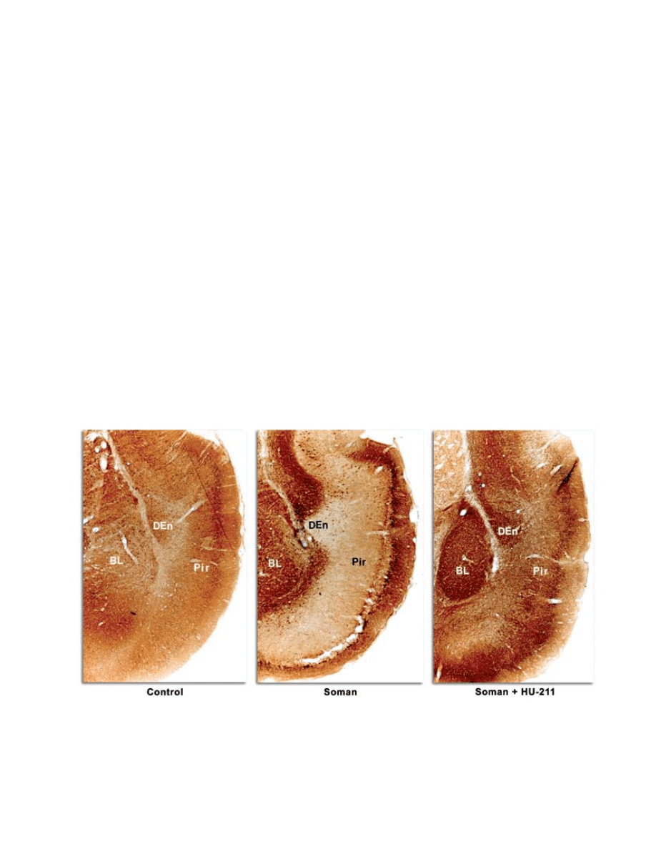
piriform cortex (Figure 6-2) when given as late as 40
minutes after the eeG-proven onset of seizures. the
drug was not an anticonvulsant and had no effect
upon the seizures, indicating that the results showed
a true neuroprotective effect and not part of an anti-
convulsant effect. Hu-211 has been reported to inhibit
nmda receptors, act as an antioxidant and free radical
scavenger, suppress nitrous oxide and tumor necrosis
factor-α generation, and stabilize calcium levels.
130–132
Hu-211 is generally well tolerated in humans.
133
when Hu-211 (25 mg/kg, iP) was administered 5
minutes after the onset of soman-induced seizures,
in conjunction with Hi-6 and amn pretreatment
and posttreatment, respectively, temporal lobe lesion
volume/necrosis (assessed at 28 h after seizure onset)
was reduced by 86%, compared with unprotected
soman-positive controls (see Figure 6-2).
134,135
Hu-211
had no effect on the strength or duration of seizure
activity, as determined by quantitative eeG analysis.
Significant neuroprotection was also observed when
Hu-211 administration was delayed 40 minutes after
seizure onset. neuroprotection by Hu-211 was most
evident in the piriform cortex and contiguous temporal
lobe structures, such as the amygdala, entorhinal, and
perirhinal cortices, but did not extend to the thalamus.
administration of Hu-211 and diazepam 40 minutes
after seizure onset did not augment the neuroprotec-
tion obtained with diazepam alone.
in analyzing the mechanisms of neuroprotection by
Hu-211 and diazepam, it is important to differentiate
between protection obtained by anticonvulsant effects
fig. 6-2. dexanabinol (Hu-211) protects against soman-induced neurological damage. microtubule-associated protein 2
(maP-2) staining is neuron-specific. maP-2 negative immunostaining indicates necrosis, except in areas of white matter.
Bl: basolateral amygdaloid nuclear group
den: dorsal endopiriform nucleus
Pir: piriform cortex.

230
Medical Aspects of Chemical Warfare
and that produced by interfering with delayed calcium
overload. in the above studies, Hu-211 was protective,
despite the continued presence of undiminished sei-
zures and Se, whereas diazepam attenuated (without
stopping) seizure intensity and thereby reduced the ini-
tial insult. the anticonvulsant action of diazepam, via
agonistic modulation of γ-aminobutyric a (GaBa[a])
receptors, is well known. these mechanisms are non-
overlapping, and neuroprotective effects should be
additive or synergistic. Hu-211 is not approved for
clinical use, and the company that owns the rights to
it (Pharmos ltd, israel) is developing it as a possible
adjunctive therapeutic for head trauma.
Gacyclidine
Gacyclidine (Gk-11) is another nmda receptor
antagonist that has shown considerable neuroprotec-
tive efficacy. when Gk-11 (0.01–0 .1 mg/kg, iv) was
given to rats 10 minutes after soman exposure (in
conjunction with PB pretreatment, and aS, 2-Pam,
and diazepam posttreatments, 1 min after soman in-
jections), it completely blocked SrBd when assessed 3
weeks after exposure.
136
in a more realistic battlefield
scenario, Gk-11 was administered 45 minutes after an
exposure of 8 times the median lethal dose (ld
50
) of so-
man in nonhuman primates. animals also received PB
pretreatment, followed by aS, 2-Pam, and diazepam
posttreatments (1 min after soman exposure) equiva-
lent to a single autoinjector of each in humans. when
brain pathology was assessed 3 weeks after exposure,
all three Gk-11–treated primates showed little or no
evidence of pathology in the frontal and entorhinal
cortices, amygdala, caudate nucleus, hippocampus,
thalamus, midbrain, pons, medulla, and cerebellum,
compared with the only surviving soman-treated ani-
mal (1 of 3) that received aS, 2-Pam, and diazepam
but not Gk-11.
137
in a study that approximates casualty
management following a terrorist attack, soman-in-
toxicated (2 times the ld
50
) primates did not receive
PB pretreatment and received delayed aS, 2-Pam,
and diazepam treatments (one human-equivalent of
each, as above) 30 minutes postexposure, followed by
Gk-11 (0.1 mg/kg, iv). in this study, the addition of
Gk-11 restored normal eeG activity and completely
prevented neuropathology (assessed 5 weeks after
exposure), compared with subjects that received aS,
2-Pam, or diazepam alone.
138
Gk-11 has a binding af-
finity for nmda receptors that is only one tenth that of
mk-801. in addition, it binds to non-nmda receptors
when interaction with nmda receptors is prevented.
For these reasons, Gk-11 is considered substantially
less neurotoxic than mk-801.
139
it is currently being
evaluated in human clinical trials for a different neu-
roprotective indication.
139,140
Ketamine
ketamine appears to be the most promising neu-
roprotectant candidate to date,
141,142
and it should be
used in combination with a benzodiazepine, such as
diazepam. ketamine is an Fda-approved anesthetic
that blocks neurotransmissions without depressing
respiratory and circulatory functions. its actions are
mediated by low-affinity binding to nmda receptor
channels and prevention of calcium influx.
142–145
ket-
amine is garnering considerable attention as a puta-
tive neuroprotectant against ischemic brain injury,
damage resulting from seizures and Se, irrespective
of etiology, and SrBd specifically resulting from
nerve-agent–induced seizures.
144–149
Fujikawa
147
re-
ported remarkable neuroprotection in 21 of 24 brain
regions in rats when 100 mg/kg of ketamine was ad-
ministered (iP) 15 minutes after lithium-pilocarpine-
induced Se onset. Similarly, 100 mg/kg of ketamine
(iP) prevented learning impairment in rats when
administered immediately after lithium-pilocarpine-
induced Se.
150
Borris et al
151
report that ketamine (58
mg/kg, the effective dose in 50% of those taking it
[ed
50
]) can control prolonged Se in rats when admin-
istered 1 hour after onset. cumulative evidence for
the beneficial effects of ketamine following Se onset
has led to its recommended use in humans when Se
cannot be alleviated by conventional anticonvulsant
therapy.
148
Based on its neuroprotective and anticonvulsant
properties, mion et al
145
recommend ketamine for vic-
tims of nerve agent exposure. more recently, dorandeu
et al
149
reported that ketamine proved effective in stop-
ping seizures, highly reducing SrBd, and improving
guinea pig survival when administered between 30
minutes and 2 hours after soman poisoning. increasing
dosages of ketamine (ie, 10–60 mg/kg, im) were re-
quired as post-Se onset delay increased, and ketamine
was always administered with atropine sulfate (2–10
mg/kg); in addition, guinea pigs received pyridostig-
mine (26 mg/kg, im) 30 minutes prior to soman and
amn (4 mg/kg, im) within 1 minute following the
soman injection. their study also provided compelling
evidence of neuroprotection by ketamine at dosages
that did not modify seizures (ie, 2–10 mg/kg), and
suggested combining ketamine and benzodiazepine
treatments when treatment is delayed 2 hours.
results from the authors’ laboratory corroborate
reports of neuroprotection by ketamine following
soman-induced Se. the authors observed that neuro-
protection was greatly augmented by administering
ketamine plus diazepam, compared to diazepam
alone. when soman-exposed (1.6 times the ld
50
) rats
were administered 20 mg/kg diazepam (im) and 25
mg/kg ketamine (iP), 40 minutes after seizure onset,

231
Neuroprotection as a Treatment for Nerve Agent Survivors
the mean cross-sectional area of temporal lobe necro-
sis (ie, piriform cortex and surrounding structures)
was reduced by 85.5% compared to soman-positive
controls (P = 0.018). the mean reduction produced by
diazepam alone was only 39.9% and was not signifi-
cant. in the lateral dorsal thalamus and surrounding
thalamic nuclei, diazepam plus ketamine reduced
severe damage by 91.4% compared to soman controls
(P < 0.001). the reduction in lateral dorsal thalamus
damage by diazepam alone was only 27.4% and was
not significant. neuronal pathological assessments,
using haematoxylin and eosin stain, confirmed these
quantitative findings. it is likely that reduced seizure
intensities contributed to the observed neuroprotec-
tion; however, this speculation is unconfirmed because
eeGs were not obtained from these animals.
taken together, the preponderance of evidence
indicates that ketamine is a viable neuroprotectant can-
didate against nerve-agent–induced SrBd. However,
ketamine is not Fda approved for this purpose. there
have been no human or nonhuman primate studies to
determine the optimal dose of ketamine to be used in
combination with diazepam or other benzodiazepines
to alleviate nerve-agent–induced Se. on the other
hand, several case reports describe the effectiveness
of ketamine, following benzodiazepine therapy, for
refractory human Se from different causes. therefore,
off-label use of ketamine, as adjunct neuroprotective
therapy following nerve agent intoxication, should be
undertaken with caution and consideration of the best
available evidence.
Because ketamine would be administered in con-
junction with diazepam, and because of an increased
risk of respiratory insufficiency by the combined treat-
ments (see below), it is important to review treatment
recommendations for diazepam. the autoinjector is-
sued by the uS military contains 10 mg diazepam. For
a 70-kg (154-lb) individual, one autoinjector delivers a
dose (0.14 mg/kg, im) consistent with the diazepam
loading dosage (0.15 mg/kg, iv) recommended by the
recent Belgian consensus on Se.
148
the autoinjector
dose is also consistent with the diazepam dose (5–20
mg/70 kg) recommended by durham
152
as initial treat-
ment for Se, and is in agreement with the 20-mg diaz-
epam dose (per rectum) recommended in “treatment
of Status epilepticus in adults: columbia university
Protocol,” as first line therapy when iv access is not
available.
153
the Belgian consensus
148
further recom-
mends 4 to 8 mg per hour iv maintenance dosing
with diazepam. on the battlefield, medics and unit
lifesavers are permitted to administer two additional
10-mg dosages of diazepam. overall there is regular-
ity in the recommended use of diazepam in the initial
treatment of adult Se, regardless of cause. the main
adverse effects of diazepam, and benzodiazepines in
general, are respiratory depression, hypotension, and
decreased consciousness.
148
For intractable Se, the Belgian consensus advocates
an adult dosage of 50 to 100 mg ketamine as a follow
up to diazepam for its “theoretical neuroprotective
effects.”
148
this dosage is consistent with durham’s
152
recommendation of 50 to 100 mg ketamine followed
by 50 to 100 mg per hour, as a “second-line” treatment
for refractory Se. walker et al
154
report successfully
treating an adult patient exhibiting “partial motor Se”
with an anesthetic dosage of ketamine (ie, 100 mg/h).
in a 13-year-old girl whose Se failed to respond to all
standard treatments, control of clinical and electro-
graphic Se was obtained within 90 seconds following
a bolus injection (iv) of 2 mg/kg ketamine; control was
maintained by continuous infusion of ketamine up to
a maximum of 7.5 mg/kg per hour.
155
adverse effects of ketamine include a transient
decrease in respiratory rate with bolus administration
(ie, ≥ 2 mg/kg, IV), pulmonary secretions (controllable
with atropine), transient cardiovascular stimulation
and possible tachycardia, intracranial hypertension
(making it contraindicated for closed head injury),
and undesired psychic effects.
148,156
in field situations,
ketamine is preferred above other anesthetics because
it is relatively unlikely to cause respiratory depression.
it is generally accepted that ketamine does not produce
significant ventilatory depression in humans.
156
ketamine may also produce neurotoxicity typical
of nmda receptor antagonists. as mentioned above,
nmda receptor antagonists have been shown to cause
neurotoxicity in the cingulate and retrosplenial cortices
as well as cerebellar Purkinje cells.
117,118,157,158
a case of
possible ketamine toxicity was seen in a 44-year-old
man treated for refractory Se.
158
control of his Se was
achieved with an initial bolus injection of 2 mg/kg
ketamine (iv, over 2 min), followed by a continuous
infusion of 2 mg/kg per hour. infusion dosages were
progressively increased until achieving a final dose of
7.5 mg/kg per hour after 48 hours. dosages were then
titrated down over the next 72 hours. the patient exhib-
ited diffuse cerebellar and cerebral atrophy consistent
with animal models of nmda antagonist-mediated
neurotoxicity.
158
Studies have reported that the mecha-
nism of this toxicity is indirectly mediated by exces-
sive cholinergic stimulation,
119–121
and supplemental
atropine could have an ameliorative effect. in addition,
GaBaergic stimulation is reportedly protective against
this specific form of neurotoxicity.
119–121
However, high dosages of both diazepam and ket-
amine could exacerbate respiratory distress already
present in nerve agent casualties. therefore, a conser-
vative dose range for ketamine is advisable. in humans,
a ketamine dose less than 1 mg/kg, iv, provides effec-
tive analgesia against acute and chronic pain.
146,156,159

232
Medical Aspects of Chemical Warfare
the anesthetic dose range in humans is 5 to 10 mg/
kg, iv.
146,159
For a nerve agent victim on the battlefield,
a ketamine dosage below 2 mg/kg, iv, should prove
safe in combination with the high dosages of diazepam
that are likely to be administered. while possibly not
high enough to augment the anticonvulsant effects of
diazepam and arrest Se, anesthetic or subanesthetic
dosages of ketamine should provide considerable ad-
ditional neuroprotection, compared to diazepam alone.
moreover, the ketamine dosage can be increased once
patients reach a medical facility where intubation and
ventilation can be provided.
aDDitioNal NeuroproteCtive approaChes
free radical scavengers
damage produced by reactive oxygen species or
free radicals is a component of seizure and Se-related
neurotoxicity,
47,160,161
including damage resulting from
nerve agent poisoning.
160
the liberation of catalytic iron
from extravasated hemoglobin may generate reactive
oxygen species.
160,161
reactive oxygen species could
also be generated by xanthine oxidase or impaired
mitochondrial electron transport,
161–163
offering the
hope that nerve-agent–induced neurotoxicity could be
mitigated by antioxidants or free radical scavengers.
nitrone-based free radical traps, such as alpha-
phenyl-n-tert-butylnitrone (PBn), which react with
reactive oxygen species, have proven to be neuro-
protective following cholinesterase inhibition. Pre-
treatment with PBn prevented seizures induced by
diisofluorophosphate, an organophosphonate and
nerve agent simulant.
164
moreover, PBn (150 mg/kg,
iP, 5 min after seizure onset) produced significant neu-
roprotection in the piriform cortices and other cortical
areas of rats following lithium pilocarpine-induced
Se.
165
unfortunately (and reminiscent of the findings
with Hu-211 discussed above), thalamic damage was
either exacerbated or not diminished by PBn in the
latter study. another report describes neuroprotec-
tive effects by PBn 12 hours after ischemic insult.
166
a pilot study of PBn did not show neuroprotection
against soman-induced injury.
167
a new, centrally act-
ing, nitrone-based free radical scavenger, S34176, has
shown superior neuroprotective properties compared
to PBn in stroke and other glutamate excitotoxicity
models.
168
S34176 may prove useful against nerve-
agent–induced injury.
mitochondrial permeability transition inhibitors
as mentioned above, damaging stimuli can induce
neuronal mitochondria to undergo permeability tran-
sition, forming pores that allow the release of stored
calcium into the neuronal cytoplasm. this is accompa-
nied by curtailment of atP synthesis, mitochondrial
swelling, exacerbation of calcium overload, and neu-
ronal death.
59–62
the assembly of mitochondrial transi-
tion pores can be blocked by cyclosporin a, an Fda
-approved drug used in cancer chemotherapy. there is
evidence that cyclosporin a and topiramate (another
transition pore blocker) are neuroprotective in various
models of excitotoxic brain injury.
169–174
Bauman and
colleagues
169
found that cyclosporin a dramatically
reduced brain injury in rats following seizures and
Se induced by the organophosphate paraoxon. there
is also evidence of neuroprotection by topiramate fol-
lowing pilocarpine-induced seizures and Se.
170
Neuroprotective hypothermia
total-body cooling is an effective nonpharmacologic
method of treating cerebrovascular disease. Several
stroke experts have advanced this approach as holding
great promise in reducing the amount of ischemic brain
damage, and in 2004 the Fda approved a catheter for
stroke and other specific uses that cools the blood in
a penetrating artery. less technologically complicated
approaches to total-body cooling have been successful
in limited numbers of animal studies.
175,176
whether this
approach would be practical in a battlefield situation,
especially with mass casualties, is questionable, but it
should be kept in mind as a possibility.
summary
a variety of neuroprotective compounds have prov-
en useful in alleviating brain damage caused by nerve-
agent–induced seizures and Se. of these, ketamine, me-
mantine, and dantrolene have received Fda approval
for other indications, and several other compounds are
in clinical trials. Based on the evidence, ketamine, in
combination with diazepam, is the top candidate and
most viable neuroprotectant for nerve agent survivors
exhibiting seizures and Se. a dantrolene and diazepam
combination is a viable possibility as well, though less
efficacious. in addition, free radical scavengers (eg,
S34176) and transition pore blockers (eg, cyclosporin
a) show great promise. it is conceivable that the best
possible neuroprotective approach will be a “cocktail” of
two or more agents that affect, in a synergistic fashion,
different legs of the excitotoxic pathway.
177

233
Neuroprotection as a Treatment for Nerve Agent Survivors
reFerenceS
1. Solberg Y, Belkin m. the role of excitotoxicity in organophosphorous nerve agents central poisoning. Trends Pharmacol
Sci. 1997;18:183–185.
2. Shih tm, duniho Sm, mcdonough JH. control of nerve agent-induced seizures is critical for neuroprotection and
survival. Toxicol Appl Pharm. 2003;188:69–80.
3. delorenzo rJ. management of status epilepticus. Va Med Q. 1996;123:103–111.
4. delorenzo rJ, Hauser wa, towne ar, et al. a prospective, population-based epidemiologic study of status epilepticus
in richmond, virginia. Neurology. 1996;46:1029–1035.
5. newmark J. the birth of nerve agent warfare: lessons from Syed abbas Foroutan. Neurology. 2004;62:1590–1596.
6. Sidell Fr. nerve agents. in: Sidell Fr, takafuji et, Franz dr, eds. Medical Aspects of Chemical and Biological Warfare.
in: Zajtchuk r, Bellamy rF, eds. Textbook of Military Medicine. washington, dc: department of the army, office of the
Surgeon General, Borden institute; 1997: chap 5.
7. lipp Ja. effect of diazepam upon soman-induced seizure activity and convulsions. Electroencephalogr Clin Neurophysiol.
1972;32:557–560.
8. lipp Ja. effect of benzodiazepine derivatives on soman-induced seizure activity and convulsions in the monkey. Arch
Int Pharmacodyn Ther. 1973;202:244–251.
9. mcdonough JH Jr, Jaax nk, crowley ra, mays mZ, modrow He. atropine and/or diazepam therapy protects against
soman-induced neural and cardiac pathology. Fundam Appl Toxicol. 1989;13:256–276.
10. mcdonough JH Jr, Shih tm. Pharmacological modulation of soman-induced seizures. Neurosci Biobehav Rev. 1993;17:203–
215.
11. Shih tm. anticonvulsant effects of diazepam and mk-801 in soman poisoning. Epilepsy Res. 1990;7:105–116.
12. Shih tm, koviak ta, capacio Br. anticonvulsants for poisoning by the organophosphorus compound soman: phar-
macological mechanisms. Neurosci Biobehav Rev. 1991;15:349–362.
13. capacio Br, Shih tm. anticonvulsant actions of anticholinergic drugs in soman poisoning. Epilepsia. 1991;32:604–
615.
14. Philippens iH, melchers BP, de Groot dm, wolthuis ol. Behavioral performance, brain histology, and eeG se-
quela after immediate combined atropine/diazepam treatment of soman-intoxicated rats. Pharmacol Biochem Behav.
1992;42:711–719.
15. Sparenborg S, Brennecke lH, Beers et. Pharmacological dissociation of the motor and electrical aspects of convulsive
status epilepticus induced by the cholinesterase inhibitor soman. Epilepsy Res. 1993;14:95–103.
16. Harris lw, Gennings c, carter wH, anderson dr, lennox wJ, Bowersox Sl, Solana rP. efficacy comparison of scopol-
amine (ScP) and diazepam (dZ) against soman-induced lethality in guinea pigs. Drug Chem Toxicol. 1994;17:35–50.
17. Shih t, mcdonough JH Jr, koplovitz i. anticonvulsants for soman-induced seizure activity. J Biomed Sci. 1999;6:86–96.
18. lallement G, renault F, Baubichon d, et al. compared efficacy of diazepam or avizafone to prevent soman-induced
electroencephalographic disturbances and neuropathology in primates: relationship to plasmatic benzodiazepine
pharmacokinetics. Arch Toxicol. 2000;74:480–486.
19. mcdonough JH Jr, Zoeffel ld, mcmonagle J, copeland tl, Smith cd, Shih tm. anticonvulsant treatment of nerve
agent seizures: anticholinergic versus diazepam in soman-intoxicated guinea pigs. Epilepsy Res. 2000;38:1–14.

234
Medical Aspects of Chemical Warfare
20. clement JG, Broxup B. efficacy of diazepam and avizafone against soman-induced neuropathology in brain of rats.
Neurotoxicology. 1993;14:485–504.
21. Hayward iJ, wall HG, Jaax nk, wade Jv, marlow dd, nold JB. decreased brain pathology in organophosphate-ex-
posed rhesus monkeys following benzodiazepine therapy. J Neurol Sci. 1990;98:99–106.
22. mcdonough JH Jr, dochterman lw, Smith cd, Shih tm. Protection against nerve agent-induced neuropathology, but not
cardiac pathology, is associated with the anticonvulsant action of drug treatment. Neurotoxicology. 1995;16:123–132.
23. Baze wB. Soman-induced morphological changes: an overview in the non-human primate. J Appl Toxicol. 1993;13:173–
177.
24. olney Jw, de Gubareff t, labruyere J. Seizure-related brain damage induced by cholinergic agents. Nature. 1983;301:520–
522.
25. Sloviter rS. “epileptic” brain damage in rats induced by sustained electrical stimulation of the perforant path. i. acute
electrophysiological and light microscopic studies. Brain Res Bull. 1983;10:675–697.
26. meldrum BS. concept of activity-induced cell death in epilepsy: historical and contemporary perspectives. Progress
Brain Res. 2002;135:3–11.
27. Fanzone JF, levine eS, Hursh Sr. Nerve Agent Bioscavenger Pretreatment Against Chemical Warfare Agents: Challenge,
Casualty, and Intervention Modeling Support. aberdeen Proving Ground, md: uS army medical research and materiel
command. interim report. contract no. damd17-98-d-0022; 2002.
28. delorenzo rJ, waterhouse eJ, towne ar, et al. Persistent nonconvulsive status epilepticus after the control of con-
vulsive status epilepticus. Epilepsia. 1998;39:833–840.
29. Petras Jm. Soman neurotoxicity. Fundam Appl Toxicol. 1981;1:242.
30. lemercier G, carpentier P, Sentenac-roumanou H, morelis P. Histological and histochemical changes in the central
nervous system of the rat poisoned by an irreversible anticholinesterase organophosphorus compound. Acta Neuro-
pathol (Berl). 1983;61:123–129.
31. mcleod cG Jr, Singer aw, Harrington dG. acute neuropathology in soman poisoned rats. Neurotoxicology. 1984;5:53–
57.
32. carpentier P, delamanche iS, le Bert m, Blanchet G, Bouchaud c. Seizure-related opening of the blood-brain barrier
induced by soman: possible correlation with the acute neuropathology observed in poisoned rats. Neurotoxicology.
1990;11:493–508.
33. kadar t, cohen G, Sahar r, alkalai d, Shapira S. long-term study of brain lesions following soman, in comparison
to dFP and metrazol poisoning. Hum Exp Toxicol. 1992; 11:517–523.
34. mcdonough JH Jr, mcleod cG Jr, nipwoda mt. direct microinjection of soman or vX into the amygdala produces
repetitive limbic convulsions and neuropathology. Brain Res. 1987;435:123–137.
35. Pazdernik tl, cross r, Giesler m, nelson S, Samson F, mcdonough J Jr. delayed effects of soman: brain glucose use
and pathology. Neurotoxicology. 1985;6:61–70.
36. Petrali JP, maxwell dm, lenz de, mills kr. effect of an anticholinesterase compound on the ultrastructure and func-
tion of the rat blood-brain barrier: a review and experiment. J Submicrosc Cytol Pathol. 1991;23:331–338.
37. Petras Jm. neurology and neuropathology of soman-induced brain injury: an overview. J Exp Anal Behav. 1994;61:319–
329.
38. Ballough GP, martin lJ, cann FJ, et al. microtubule-associated protein 2 (maP-2): a sensitive marker of seizure-related
brain damage. J Neurosci Methods. 1995;61:23–32.

235
Neuroprotection as a Treatment for Nerve Agent Survivors
39. lallement G, carpentier P, collet a, Baubichon d, Pernot-marino i, Blanchet G. extracellular acetylcholine changes
in rat limbic structures during soman-induced seizures. Neurotoxicology. 1992;13:557–567.
40. tonduli lS, testylier G, marino iP, lallement G. triggering of soman-induced seizures in rats: multiparametric
analysis with special correlation between enzymatic, neurochemical, and electrophysiological data. J Neurosci Res.
1999;58:464–473.
41. wade Jv, Samson Fe, nelson Sr, Pazdernik tl. changes in extracellular amino acids during soman- and kainic acid-
induced seizures. J Neurochem. 1987;49:645–650.
42. Braitman dJ, Sparenborg S. mk-801 protects against seizures induced by the cholinesterase inhibitor soman. Brain
Res Bull. 1989;23:145–148.
43. Sparenborg S, Brennecke lH, Jaax nk, Braitman dJ. dizocilpine (mk-801) arrests status epilepticus and prevents
brain damage induced by soman. Neuropharmacology. 1992;31:357–368.
44. Fosbraey P, wetherell Jr, French mc. neurotransmitter changes in guinea-pig brain regions following soman intoxica-
tion. J Neurochem. 1990;54:72–79.
45. choi dw. calcium-mediated neurotoxicity: relationship to specific channel types and role in ischemic damage. Trends
Neurosci. 1988;11:465–469.
46. Shih tm, capacio Br, cook la. effects of anticholinergic-antiparkinsonian drugs on striatal neurotransmitter levels
of rats intoxicated with soman. Pharmacol Biochem Behav. 1993;44:615–622.
47. Fujikawa dG. Prolonged seizures and cellular injury: understanding the connection. Epilepsy Behav. 2005:7(suppl 3):
S3–11.
48. carafoli e. calcium signaling: a tale for all seasons. Proc Natl Acad Sci U S A. 2002;99:1115–1122.
49. verkhratsky a, toescu ec. endoplasmic reticulum ca(2+) homeostasis and neuronal death. J Cell Mol Med. 2003;7:351–
361.
50. Parekh aB. Store-operated ca2+ entry: dynamic interplay between endoplasmic reticulum, mitochondria and plasma
membrane. J Physiol. 2003;547(pt 2):333–348.
51. Bliss tv, collingridge Gl. a synaptic model of memory: long-term potentiation in the hippocampus. Nature.
1993;361:31–39.
52. randall rd, thayer Sa. Glutamate-induced calcium transient triggers delayed calcium overload and neurotoxicity
in rat hippocampal neurons. J Neurosci.1992;12:1882–1895.
53. orrenius S, Burkitt mJ, kass Ge, dypbukt Jm, nicotera P. calcium ions and oxidative cell injury. Ann Neurol. 1992;
32(suppl):S33–42.
54. orrenius S, nicotera P. the calcium ion and cell death. J Neural Transm Suppl. 1994;43:1–11.
55. nicotera P. molecular switches deciding the death of injured neurons. Toxicol Sci. 2003;74:4–9.
56. nicholls dG. mitochondrial dysfunction and glutamate excitotoxicity studied in primary neuronal cultures. Curr Mol
Med. 2004;4:149–177.
57. Jayakar SS, dikshit m. amPa receptor regulation mechanisms: future target for safer neuroprotective drugs. Int J
Neurosci. 2004;114:695–734.
58. kulak w, Sobaniec w, wojtal k, czuczwar SJ. calcium modulation in epilepsy. Pol J Pharmacol. 2004;56:29–41.
59. duchen mr. mitochondria and calcium: from cell signaling to cell death. J Physiology. 2000;529:57–68.

236
Medical Aspects of Chemical Warfare
60. Halestrap aP, mcStay GP, clarke SJ. the permeability transition pore complex: another view. Biochimie. 2002;84:153–
166.
61. chang lk, Putcha Gv, deshmukh m, Johnson em Jr. mitochondrial involvement in the point of no return in neuronal
apoptosis. Biochimie. 2002;84:223–231.
62. mattson mP. excitotoxic and excitoprotective mechanisms: abundant targets for the prevention and treatment of
neurodegenerative disorders. Neuromolecular Med. 2003;3:65–94.
63. Hou St, macmanus JP. molecular mechanisms of cerebral ischemia-induced neuronal death. Intern Rev Cytol.
2002;221:93–149.
64. clarke PGH. apoptosis versus necrosis--how valid a dichotomy for neurons: in: koliatsos ve, ratan rr, eds. Cell
Death and Diseases of the Nervous System. totowa, nJ: Humana Press inc; 1999: 3–28.
65. Sloviter rS. apoptosis: a guide for the perplexed. Trends Pharmacol Sci 2002;23:19–24.
66. martin lJ, al-abdulla na, Brambrink am, kirsch Jr, Sieber Fe, Portera-cailliau c. neurodegeneration in excitotox-
icity, global cerebral ischemia, and target deprivation: a perspective on the contributions of apoptosis and necrosis.
Brain Res Bull. 1998;46:281–309.
67. Baille v, clarke PG, Brochier G, et al. Soman-induced convulsions: the neuropathology revisited. Toxicology. 2005;215:1–
24.
68. Ballough GP, Forster JS, makowski JP, Sordoni nc, Filbert mG. Soman-induced seizures produce neuronal apoptosis
[abstract]. Abstr Soc Neurosci. 1997;23:1936.
69. leist m, Single B, castoldi aF, kühnle S, nicotera P. intracellular adenosine triphosphate (atP) concentration: a switch
in the decision between apoptosis and necrosis. J Exp Med. 1997;185:1481–1486.
70. nicotera P, leist m, Ferrando-may e. intracellular atP, a switch in the decision between apoptosis and necrosis. Toxicol
Lett. 1998;102–103:139–142.
71. Shoulson i. experimental therapeutics of neurodegenerative disorders: unmet needs. Science. 1998;282:1072–1074.
72. Schulz, JB. neurodegeneration. in: Bahr m, ed. Neuroprotection: Models, Mechanisms, and Therapies. weinheim, Baden
württemberg, Germany: wiley-vcH verlag GmbH & co; 2004: chap 16.
73. Siesjo Bk, Bengtsson F. calcium fluxes, calcium antagonists, and calcium-related pathology in brain ischemia, hypo-
glycemia, and spreading depression: a unifying hypothesis. J Cereb Blood Flow Metab. 1989;9:127–140.
74. Silver B, weber J, Fisher m. medical therapy for ischemic stroke. Clin Neuropharmacol. 1996;19:101–128.
75. costa e, armstrong dm, Guidotti a, et al. Gangliosides in the protection against glutamate excitotoxicity. Prog Brain
Res. 1994;101:357–373.
76. washington university. the internet Stroke center. available at: www.strokecenter.org. accessed march 21, 2007.
77. orbach J. The Neuropsychological Theories of Lashley and Hebb: Contemporary Perspectives Fifty Years After Hebb’s the
organization of Behavior, Vanuxem Lectures and Selected Theoretical Papers of Lashley. lanham, md: university Press of
america; 1998.
78. choi dw. ionic dependence of glutamate neurotoxicity. J Neurosci. 1987;7:369–379.
79. lallement G, carpentier P, collet a, Pernot-marino i, Baubichon d, Blanchet G. effects of soman-induced seizures on
different extracellular amino acid levels and on glutamate uptake in rat hippocampus. Brain Res. 1991;563:234–240.
80. Shih tm, mcdonough JH Jr. neurochemical mechanisms in soman-induced seizures. J Appl Toxicol. 1997;17:255–264.

237
Neuroprotection as a Treatment for Nerve Agent Survivors
81. mcdonough JH Jr, Shih tm. neuropharmacological mechanisms of nerve agent-induced seizure and neuropathology.
Neurosci Biobehav Rev. 1997;21:559–579.
82. manev H, Guidotti a, costa e. Protection by gangliosides against glutamate excitotoxicity. Adv Lipid Res. 1993;25:269–
288.
83. tubaro e, Santiangeli c, cavallo G, et al. effect of a new de-n-acetyl-lysoglycosphingolipid on chemically-induced
inflammatory bowel disease: possible mechanism of action. Naunyn Schmiedebergs Arch Pharmacol. 1993;348:670–678.
84. otani S, daniel H, takita m, crepel F. long-term depression induced by postsynaptic group ii metabotropic glutamate
receptors linked to phospholipase c and intracellular calcium rises in rat prefrontal cortex. J Neurosci. 2002;22:3434–
3444.
85. monnet FP, morin-Surun mP, leger J, combettes l. Protein kinase c-dependent potentiation of intracellular calcium
influx by sigma1 receptor agonists in rat hippocampal neurons. J Pharmacol Exp Ther. 2003;307:705–312.
86. chaban vv, li J, ennes HS, nie J, mayer ea, mcroberts Ja. n-methyl-d-aspartate receptors enhance mechanical
responses and voltage-dependent ca2+ channels in rat dorsal root ganglia neurons through protein kinase c. Neuro-
science. 2004;128:347–357.
87. niebauer m, Gruenthal m. neuroprotective effects of early vs. late administration of dantrolene in experimental status
epilepticus. Neuropharmacology. 1999;38:1343–1348.
88. Ballough GPH, Filbert mG. A Viable Neuroprotection Strategy Following Soman-Induced Status Epilepticus. aberdeen
Proving Ground, md: uS army medical research institute of chemical defense; 2003. uSamricd technical report
03-09, ad a443565.
89. krause t, Gerbershagen mu, Fiege m, weisshorn r, wappler F. dantrolene–a review of its pharmacology, therapeutic
use, and new developments. Anaesthesia. 2004;59:364–373.
90. Ballough GP, cann FJ, Smith cd, Forster JS, kling ce, Filbert mG. Gm1 monosialoganglioside pretreatment protects
against soman-induced seizure-related brain damage. Mol Chem Neuropathol. 1998;34:1–23.
91. ando S. Gangliosides in the nervous system. Neurochem Int. 1983;5:507–537.
92. ledeen rw. Biology of gangliosides: neuritogenic and neuronotrophic properties. J Neurosci Res. 1984;12:147–159.
93. Yu rk, Goldenring Jr, kim JYH, delorenzo rJ. Gangliosides as differential modulators of membrane-bound protein
kinase systems. in: tettamanti G, ledeen rw, Sandhoff k, nagai Y, toffano G, eds. Fidia Research Series: Gangliosides
and Neuronal Plasticity. vol 6. Padova, italy: liviana Press; 1986: 95–104.
94. vaccarino F, Guidotti a, costa e. Ganglioside inhibition of glutamate-mediated protein kinase c translocation in
primary cultures of cerebellar neurons. Proc Natl Acad Sci U S A. 1987;84:8707–8711.
95. manev H, Favaron m, vicini S, Guidotti a. Ganglioside-mediated protection from glutamate-induced neuronal death.
Acta Neurobiol Exp (Wars). 1990;50:475–488.
96. wagey r, Hu J, Pelech Sl, raymond la, krieger c. modulation of nmda-mediated excitotoxicity by protein kinase
c. J Neurochem. 2001;78:715–726.
97. koponen S, kurkinen k, akerman ke, mochly-rosen d, chan PH, koistinaho J. Prevention of nmda-induced death
of cortical neurons by inhibition of protein kinase czeta. J Neurochem. 2003;86:442–450.
98. Zhang l, rzigalinski Ba, ellis eF, Satin lS. reduction of voltage-dependent mg2+ blockade of nmda current in
mechanically injured neurons. Science. 1996;274:1921–1923.
99. tubaro e, croce c, cavallo G, Belogi l, Guida G, Santiangeli c, cifone mG, Santoni a, mainiero F. in vitro and in vivo
impact of a new glycosphingolipid on neutrophils. Agents Actions. 1994;42:107–113.

238
Medical Aspects of Chemical Warfare
100. mattei v, Garofalo t, misasi r, Gizzi c, mascellino mt, dolo v, Pontieri Gm, Sorice m, Pavan a. association of cellular
prion protein with gangliosides in plasma membrane microdomains of neural and lymphocytic cells. Neurochem Res.
2002;27:743–749.
101. eliasson mJ, Sampei k, mandir aS, et al. Poly(adP-ribose) polymerase gene disruption renders mice resistant to
cerebral ischemia. Nat Med. 1997;3:1089–1095.
102. mandir aS, Poitras mF, Berliner ar, et al. nmda but not non-nmda excitotoxicity is mediated by Poly(adP-ribose)
polymerase. J Neurosci. 2000;20:8005–8011.
103. whalen mJ, clark rS, dixon ce, et al. traumatic brain injury in mice deficient in poly-adP(ribose) polymerase: a
preliminary report. Acta Neurochir Suppl. 2000;76:61–64.
104. abdelkarim Ge, Gertz k, Harms c, et al. Protective effects of PJ34, a novel, potent inhibitor of poly(adP-ribose)
polymerase (ParP) in in vitro and in vivo models of stroke. Int J Mol Med. 2001;7:255–260.
105. kamanaka Y, kondo k, ikeda Y, et al. neuroprotective effects of ono-1924H, an inhibitor of poly adP-ribose poly-
merase (ParP), on cytotoxicity of Pc12 cells and ischemic cerebral damage. Life Sci. 2004;76:151–162.
106. Sharma SS, munusamy S, thiyagarajan m, kaul cl. neuroprotective effect of peroxynitrite decomposition catalyst
and poly(adenosine diphosphate-ribose) polymerase inhibitor alone and in combination in rats with focal cerebral
ischemia. J Neurosurg. 2004;101:669–675.
107. nakajima H, kakui n, ohkuma k, ishikawa m, Hasegawa t. a newly synthesized poly(adP-ribose) polymerase
inhibitor, dr2313 [2-methyl-3,5,7,8-tetrahydrothiopyrano[4,3-d]-pyrimidine-4-one]: pharmacological profiles, neuro-
protective effects, and therapeutic time window in cerebral ischemia in rats. J Pharmacol Exp Ther. 2005;312:472–481.
108. Ying w, alano cc, Garnier P, Swanson ra. nad+ as a metabolic link between dna damage and cell death. J Neurosci
Res. 2005;79:216–223.
109. Szabo c, dawson vl. role of poly(adP-ribose) synthetase in inflammation and ischaemia-reperfusion. Trends Phar-
macol Sci. 1998;19:287–298.
110. virag l, Szabo c. the therapeutic potential of poly(adP-ribose) polymerase inhibitors. Pharmacol Rev. 2002;54:375–
429.
111. Park em, cho S, Frys k, et al. interaction between inducible nitric oxide synthase and poly(adP-ribose) polymerase
in focal ischemic brain injury. Stroke. 2004;35:2896–2901.
112. wang H, Yu Sw, koh dw, et al. apoptosis-inducing factor substitutes for caspase executioners in nmda-triggered
excitotoxic neuronal death. J Neurosci. 2004;24:10963–10973.
113. meier Hl, Ballough GP, Forster JS, Filbert mG. Benzamide, a poly(adP-ribose) polymerase inhibitor, is neuroprotec-
tive against soman-induced seizure-related brain damage. Ann N Y Acad Sci. 1999;890:330–335.
114. iwashita a, tojo n, matsuura S, et al. a novel and potent poly(adP-ribose) polymerase-1 inhibitor, Fr247304 (5-chloro-
2-[3-(4-phenyl-3,6-dihydro-1(2H)-pyridinyl)propyl]-4(3H)-quinazolinone), attenuates neuronal damage in vitro and
in vivo models of cerebral ischemia. J Pharmacol Exp Ther. 2004;310:425–436.
115. Frandsen a, Schousboe a. dantrolene prevents glutamate cytotoxicity and ca2+ release from intracellular stores. J
Neurochem. 1991;56:1075–1078.
116. Popescu Bo, oprica m, Sajin m, et al. dantrolene protects neurons against kainic acid induced apoptosis in vitro and
in vivo. J Cell Mol Med. 2002;6:555–569.
117. olney Jw, labruyere J, Price mt. Pathological changes induced in cerebrocortical neurons by phencyclidine and related
drugs. Science. 1989;244:1360–1362.

239
Neuroprotection as a Treatment for Nerve Agent Survivors
118. Fix aS, Horn Jw, wightman ka, et al. neuronal vacuolization and necrosis induced by the noncompetitive n-methyl-
d-aspartate (nmda) antagonist mk(+)801 (dizocilpine maleate): a light and electron microscopic evaluation of the
rat retrosplenial cortex. Exp Neurol. 1993;123:204–215.
119. corso td, Sesma ma, tenkova ti, et al. multifocal brain damage induced by phencyclidine is augmented by pilocar-
pine. Brain Res. 1997;752:1–14.
120. olney Jw, labruyere J, wang G, wozniak dF, Price mt, Sesma ma. nmda antagonist neurotoxicity: mechanism and
prevention. Science. 1991;254:1515–1518.
121. wozniak dF, dikranian k, ishimaru mJ, et al. disseminated corticolimbic neuronal degeneration induced in rat brain
by mk-801: potential relevance to alzheimer’s disease. Neurobiol Dis. 1998;5:305–322.
122. Bormann J. memantine is a potent blocker of n-methyl-d-aspartate (nmda) receptor channels. Eur J Pharmacol.
1989;166:591–592.
123. chen HS, Pellegrini Jw, aggarwal Sk, et al. open-channel block of n-methyl-d-aspartate (nmda) responses by
memantine: therapeutic advantage against nmda receptor-mediated neurotoxicity. J Neurosci. 1992;12:4427–4436.
124. lipton Sa. excitotoxicity. in: Bahr m, ed. Neuroprotection: Models, Mechanisms, and Therapies. weinheim, Baden würt-
temberg, Germany: wiley-vcH verlag GmbH & co; 2004: chap 14.
125. mclean mJ, Gupta rc, dettbarn wd, wamil aw. Prophylactic and therapeutic efficacy of memantine against seizures
produced by soman in the rat. Toxicol Appl Pharmacol. 1992;112:95–103.
126. koplovitz i, Schulz S, Shutz m, railer r, Smith F, okerberg c, Filbert m. memantine effects on soman-induced seizures
and seizure-related brain damage. Toxicol Meth 1997;7:227–239.
aQ2
127. Parsons cG, danysz w, Quack G. memantine is a clinically well tolerated n-methyl-d-aspartate (nmda) receptor
antagonist--a review of preclinical data. Neuropharmacology. 1999;38:735–767.
128. chen HS, wang YF, rayudu Pv, et al. neuroprotective concentrations of the n-methyl-d-aspartate open-channel
blocker memantine are effective without cytoplasmic vacuolation following post-ischemic administration and do not
block maze learning or long-term potentiation. Neuroscience. 1998;86:1121–1132.
129. Filbert mG, Forster JS, Smith cd, Ballough GP. neuroprotective effects of Hu-211 on brain damage resulting from
soman-induced seizures. Ann N Y Acad Sci. 1999;890:505–514.
130. Shohami e, novikov m, mechoulam r. a nonpsychotropic cannabinoid, Hu-211, has cerebroprotective effects after
closed head injury in the rat. J Neurotrauma. 1993;10:109–119.
131. Biegon a, Joseph aB. development of Hu-211 as a neuroprotectant for ischemic brain damage. Neurol Res. 1995;17:275–
280.
132. lavie G, teichner a, Shohami e, ovadia H, leker rr. long term cerebroprotective effects of dexanabinol in a model
of focal cerebral ischemia. Brain Res. 2001;901:195–201.
133. darlington cl. dexanabinol: a novel cannabinoid with neuroprotective properties. IDrugs. 2003;6:976–979.
134. kitagawa k, matsumoto m, niinobe m, et al. microtubule-associated protein 2 as a sensitive marker for cerebral
ischemic damage—immunohistochemical investigation of dendritic damage. Neurosci 1989;31:401–411.
135. Gilland e, Bona e, Hagberg H. temporal changes of regional glucose use, blood flow and microtubule-associated pro-
tein 2 immunostaining after hypoxia ischemia in the immature rat brain. J Cereb Blood Flow Metab. 1998:18:222–228.
136. lallement G, mestries Jc, Privat a, et al. Gk 11: promising additional neuroprotective therapy for organophosphate
poisoning. Neurotoxicology. 1997;18:851–856.

240
Medical Aspects of Chemical Warfare
137. lallement G, clarencon d, masqueliez c, et al. nerve agent poisoning in primates: antilethal, anti-epileptic, and
neuroprotective effects of Gk-11. Arch Toxicol. 1998;72:84–92.
138. lallement G, clarencon d, Galonnier m, Baubichon d, Burckhart mF, Peoc’h m. acute soman poisoning in primates
neither pretreated nor receiving immediate therapy: value of gacyclidine (Gk-11) in delayed medical support. Arch
Toxicol. 1999;73:115–122.
139. Hirbec H, Gaviria m, vignon J. Gacyclidine: a new neuroprotective agent acting at the n-methyl-d-aspartate receptor.
CNS Drug Rev. 2001;7:172–198.
140. lepeintre JF, d’arbigny P, mathe JF, et al. neuroprotective effect of gacyclidine. a multicenter double-blind pilot trial
in patients with acute traumatic brain injury. Neurochirurgie. 2004;50(Part 1):83–95.
141. lallement G. centre de recherches du Service de Sante des armees, la tronche, France. Personal communication
with Ballough G, June 2005.
142. dorandeu F. centre de recherches du Service de Sante des armees, la tronche, France. Personal communication with
Ballough G, august 2006.
143. macdonald JF, nowak lm. mechanisms of blockade of excitatory amino acid receptor channels. Trends Pharmacol Sci.
1990;11:167–172.
144. werner c, reeker w, engelhard k, lu H, kochs e. ketamine racemate and S-(+)-ketamine. cerebrovascular effects
and neuroprotection following focal ischemia. Anaesthesist. 1997;46(supp; 1):S55–S60.
145. mion G, tourtier JP, Petitjeans F, dorandeu F, lallement G, ruttimann m. neuroprotective and antiepileptic activities
of ketamine in nerve agent poisoning. Anesthesiology. 2003;98:1517.
146. ivani G, vercellino c, tonetti F. ketamine: a new look to an old drug. Minerva Anestesiol. 2003;69:468–471.
147. Fujikawa dG. neuroprotective effect of ketamine administration after status epilepticus onset. Epilepsia. 1995;36:186–
195.
148. van rijckevorsel k, Boon P, Hauman H, et al. Standards of care for adults with convulsive status epilepticus: Belgian
consensus recommendations. Acta Neurol Belg. 2005;105:111–118.
149. dorandeu F, carpentier P, Baubichon d, et al. efficacy of the ketamine-atropine combination in the delayed treatment
of soman-induced status epilepticus. Brain Res. 2005;1051:164–175.
150. Stewart lS, Persinger ma. ketamine prevents learning impairment when administered immediately after status
epilepticus onset. Epilepsy Behav. 2001;2:585–591.
151. Borris dJ, Bertram eH, kapur J. ketamine controls prolonged status epilepticus. Epilepsy Res. 2000;42:117–122.
152. durham d. management of status epilepticus. Crit Care Resusc. 1999;1:344–353.
153. robakis tk, Hirsch lJ. literature review, case report, and expert discussion of prolonged refractory status epilepticus.
Neurocrit Care. 2006;4:35–46.
154. walker mc, Howard rS, Smith SJ, miller dH, Shorvon Sd, Hirsch nP. diagnosis and treatment of status epilepticus
on a neurological intensive care unit. QJM. 1996;89:913–920.
155. Sheth rd, Gidal Be. refractory status epilepticus: response to ketamine. Neurology. 1998;51:1765–1766.
156. annetta mG, iemma d, Garisto c, tafani c, Proietti r. ketamine: new indications for an old drug. Curr Drug Targets.
2005;6:789–794.
157. nakki r, koistinaho J, Sharp Fr, Sagar Sm. cerebellar toxicity of phencyclidine. J Neurosci. 1995;15(pt 2):2097–2108.

241
Neuroprotection as a Treatment for Nerve Agent Survivors
158. ubogu ee, Sagar Sm, lerner aJ, maddux Bn, Suarez Ji, werz ma. ketamine for refractory status epilepticus: a case
of possible ketamine-induced neurotoxicity. Epilepsy Behav. 2003;4:70–75.
159. wolff k, winstock ar. ketamine: from medicine to misuse. CNS Drugs. 2006;20:199–218.
160. Pazdernik tl, emerson mr, cross r, nelson Sr, Samson Fe. Soman-induced seizures: limbic activity, oxidative stress
and neuroprotective proteins. J Appl Toxicol. 2001:21(suppl 1):S87–S94.
161. Pagni ca, Zenga F. Posttraumatic epilepsy with special emphasis on prophylaxis and prevention. Acta Neurochir Suppl.
2005;93:27–34.
162. Yokoi i, toma J, liu J, kabuto H, mori a. adenosines scavenged hydroxyl radicals and prevented posttraumatic
epilepsy. Free Radic Biol Med. 1995;19:473–479.
163. davies kJ. oxidative stress: the paradox of aerobic life. Biochem Soc Symp. 1995;61:1–31.
164. Zivin m, milatovic d, dettbarn wd. nitrone spin trapping compound n-tert-butyl-alpha-phenylnitrone prevents
seizures induced by anticholinesterases. Brain Res. 1999;850:63–72.
165. Peterson Sl, Purvis rS, Griffith Jw. comparison of neuroprotective effects induced by alpha-phenyl-n-tert-butyl
nitrone (PBn) and n-tert-butyl-alpha-(2 sulfophenyl) nitrone (S-PBn) in lithium-pilocarpine status epilepticus. Neu-
rotoxicology. 2005;26:969–979.
166. cao X, Phillis Jw. alpha-Phenyl-tert-butyl-nitrone reduces cortical infarct and edema in rats subjected to focal ischemia.
Brain Res. 1994;644:267–272.
167. meyerhoff Jl. division of neurosciences, walter reed army institute of research, Silver Spring, md 20910-7500, uSa.
Personal communication with newmark J, march 2006.
168. lockhart B, roger a, Bonhomme n, Goldstein S, lestage P. in vivo neuroprotective effects of the novel imidazolyl
nitrone free-radical scavenger (Z)-alpha-[2-thiazol-2-yl)imidazol-4-yl]-n-tert-butylnitrone (S34176). Eur J Pharmacol.
2005;511:127–136.
169. Bauman ra, Yourick dl, wessner k, et al. a model for studying neuroprotection from seizure-induced brain injury
resulting from exposure to organophosphate (oP) nerve agents. in: Proceedings of the nato task Group on Prophy-
laxis and therapy against chemical agents; may 23–27, 2005. Hradec kralove, czech republic.
170. kudin aP, debska-vielhaber G, vielhaber S, elger ce, kunz wS. the mechanism of neuroprotection by topiramate
in an animal model of epilepsy. Epilepsia. 2004;45:1478–1487.
171. Setkowicz Z, ciarach m, Guzik r, Janeczko k. different effects of neuroprotectants Fk-506 and cyclosporin a on sus-
ceptibility to pilocarpine-induced seizures in rats with brain injured at different developmental stages. Epilepsy Res.
2004;61:63–72.
172. Santos JB, Schauwecker Pe. Protection provided by cyclosporin a against excitotoxic neuronal death is genotype
dependent. Epilepsia. 2003;44:995–1002
173. okonkwo do, melon de, Pellicane aJ, et al. dose-response of cyclosporin a in attenuating traumatic axonal injury
in rat. Neuroreport. 2003;14:463–466.
174. Friberg H, wieloch t. mitochondrial permeability transition in acute neurodegeneration. Biochimie. 2002;84:241–250.
175. de keyser J, uyttenboogaart m, koch mw, et al. neuroprotection in acute ischemic stroke. Acta Neurol Belg.
2005;105:144–148.
176. li J, luan X, lai Q, et al. long-term neuroprotection induced by regional brain cooling with saline infusion into isch-
emic territory in rats: a behavioral analysis. Neurol Res. 2004;26:677–683.

242
Medical Aspects of Chemical Warfare
177. Filbert m, levine e, Ballough G. neuroprotection for nerve agent-induced brain damage by blocking delayed calcium
overload: a review. Journal of Medical Chemical Biological and Radiological Defense. 2005;3:1–21. available at http://jmed-
cbr.org/issue_0301/Filbert/Filbert_1105.pdf.
Wyszukiwarka
Podobne podstrony:
ch6 030702
CH6
242 Manuskrypt przetrwania
Ch6 E4
ch6
ch6
cisco2 ch6 focus B5BPVOCQFZHFLVQCHJBGE44PTKTAEUAFQWKNNWQ
242.Twórczosc Horacego i motywy zawarte w jego utworach - filozofia piesni (stoicyzm, epikureizm).
242 , -
komp wspom konstruowania id 242 Nieznany
Beginning smartphone development CH6
242(1), hackersrussia.org
cisco2 ch6 vocab IWY4ZIDR4ERMF3TLNEL6RP6FA7AK2SQL7RQWQBY
kp, ART 18(3d) KP, Wyrok Sądu Najwyższego - Izba Pracy, Ubezpieczeń Społecznych i Spraw Publicznych
kp, ART 11(3) KP, Wyrok Sądu Najwyższego - Izba Pracy, Ubezpieczeń Społecznych i Spraw Publicznych z
2015 06 23 Dec nr 242 MON WKU Białystok odznaka pamiątkowa
ch6
więcej podobnych podstron