
© 2014 Qi et al. This work is published by Dove Medical Press Limited, and licensed under Creative Commons Attribution – Non Commercial (unported, v3.0)
License. The full terms of the License are available at http://creativecommons.org/licenses/by-nc/3.0/. Non-commercial uses of the work are permitted without any further
permission from Dove Medical Press Limited, provided the work is properly attributed. Permissions beyond the scope of the License are administered by Dove Medical Press Limited. Information on
how to request permission may be found at: http://www.dovepress.com/permissions.php
Neuropsychiatric Disease and Treatment 2014:10 499–506
Neuropsychiatric Disease and Treatment
submit your manuscript
499
O r i g i N a l r e s e a r c h
open access to scientific and medical research
Open access Full Text article
//dx.doi.org/10.2147/NDT.S52517
A novel five-category multimodal T1-weighted and
T2-weighted magnetic resonance imaging-based
stratification system for the selection of spinal
arachnoid cyst treatment: a 15-year experience
of 81 cases
Ji Qi
Jun Yang
Guihuai Wang
Department of Neurosurgery, Beijing
Tiantan Hospital, Capital Medical
University, Beijing, People’s Republic
of China
Correspondence: Jun Yang
Department of Neurosurgery, Beijing
Tiantan Hospital, Capital Medical
University, 6 Tiantan Xili, Dongcheng
District, Beijing 100050, People’s
Republic of China
Tel
+86 139 1050 1302
Fax
+86 10 6879 2431
Background: Idiopathic spinal arachnoid cysts are rare cystic masses of the spinal canal
generally classified as intra- or extradural, based on anatomical presentation. However, this
system may not effectively indicate treatment.
Objective: To investigate the incidence, resection modality, and prognosis of spinal arachnoid
cyst in a 15-year case series.
Patients and methods: A retrospective study was conducted in 81 spinal arachnoid cyst
patients (male:female 34:47, mean age 36.5 years, age range 6–66 years) classified using a novel
five-category T1-weighted and T2-weighted magnetic resonance imaging (MRI) classification
system (intramedullary, subdural extramedullary, subdural/epidural, intraspinal epidural, or
intraspinal/extraspinal). Conservative treatment failed in all patients. They underwent spinal
surgery between January 1995 and December 2010 and were followed up for 69 (range 3–187)
months. Performance outcomes were assessed using the Fugl-Meyer (FM) scale 90 days after
operation. Recurrences and deaths were recorded.
Results: Subdural/epidural and intraspinal epidural cysts accounted for 66.7% (54 of 81) of
patients, but exhibited relatively lower rates of postsurgical improvement using FM, with only
66.7% (36 of 54) of patients showing improvements. Excellent outcomes using the FM scale
were reached in 100% (eight of eight) of intramedullary, intraspinal/extraspinal, and subdural
extramedullary cyst patients, 86.7% (13 of 15) of subdural extramedullary cyst patients, and
66.7% (36 of 54) of epidural intraspinal cyst patients.
Conclusion: The proposed five-category multimodal MRI-based stratification system for spinal
arachnoid cyst patients may more effectively allow clinicians to select the appropriate surgical
intervention, and may help to predict outcomes.
Keywords: spinal arachnoid cyst, classification, intramedullary, extramedullary, subdural,
epidural, spinal surgery
Introduction
Spinal arachnoid cysts are relatively rare, variable, nonspecific, and nonmalignant cystic
masses that occur in the spinal canal, generally classified as either intra- or extradural,
based on anatomical presentation.
1
The incidence of spinal arachnoid cysts is low, with
most cases being incidentally detected by magnetic resonance imaging (MRI) before or
after manifestation of pain or neuropathy due to spinal compression.
2,3
In many cases,
Number of times this article has been viewed
This article was published in the following Dove Press journal:
Neuropsychiatric Disease and Treatment
19 March 2014

Neuropsychiatric Disease and Treatment 2014:10
submit your manuscript
500
Qi et al
the disease remains undetected for a long period of time in
asymptomatic patients, and is only treated when symptoms
emerge, such as radiculalgia, limb spasm, weakness, upper-
limb pain, and defecation and urination dysfunction.
4
Thus,
the relatively little available information pertaining to spinal
arachnoid cyst treatment is generally based on reviews of
isolated case reports that are not widely representative of the
majority of spinal arachnoid cyst patients.
5,6
Unfortunately,
for many patients, anatomical presentation at symptoms’
onset does not fully indicate the effects on the subarachnoid
space,
7
resulting in selection of suboptimal surgical interven-
tion strategies that can lead to poor prognosis and failure to
alleviate symptoms.
The etiology of spinal arachnoid cysts is complex,
involving congenital, idiopathic, and acquired cases that
are secondary to bleeding, inflammation, infections, or
puncture-related traumas.
8
Incidental asymptomatic cysts
are usually treated with conservative methods. However,
in cases of failure of conservative treatment, a surgery
may be selected. To better select treatment strategies,
several systems have been designed for classification of
these patients based on anatomical characteristics of the
lesions, including intra/extradural,
1
subdural/epidural,
9,10
and Nabors’ classification.
11
Of these, the Nabors clas-
sification was developed in 1988 and remains the most
widely used strategy for classifying spinal arachnoid cyst
patients, defining type I as extradural meningeal cysts
without neural tissue, type II as extradural meningeal cysts
containing neural tissue, and type III as intradural spinal
arachnoid cysts.
12
However, each of these systems makes
basic assumptions about the formation of spinal subdural
cysts, failing to consider abnormalities due to defects,
spinal protrusion, endorrhachis, and cysticercosis.
10
Thus,
many practitioners select the surgery based on a type that
does not accurately consider all factors of the patient’s
status, necessitating the development of more accurate,
individualized, and comprehensive treatment-selection
strategies for these patients who consider both anatomical
and pathological classifications.
In order to evaluate a novel five-category system for
classification of spinal arachnoid cyst patients, an extensive
case series spanning a 15-year period was retrospectively
examined. Outcomes of patients with spinal arachnoid
cysts classified as intramedullary, subdural extramedul-
lary, subdural/epidural, intraspinal epidural, or intraspinal/
extraspinal, based on the anatomical location and abnormali-
ties detected by MRI were examined. This strategy fills a criti-
cal need for an improved classification of spinal arachnoid
cyst patients, potentially improving treatment selection and
overall prognosis.
Patients and methods
Study design
A total of 81 spinal arachnoid cyst patients (male:female
34:47, mean age 32 years) undergoing surgery in Beijing
Tiantan Hospital from January 1995 to December 2010 were
retrospectively studied. The study protocol was approved by
the Ethics Committee of Beijing Tiantan Hospital. Written
informed consent was obtained from all patients or from
guardians for patients less than 18 years old.
Patients
Inclusion criteria were: 1) diagnosis of idiopathic or con-
genital spinal arachnoid cyst based on MRI and clinical
features, using the diagnostic criteria provided by Hughes
et al
1
; 2) aged 6–70 years at the time of treatment; and
3) conservative-treatment failure, and patient still exhibiting
a baseline preoperative Fugl-Meyer (FM) score of ,50 after
conservative treatment. Exclusion criteria were: 1) diagnosis
of another disease requiring clinical intervention or impair-
ing routine operative care, including spinal tuberculosis or
tumor; 2) undergoing treatment for diabetes mellitus or other
chronic diseases; or 3) had been diagnosed with arachnoid
cysts secondary to trauma, including hemorrhage, inflam-
mation, surgery, or lumbar puncture.
Preoperative examinations
All patients underwent routine MRI examinations. Spinal
arachnoid cysts were identified based on apparent low-signal
regions in
T1
-weighted images. Similarly, high signals were
used to indicate cerebrospinal fluid without enhancement in
T2
-weighted images. All examinations were conducted in
accordance with previously published guidelines.
1
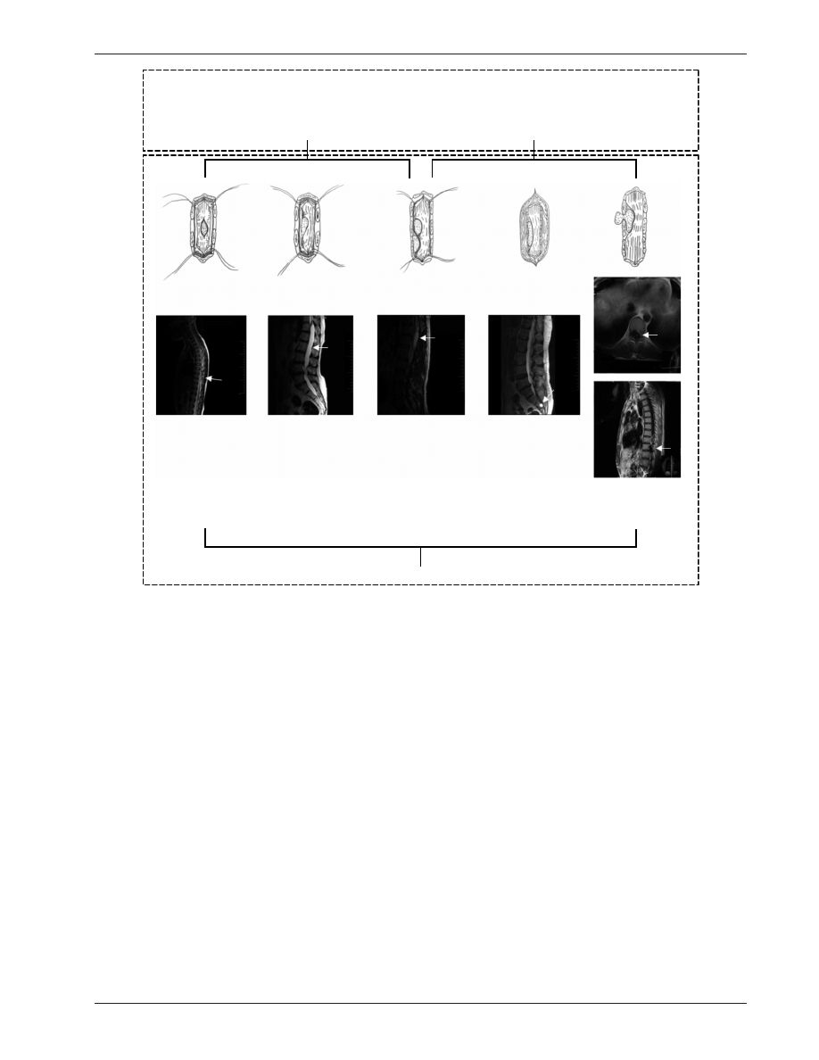
Classification using the five-category
system
Both anatomical location and abnormalities observed by MRI
were assessed for each patient. Surgical procedure of spinal
arachnoid cysts was determined before the year 1995 in our
department. Spinal arachnoid cysts were subdivided into
five types: 1) intramedullary cysts/syrinxes, 2) subdural
extramedullary, 3) subdural/epidural, 4) intraspinal epidural,
or 5) intraspinal/extraspinal (Figure 1). If surgical observa-
tion was inconsistent with preoperative evaluation by MRI,
the surgical procedure was modified according to intraopera-
tive observations.
Neuropsychiatric Disease and Treatment 2014:10
submit your manuscript
501
Spinal arachnoid cyst stratification
Surgical procedures
Surgical treatment was selected based on spinal arachnoid
cyst type and conducted by a team of two trained surgeons
and two assistants. All surgeries were conducted at the same
facility. The use of total or partial resection was recorded
for each patient.
intramedullary cyst treatment
For patients with this kind of cyst, experimental puncture was
performed to identify the location of the cysts, then an inci-
sion along the posterior median sulcus was made to achieve
cyst opening (the length of the incision varied according to
the size of the cyst). For some patients with dense adhesion
between the cyst wall and spinal cord, the separation of the
cyst was not continued if it was very difficult to separate. On
the contrary, the clinicians removed the parts that could be
separated or sutured the pia mater to the cyst wall and ensured
the connection of the cyst cavity and the subarachnoid space
to prevent the recurrence of the cyst.
Subdural extramedullary cyst treatment
Subdural extramedullary spinal arachnoid cysts feature
abnormal thickening and adhesion of the arachnoid caused
by congenital aplasia and/or inflammatory responses.
These abnormalities are commonly found at the ventral and
ventrolateral spinal cord. Posterior shifting of the spinal
cord is generally found in patients with cysts at the ventral
and ventrolateral spinal cord, and thickening and adhesion
of the arachnoid that could lead to dense adhesion between
the spinal cord and the endorrhachis. Thus the incision of
the endorrhachis should be carefully performed with the
assistance of endoscopy to avoid injuries to the spinal cord.
The long-term pressure on the spinal cord could result in the
adhesion and thickening of the adjacent arachnoid and poor
Subdural cyst
Epidural cyst
A
B
Intramedullary
cyst
Subdural
extramedullary
cyst
Subdural
epidural
cyst
Intraspinal multiple cyst
Intraspinal
epidural
cyst
Intraspinal
extraspinal
cyst
Figure 1 (A and B) Classification of spinal arachnoid cysts. (A) Two anatomical types of spinal arachnoid cysts; (B) novel five-type classification system determined by
magnetic resonance imaging (MRI).

Neuropsychiatric Disease and Treatment 2014:10
submit your manuscript
502
Qi et al
spinal cord pulsation. Thus the adhesive arachnoid between
the spinal cord and the adjacent endorrhachis was separated
carefully and removed as much as possible to release the
spinal cord. However, the operational view of the surgical
procedures on the cysts at the ventral spinal cord is generally
limited, and traction of the spinal cord should be avoided as
much as possible to avoid damage to the spinal cord nerves
and adjacent vessels.
Subdural/epidural cyst treatment
Subdural/epidural cysts were treated by resection of the cyst
wall. If no dense adhesion between the cyst wall and the end-
orrhachis or nerve root was found, the cyst was separated until
the neck of the cyst, and then tight suturing was performed
after resection of the cyst. In contrast, if dense adhesion
between the cyst wall and nerve root made the separation
of the cyst very difficult, partial removal of the cyst was
performed, and then tight suturing was performed after the
connecting hole had been filled with a section of free muscle
mass. For cysts not connected with the subarachnoid space,
the cyst wall was removed as much as possible. In cases of
dense adhesion between nerve root and the cyst, the cyst
wall was partially resected for drainage and decompression
before tight overlapping suturing. A section of free muscle
mass was then positioned on the dura at the site of the cyst
and fixed by suture and inward pressure to prevent new cyst
formation.
intraspinal epidural cyst treatment
Intraspinal epidural cysts were treated by ligation of the
cervix. In some cases, the muscle mass was isolated and used
to plug the access hole prior to suturing of the cyst walls.
If the nerve root or dural adhesion was weak, the cyst was
mobilized to the neck, resected, and sutured tightly. However,
in cases of very strong nerve root or dural adhesion the cyst
wall was partially resected and a piece of free muscle mass
was used to plug the access hole prior to tight suturing.
Intraspinal/extraspinal cyst treatment
Intraspinal and extraspinal cysts were removed through
enlarged intervertebral foramina. Extraspinal cysts are easy
to be treated. Operation of intraspinal cysts is similar to
subdural/epidural cysts. If no dense adhesion between the
cyst wall and the endorrhachis or nerve root was found, the
cyst was separated until the neck of the cyst, and then tight
suturing was performed after resection of the cyst. If dense
adhesion between the cyst wall and nerve root made the
separation of the cyst difficult, partial removal of the cyst
was performed, and then tight suturing was performed with
a section of free muscle mass. For cysts not connected with
the subarachnoid space, the cyst wall was removed.
Postoperative follow-up
All patients were followed up, with a mean period of
69 months (range 3–187 months). Cases of recurrence, death,
or second surgery were recorded.
Fugl-Meyer scale assessments
FM scores were assessed preoperatively and at postoperative
day 90 on a 0- to 99-point scale. As previously described,
13,14
FM scores were assessed as severe or marked motor impair-
ment (0–84), moderate motor impairment (85–95 points),
and slight motor impairment (96–99 points).
Preoperative FM scores of all patients were less than
50 points after conservative therapy. Significant improvement
was determined when postoperative FM score achieved 96–99,
and clinical symptoms disappeared with incident pain or numb-
ness; patients had a normal life and work. Improvement was
determined when postoperative FM score achieved 85–95,
and parts of symptoms disappeared but some moderate motor
impairment still remained. No improvement was determined
when postoperative FM score remained 0–84.
Results
Patients’ demographic and clinical
characteristics
Among the 81 included patients, 34 were males and 47 were
females, with a mean age of 36.5 years (ranging from 6 to
66 years) (Table 1). Intraspinal epidural cysts were signifi-
cantly more frequent than other types – 66.7% (54 of 81).
Ten (12.4%) patients developed multiple lumbar and lum-
bosacral segment intraspinal cysts. Intraspinal epidural cysts
were predominantly located in the lumbar and lumbosacral
segments (83.3%, 45 of 54) and in the thoracic and thora-
columbar segments (18.5%, 15 of 81). The incidence of
subdural extramedullary cysts was 18.5% (15 of 81), with
cysts located primarily in the thoracic and thoracolumbar seg-
ments (93.3%, 14 of 15). Eight patients had intramedullary
cysts in the cervical and cervicothoracic segments (62.5%,
five of eight) and the thoracic and thoracolumbar segments
(37.5%, three of eight). Intraspinal/extraspinal cyst (three
of 81) occurrence was rare, occurring in significantly fewer
patients than other types (Table 1).
The clinical symptoms of the patients with spinal arach-
noid cyst varied with the location of the cyst. For patients with
the cyst at the cervical level, the symptoms were mainly pain
Neuropsychiatric Disease and Treatment 2014:10
submit your manuscript
503
Spinal arachnoid cyst stratification
Table
1
Incidence, resection modality, and prognosis of the five types of spinal arachnoid cysts
Patients, n
Sex (male), n
Mean age
Resection modality
Prognosis (FM scale)*
Subtotal
resection, n (%)
Total
resection, n
Significant
improvement, n
Improvement,
n
No
improvement, n
Intramedullary cysts/syrinxes
8 (100%)
3 (37.5%)
33.0
6 (75%)
2 (25%)
8 (100%)
0 (0%)
0 (0%)
c
1–T2
5 (62.5%)
2 (40%)
32.4
4 (80%)
1 (20%)
5 (100%)
0 (0%)
0 (0%)
T3–L1
3 (37.5%)
1 (33.3%)
34.0
2 (66.7%)
1 (33.3%)
3 (100%)
0 (0%)
0 (0%)
l2–
s1
0
0
0
0
0
0
0
0
Subdural extramedullary
15 (100%)
6 (40.0%)
30.3
10 (66.7%)
5 (33.3%)
13 (86.7%)
2 (13.3%)
0 (0%)
c
1–T2
1 (6.7%)
0 (0%)
29.0
1 (100%)
0 (0%)
1 (100%)
0 (0%)
0 (0%)
T3–L1
14 (93.3%)
6 (42.9%)
30.4
9 (64.3%)
5 (35.7%)
12 (85.7%)
2 (14.3%)
0 (0%)
l2–
s1
0
0
0
0
0
0
0
0
Subdural/epidural
1 (100%)
1 (100%)
38.0
0 (0%)
1 (100%)
1 (100%)
0 (0%)
0 (0%)
c
1–T2
0
0
0
0
0
0
0
0
T3–L1
1 (100%)
1 (100%)
38.0
0 (0%)
1 (100%)
1 (100%)
0 (0%)
0 (0%)
l2–
s1
0
0
0
0
0
0
0
0
intraspinal epidural
54 (100%)
23 (42.6%)
31.2
19 (35.3%)
35 (64.8%)
36 (66.7%)
13 (24.1%)
5 (9.26%)
c
1–T2
0
0
0
0
0
0
0
0
T3–L1
9 (16.7%)
4 (44.4%)
31.1
0 (0%)
9 (100%)
5 (55.6%)
4 (44.4%)
0 (0%)
l2–
s1
45 (83.3%)
19 (42.2%)
31.2
19 (42.2%)
26 (57.8%)
31 (68.9%)
9 (20%)
5 (11.1%)
Intraspinal/extraspinal
3 (100%)
1 (33.3%)
39.6
0 (0%)
3 (100%)
3 (100%)
0 (0%)
0 (0%)
c
1–T2
0
0
0
0
0
0
0
0
T3–L1
2 (66.7%)
1 (50%)
40.5
0 (0%)
2 (100%)
2 (100%)
0 (0%)
0 (0%)
l2–
s1
1 (33.3%)
0 (0%)
38.0
0 (0%)
1 (100%)
1 (100%)
0 (0%)
0 (0%)
Notes:
*Fugl-Meyer
(FM)
scores
were
evaluated
at
postoperative
day
90.
FM
scores
in
preoperation
patients
were
less
than
50
points.
Significant
improvement:
slight
motor
impairment
(96–99
points).
Improvement:
moderate
motor
impairment (85–95 points). No improvement: severe or marked motor impairment (0–84 points).

Neuropsychiatric Disease and Treatment 2014:10
submit your manuscript
504
Qi et al
at the neck, shoulder, and upper limbs; weakness of the upper
limbs could also be observed. For patients with the cyst at
the thoracic level, the major symptoms were pain in the chest
and back, as well as spastic paralysis of the lower limbs. For
patients with the cyst at the lumbosacral level, lumbosacral
pain and lower-limb pain could occur when abdominal
pressure increased. As some cysts are connected with the
subarachnoid space, the cerebrospinal fluid could flow into
the cyst cavity when the abdominal pressure increased and
caused enlargement of the cyst, which could compress the
nerve root and aggravate the symptoms. When the patients
were put in the horizontal position, the cyst shrank, relieving
the symptoms. Sphincter dysfunction could be observed in
the late period of the disease, and urination- and defecation-
function impairment could also occur in some cases that
needed more time to recover after the operation.
Surgical difficulty varied according
to type and anatomical location
Total resection was not performed in 55.6% (45 of 81) of
patients due to surgical difficulties. Compared to all other
types, patients with intramedullary and subdural extramedul-
lary cysts were more difficult to surgically handle. For these
patients, subtotal resection was significantly more common
than total resection (intramedullary, six versus two; subdural
extramedullary, ten versus five). Notably, 100% (ten of ten) of
patients with multiple intraspinal cysts experienced surgical dif-
ficulties and thus underwent subtotal resection. Total resection
was performed in 100% (ten of ten) of patients with subdural/
epidural cysts. In patients with intraspinal epidural cysts,
total resection was significantly more common than subtotal
resection, applicable in 100% (nine of nine) of cases affecting
thoracic and thoracolumbar segments and in 57.8% (26 of 45)
of cases affecting lumbar and lumbosacral segments.
FM scores
All patients (100%, eight of eight) with intramedullary cysts/
syrinxes reported slight FM-score improvement. The major-
ity of subdural extramedullary cyst patients experienced
slight improvements (13 of 15, 86.7%), and none of these
patients reported no improvement. All subdural/epidural
(100%, one of one) and intraspinal/extraspinal (100%, three
of three) reported improvements. Notably, in the largest
patient group, intraspinal epidural cyst patients, only 66.7%
(36/54) of patients reported slight improvements, and 9.26%
(five of 54) reported no improvement, making these patients
significantly more likely to experience no postoperative
improvement in FM score.
Outcomes and recurrence
Two patients suffered from an intramedullary cyst recurrence.
Their first surgery was posterior myelotomy and cyst
opening. Recurrence was observed 2 months after operation
in both patients. During the second operation, the incision of
the spinal cord was found to be connected by scars, which led
to the recurrence of the cyst. These two patients recovered
after the cysts were totally removed, and no recurrence was
identified by the end of the follow-up. No death was observed
in any patient.
Discussion
The current study used a novel five-category classifica-
tion system for spinal arachnoid cysts. Intraspinal epidural
spinal arachnoid cysts were significantly more common
than other cyst types, followed by subdural extramedullary
and intramedullary cysts/syrinxes. Notably, conventional
classification systems that only use anatomical location for
diagnosis fail to consider intraspinal epidural spinal arach-
noid cysts as a distinct type, despite the current indications
that these patients are much more likely to have limited or
no improvement following routine surgical intervention.
Thus, the use of the five-category classification system for
spinal arachnoid cysts clearly indicated that some patients
were at greater risk for poor outcomes following surgery,
requiring more careful treatment planning. Wider use of
the five-category classification may allow a better clinical
assessment of patients at risk for poor surgical outcomes after
failure of a conservative treatment.
Though conventional classification of spinal arachnoid
cysts may be effective in most patients, the present study indi-
cates that there is a distinct subpopulation of patients that will
have no evidence of symptom improvement after surgery.
A number of previous clinical studies have identified charac-
teristics of this at-risk population,
14–19
including patients with
slight adherence of the nerve root and cyst, separation of the
cervical region of the cyst, and close adherence between the
nerve root and cyst during resection, complicating separation
or leading to partial resection. Hamamcioglu et al
20
reported
a case of extradural cyst, unusual in size, location, and clini-
cal features, that complicated a routine surgery, due to the
required movement of muscle mass to close the dural defect
after excision. Similarly, Lee and Cho
21
reported that while
complete surgical excision was the best treatment for a vari-
ety of spinal arachnoid cyst types, the pleural cavity or right
atrium modality should be based on MRI findings rather than
on anatomical classifications. Thus, the additional benefits of
identifying and stratifying spinal arachnoid cysts based on

Neuropsychiatric Disease and Treatment 2014:10
submit your manuscript
505
Spinal arachnoid cyst stratification
both anatomical classification and MRI findings have been
increasingly reported, and the current study offers the first
system for standardizing these recommendations.
The current study observed that intraspinal epidural
spinal arachnoid cysts were the most common. Though
intraspinal cyst cases have been previously reported,
22
the
intraspinal epidural classification is unique to the proposed
system. The present study demonstrated that these cases are
more likely to experience poor surgical outcomes as well as
limited functional improvements and symptom alleviation,
consistent with a previous report that these cases are often
afflicted with complications, such as spinal disk herniation.
22
Additionally, though subdural/epidural cysts (one patient)
and intraspinal multiple cysts (ten patients) were relatively
rare in this study, examination of larger cohorts of these
patients may reveal that they are similarly at risk, due to the
unique nature of these lesions and potential for complica-
tions, particularly when they occur in ventral locations.
23
Additionally, trauma and labor in females can result in
multiple spinal arachnoid cysts, where the presence of more
than one lesion complicates treatment even further.
24
Thus,
classification schemes that use only anatomical location
may increase the risk of poor surgical outcomes in many
patient subpopulations. Therefore, these patients should be
identified early in the treatment process, and specific surgical
procedures and treatment plans should be developed based
on MRI as well as anatomical findings.
While the proposed five-category classification sys-
tem is based on MRI findings of cyst manifestation, other
researchers have employed stratification by clinical signs
and symptoms.
24,25
Wang et al
23
reported that patients with
preoperative neuropathic pain or numbness were much less
likely to benefit from surgery than those with myelopathy
or weakness, suggesting that surgical success could also be
improved by the use of intraoperative ultrasound to guide
aggressive surgical treatment. Based on size and clinical
symptoms, it may be possible to determine more readily
whether complete resection, shunting, or duraplasty will most
optimally result in a high rate of cyst and syrinx oblitera-
tion without symptom recurrence,
24
particularly in the case
of giant spinal arachnoid cysts.
25
Thus, further study will
be required to determine whether the current five-category
system is significantly related to alleviation of certain symp-
toms, requiring investigation before wide implementation of
these findings.
Cysts could also be classified according to their etiology.
However, such classifications need to be validated, because
cysts at different locations could have the same causes, and
the same kinds of cysts could also have different causes.
Nevertheless, the predilection sites vary with the types of
cysts. In the present study, all the subdural extramedullary
cysts were found at the cervicothoracic level, while the
extradural cysts were mainly located at the lumbosacral
level. However, there were nine patients with extradural cysts
located at the cervicothoracic level. Several similar clinical
symptoms were found between the patients with subdural
extramedullary and extradural cysts at the cervicothoracic
vertebra. For patients with the cysts located at the thoracic
or thoracolumbar level, numbness, weakness, and pain of
lower extremities were generally observed. For patients with
cysts located at the lumbosacral level, radiculalgia, extrem-
ity spasticity, and weakness were generally observed, and
urination- and defecation-function disturbances were also
found in some patients.
Considering these findings, the retrospective nature, wide
time intervals between treatments, and the possibility of
unrecognized selection and recall biases must be considered.
A selection bias might be due to the fact that all included
patients did not respond to conservative treatment and had to
undergo surgery. In addition, due to the retrospective nature
of the study, FM scores were not available for all patients,
since scores were assessed in patients with obvious symptoms
at admission. Furthermore, considerable changes in surgi-
cal technology from 1995 to now may further complicate
the interpretation of these findings. However, the rarity of
the condition necessitates a long study period to achieve a
relevant number of cases. In addition, the group of selected
patients all had a preoperative FM score ,50 after conserva-
tive treatment. Surgery is only an alternative treatment, and
many patients did not improve their FM score after surgery,
indicating that the surgical strategies could still be improved
and that a careful selection of patients for surgery could
improve surgical outcomes. Nevertheless, validation in larger
sample sizes is required, potentially as a multicenter effort.
After failure of a conservative treatment, patients with the
most common cyst type, intraspinal epidural cysts, were the
most likely to exhibit poor outcomes following conventional
resection surgery. By using combined MRI imaging and
anatomical findings, the proposed five-category classifica-
tion provides a critically needed means for classifying spinal
arachnoid cyst patients at risk for poor treatment outcomes or
symptom alleviation, thereby allowing for divergent surgical
intervention for specific cyst types. This may lead to a greater
standardization of care and improved overall prognoses
for spinal arachnoid cyst patients. Despite these positive
preliminary indications of the effectiveness of this system,

Neuropsychiatric Disease and Treatment
Publish your work in this journal
Submit your manuscript here: http://www.dovepress.com/neuropsychiatric-disease-and-treatment-journal
Neuropsychiatric Disease and Treatment is an international, peer-
reviewed journal of clinical therapeutics and pharmacology focusing
on concise rapid reporting of clinical or pre-clinical studies on a
range of neuropsychiatric and neurological disorders. This journal
is indexed on PubMed Central, the ‘PsycINFO’ database and CAS.
The manuscript management system is completely online and includes
a very quick and fair peer-review system, which is all easy to use. Visit
http://www.dovepress.com/testimonials.php to read real quotes from
published authors.
Neuropsychiatric Disease and Treatment 2014:10
submit your manuscript
506
Qi et al
larger multicenter prospective studies will be required to
confirm these observations, particularly in rarely reported
spinal arachnoid cyst subtypes.
Disclosure
The authors report no conflicts of interest in this work.
References
1. Hughes G, Ugokwe K, Benzel EC. A review of spinal arachnoid cysts.
Cleve Clin J Med. 2008;75:311–315.
2. Pradilla G, Jallo G. Arachnoid cysts: case series and review of the
literature. Neurosurg Focus. 2007;22:E7.
3. Bitaraf MA, Zeinalizadeh M, Meybodi AT, Meybodi KT, Habibi Z.
Multiple extradural spinal arachnoid cysts: a case report and review of
the literature. Cases J. 2009;2:7531.
4. Zhou JY, Pu JL, Chen S, Hong Y, Ling CH, Zhang JM. Mirror-image
arachnoid cysts in a pair of monozygotic twins: a case report and review
of the literature. Int J Med Sci. 2011;8:402–405.
5. Oberbauer RW, Haase J, Pucher R. Arachnoid cysts in children:
a European co-operative study. Childs Nerv Syst. 1992;8:281–286.
6. Rengachary SS, Watanabe I. Ultrastructure and pathogenesis of intracranial
arachnoid cysts. J Neuropathol Exp Neurol. 1981;40:61–83.
7. Choi JY, Kim SH, Lee WS, Sung KH. Spinal extradural arachnoid cyst.
Acta Neurochir (Wien). 2006;148:579–585; discussion 585.
8. Brant WE, Helms CA. The Brant and Helms Solution: Fundamentals
of Diagnostic Radiology. 3rd ed. Philadelphia: Lippincott Williams and
Wilkins; 2006.
9. Go KG, Hew JM, Kamman RL, Molenaar WM, Pruim J, Blaauw EH.
Cystic lesions of the brain. A classification based on pathogenesis, with
consideration of histological and radiological features. Eur J Radiol.
1993;17:69–84.
10. Galassi E, Tognetti F, Gaist G, Fagioli L, Frank F, Frank G. CT scan
and metrizamide CT cisternography in arachnoid cysts of the middle
cranial fossa: classification and pathophysiological aspects. Surg
Neurol. 1982;17:363–369.
11. Nabors MW, Pait TG, Byrd EB, et al. Updated assessment and current
classification of spinal meningeal cysts. J Neurosurg. 1988;68:
366–377.
12. Neurosurgical Video Gallery. Intraspinal cysts. Available from: http://
www.neurosurgery.tv/intraspinalcysts.html. Accessed March 27,
2013.
13. Clancey JK. Karnofsky performance scale. J Neurosci Nurs.
1995;27:220.
14. Mor V, Laliberte L, Morris JN, Wiemann M. The Karnofsky
Performance Status Scale. An examination of its reliability and validity
in a research setting. Cancer. 1984;53:2002–2007.
15. Raeder MB, Helland CA, Hugdahl K, Wester K. Arachnoid cysts cause
cognitive deficits that improve after surgery. Neurology. 2005;64:
160–162.
16. Jallo GI, Woo HH, Meshki C, Epstein FJ, Wisoff JH. Arachnoid cysts
of the cerebellopontine angle: diagnosis and surgery. Neurosurgery.
1997;40:31–37; discussion 37–38.
17. Koch CA, Voth D, Kraemer G, Schwarz M. Arachnoid cysts: does
surgery improve epileptic seizures and headaches? Neurosurg Rev.
1995;18:173–181.
18. Gangemi M, Maiuri F, Colella G, Sardo L. Endoscopic surgery for large
posterior fossa arachnoid cysts. Minim Invasive Neurosurg. 2001;44:
21–24.
19. Kunz U, Mauer UM, Waldbaur H. Lumbosacral extradural arachnoid
cysts: diagnostic and indication for surgery. Eur Spine J. 1999;8:
218–222.
20. Hamamcioglu MK, Kilincer C, Hicdonmez T, Simsek O, Birgili B,
Cobanoglu S. Giant cervicothoracic extradural arachnoid cyst: case
report. Eur Spine J. 2006;15 Suppl 5:595–598.
21. Lee HJ, Cho DY. Symptomatic spinal intradural arachnoid cysts in the
pediatric age group: description of three new cases and review of the
literature. Pediatr Neurosurg. 2001;35:181–187.
22. Kono K, Nakamura H, Inoue Y, Okamura T, Shakudo M, Yamada R.
Intraspinal extradural cysts communicating with adjacent herniated
disks: imaging characteristics and possible pathogenesis. AJNR Am J
Neuroradiol. 1999;20:1373–1377.
23. Wang MY, Levi AD, Green BA. Intradural spinal arachnoid cysts in
adults. Surg Neurol. 2003;60:49–55; discussion 55–46.
24. Ergun T, Lakadamyali H. Multiple extradural spinal arachnoid
cysts causing diffuse myelomalacia of the spinal cord. Neurologist.
2009;15:347–350.
25. Kumar S, Chauresia P, Singh D, Singh H. Giant thoracolumbar intradural
multilobulated arachnoid cyst. Neurol India. 2012;60:134–135.
Wyszukiwarka
Podobne podstrony:
A Comparison between Genetic Algorithms and Evolutionary Programming based on Cutting Stock Problem
Using Predators to Combat Worms and Viruses A Simulation Based Study
Parlour And Seppi Liquidity Based Competition For Order Flow
racismz int (2) , Racism has become one of the many burdens amongst multi-cultural worlds like Canad
15 Multi annual variability of cloudiness and sunshine duration in Cracow between 1826 and 2005
Comprehending conventional and novel metaphors An ERP study
Of Mice and Man Emotional Reaction to the Novel
Pride and Prejudice The Theme of Pride in the Novel
Pride and Prejudice Analysis of the Theme of the Novel
Jakobsson, Vampires and Watchmen Categorizing the Mediaeval Icelandic Undead
Energy and Fuels 2006, 20, 155 158, Novel Process for Recycling Waste Plastics To Fuel Gas Using a M
Multi objective thermodynamic optimization of combined Brayton and inverse Brayton cycles using gene
Dracula s Women The Representation of Female Characters in a Nineteenth Century Novel and a Twentie
Sean Michael A Hammer Novel 18 Tanny and the Cage Toy Box Cage
Design and construction of three phase transformer for a 1 kW multi level converter
Jeff Mann Fog ( A Novel of Desire and Reprisal )
Ivan to Make You Laugh Jokes and Novel, Scott K Peterson
więcej podobnych podstron