
24
Surface Biology : Analysis of Biomolecular Structure by Atomic Force
Microscopy and Molecular Pulling
Emin Oroudjev, Signe Danielsen, and Helen G. Hansma
24.1
Introduction
During the past decade, the atomic force microscope has been developed from an exotic
and sometimes home-made instrument into a relatively widespread surface-imaging and
probing instrument. Atomic force microscopy (AFM) and its related techniques have pro-
liferated successfully into many different fields, including biology. Today, the method is
used to obtain high-resolution static and dynamic images in investigations of the physical
and mechanical properties of biological macromolecules. As a result, the number of AFM-
related articles listed in PubMed has mushroomed such that the recommended number of
references for a minireview constitutes little more than 10 % of the total number referred
to in PubMed for a single year. This review will therefore, of necessity, neglect many ex-
cellent reports on the subject. Reviews published during the past year on AFM include
Refs. [1–12], but other recent reviews are available in Refs. [13–20].
The atomic force microscope and other scanning probe microscopes have been used to
pioneer a new field, that of surface biology. This is a logical advance in biological method-
ology, as we now know that cells are not simply bags of cytoplasm but are bounded by
surface-membranes which comprise incredible arrangements of macromolecular com-
plexes for signaling, communicating, and regulating cellular functions. Furthermore,
we now know that cells are filled with complex structures and membrane-bound orga-
nelles, such that most of life’s processes actually occur on the cell surfaces. This new biol-
ogy – surface biology – goes beyond the realms of test-tube biology which have been so
successful in our learning about ourselves. Research into surface biology is indeed per-
formed at surfaces which sometimes are physiologically relevant (e. g., lipid bilayers)
and sometimes are physiologically irrelevant (e. g., mica). Mica serves as a flat support
upon which biomaterials can be observed, either by direct imaging or by pulling or
through the use of other techniques to probe the properties of the biomaterials.
AFM is now typically used in conjunction with other techniques to probe biomaterials.
This is a continuation of the progression first noted in 1998, at which time a large propor-
tion of biological reports included the term ‘AFM’ in their title, to a present-day situation
where this is limited to 50 % or fewer cases [21]. Today, the use of AFM in conjunction
387
Nanobiotechnology. Edited by Christof Niemeyer, Chad Mirkin
Copyright
c 2004 WILEY-VCH Verlag GmbH & Co. K aA, Weinheim
ISBN 3-527-30658-7
G

with other techniques is a further indication of the technique having matured from a no-
velty to one which generates significant amounts of scientific material. Indeed, nowadays
AFM is used in conjunction with other analytical methods and with theoretical analyses
on a highly frequent basis.
24.2
Recent Results
Research investigations into many biological macromolecules, including those for DNA,
RNA, and proteins, as well as other biopolymers and their corresponding supramolecular
structures such as viruses, living cells and cell colonies, have all benefited from recent de-
velopments in the field of AFM.
24.2.1
DNA
DNA was one of the first biological objects to be visualized by AFM, and the imaging tech-
nique has been used not only for DNA mapping and sizing but and also examining the
structural changes induced by interactions with enzymes, DNA-binding proteins and con-
densing agents. By using AFM to image DNA–protein interactions, the location of pro-
tein-binding sites and the conformation of DNA at these sites have been identified. In
fact, in some cases it has been possible to determine the stoichiometry of these interac-
tions. For a recent review of AFM studies of DNA, see Ref. [18].
Detailed information can often be obtained about the conformations and organization
of DNA and RNA molecules, as well as the native and artificial nanostructures con-
structed from these nucleic acids. DNA sizing, fingerprinting and/or mapping can be per-
formed by imaging DNA restriction fragments and using automated software packages to
measure their lengths (e. g., Ref. [22]). Some other methods of mapping sequences on
large DNA molecules are based on the ability of these sequences to interact specifically
with complementary oligonucleotides or with certain proteins. The hybridization area be-
tween DNA and the probe will usually display an increased width and height, or some
other substructure resulting from DNA–probe interactions (loops, hairpins, kinks, etc.),
and these effects can be further enhanced by tagging the probe with nanoparticles
(gold nanospheres, biotin–avidin complex, etc). In one example of DNA mapping, a
300 nucleotide-long RNA probe was hybridized to double-stranded DNA (ds-DNA), where-
upon the site of hybridization had a distinctive appearance due to presence of the double-
stranded RNA–DNA hybrid and unpaired single-stranded DNA [23]. Triple-helix-forming
DNA oligonucleotides can also be used as probes in these experiments.
24.2.1.1
2.1.1 DNA Condensation
DNA condensation is another area of the DNA field where AFM plays a significant role.
(Figure 24.1). Nonviral methods of condensing long DNA molecules, to a size that is ap-
propriate for gene delivery into cells, are now becoming increasingly important as health
complications and even human deaths have been reported recently as a result of viral-
based gene therapy.
388
24 Surface Biology : Analysis of Biomolecular Structure by Atomic Force Microscopy and Molecular Pulling
Of special interest has been the use of polycations for the development of nonviral gene
delivery systems. One of the present authors (S. D.) has been using the linear polymer
chitosan, a polycation derived from the polysaccharide chitin. Chitosan has emerged as
a promising candidate for this purpose as it is nontoxic, biodegradable, and has been
shown to yield high transfection efficiency [24]. The ability of various chitosans to compact
DNA has been studied with AFM imaging (Danielsen and Stokke, in preparation), show-
ing that chitosan effectively compacts DNA into toroidal, rod-like, and globular structures.
This distribution of geometries was found to depend upon both the charge density and
the degree of polymerization (DP) of the chitosan, with the low-DP chitosans resulting
in less well-defined structures.
The AFM technique called chemical force microscopy was proposed to improve current
high-throughput DNA micro-array screening analyses. DNA arrays are imaged in friction
force mode with chemically modified probes that have complementary DNA probes at-
tached to the tip. Increases in friction forces are detected as the tip reaches spots on
389
24.2 Recent Results
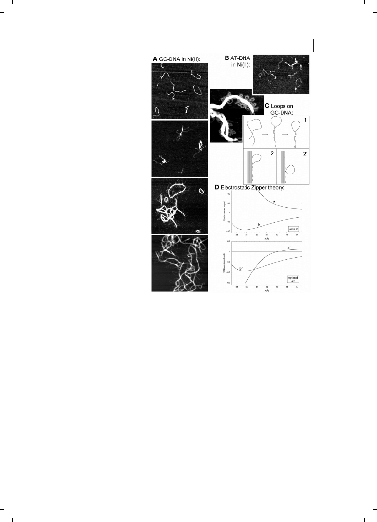
Figure 24.1
Sequence-dependent
DNA condensation [86] (A) Poly (dG-
dC)
p(dC-dG) (GC-DNA) condenses in
1 mM NiCl
2
. Times in Ni(II), top to
bottom, = 5 sec, 1 min, 3–7 min, 2
months. (B) Poly (dA-dT)
p(dT-dA)
(AT-DNA) barely condenses after
3–5 min, even in 6 mM NiCl
2
.
Images are 1 mm wide; DNA con-
centration = 2.5 ng mL
–1
in (A) and
(B). (C) ds-DNA loops are typically
seen on bundles of condensed GC-
DNA : AFM image (left) and models
(below) for formation of loops on
bundles, progressing from stage 1 to
2 and 2
l. (D) Electrostatic Zipper
theory [87] extended to condensation
of GC-DNA and AT-DNA in Ni(II).
Graphs show force per unit length
between parallel ds-DNA molecules
versus intermolecular separation, at
low ionic strength, as predicted by
Eq. 2 in [sitko. Upper graph is for
zero axial shift between parallel ds-
DNA molecules; lower graph is for
optimal axial shift, to maximize
electrostatic attraction between par-
allel ds-DNA molecules. Curves a
and a
l correspond to AT-DNA, which
is in the B-DNA conformation in
Ni(II); curves b and b
l correspond to
GC-DNA, which is in the Z-DNA conformation in Ni(II). Attraction (negative forces) at zero axial shift, as
in curve b, is indicative of DNA condensation.

the DNA array that contain DNA complementary to DNA on the tip. Enhanced sensitivity
and spatial resolution of this method relative to current micro-array technology may make
it possible to create significantly smaller and more densely packed micro-arrays or nano-
arrays of DNA.
By using AFM to pull on single DNA molecules, information about single molecular
mechanics and thermodynamics has been obtained. The forces required for DNA’s B-S
transition [25] and the attraction force between DNA molecules and cationic lipid bilayers
have been measured [26].
24.2.1.2
DNA Sequences Recognized by Mica
Some intrinsically curved DNA sequences contain phased A-tracts, such that half of each
helix turn has a run of As, paired with a run of Ts. Sequence-dependent DNA curvature
such as this is an important element in specific DNA–protein interactions.
A-rich intrinsically curved DNA sequences were joined in head-to-head and head-to-tail
palindromes to form ‘s’ or backwards ‘s’ conformations, depending on which side faced
upwards. When these DNAs were deposited onto mica, the T-rich side bound preferen-
tially to mica [27] – a finding which may have potential application(s) in the field of na-
nobiotechnology.
24.2.1.3
Drug-binding to Single ds-DNA Molecules
Molecular pulling may also become a valuable technique for monitoring the binding of
drugs to DNA, and preliminary results in this area have recently been published [28]. Re-
cently, a minor-groove binding drug (berenil), a cross-linking drug (cisplatin) and an inter-
calator (ethidium bromide) were each tested for their effects on the DNA pulling curves.
At high levels, all three drugs completely or mostly abolished the B-to-S transition, but
cisplatin also reduced the hysteresis between pulling and relaxing the DNA, even at
short incubation times [28].
24.2.2
Proteins
Proteins are the second largest class of biological macromolecules to have benefited from
developments in AFM methods [9]. Methods to image both separate protein molecules
and protein layers with different degree of organization have been developed, and these
utilize native or modified mica surfaces (see section 24.3.2). Isolated protein molecules,
when applied to a surface, often are too mobile and too soft to be imaged with resolution
sufficient for observing any submolecular features. Immunoglobulin G (IgG) and IgM are
examples of submolecular resolution in the AFM imaging of isolated protein molecules.
With IgG, the three-lobed shape (as identified by X-ray crystallography) was also observed
by Anafi using AFM (see Ref. [29]) and imaging in aqueous buffer (see Ref. [30]). New
information about IgM substructure was obtained by using cryo-AFM, which showed a
tendency for IgM molecules to adopt a compact conformation with a raised center [31].
On the other hand, proteins deposited as a densely packed layer often produce images
with very high resolution [32, 33]. Some such protein arrays occur naturally in specialized
membranes such as the bacteriorhodopsin-containing purple membranes. To create these
390
24 Surface Biology : Analysis of Biomolecular Structure by Atomic Force Microscopy and Molecular Pulling
layers with other types of proteins, the proteins are a deposited onto artificial lipid bilayers
[34], the latter being prepared by vesicle fusion onto mica or silicon surfaces or by using a
Langmuir trough. The lipid monolayer that faces mica or silicon is usually made from
phosphatidylcholine or related lipids. The opposite monolayer, which is used to bind
and/or incorporate macromolecules of interest, can be constructed from different lipids
that have the desired chemistry at their headgroups. Biological macromolecules (proteins
and DNA) adsorbed onto these membrane-like surfaces are typically imaged with a reso-
lution that is superior to that achieved on conventional surfaces such as native or modified
mica.
AFM can be used to study different protein functions. For example, it was used to mea-
sure DNase I, DNA polymerase and RNA polymerase enzymes kinetics on surface-bound
substrate (DNA) [35–37]. The dynamics of interactions between large and small subunits
GroE chaperonin protein from Escherichia coli were studied using AFM at the level of in-
dividual protein molecules [38].
24.2.2.1
Prion Proteins
AFM analysis of a yeast prion protein, combined with Fourier transform infrared (FTIR)
spectroscopy, indicates that fibrils can form from the prion protein in its native helical
conformation and do not contain the crossed-beta structures typical of amyloid fibrils [39].
391
24.2 Recent Results
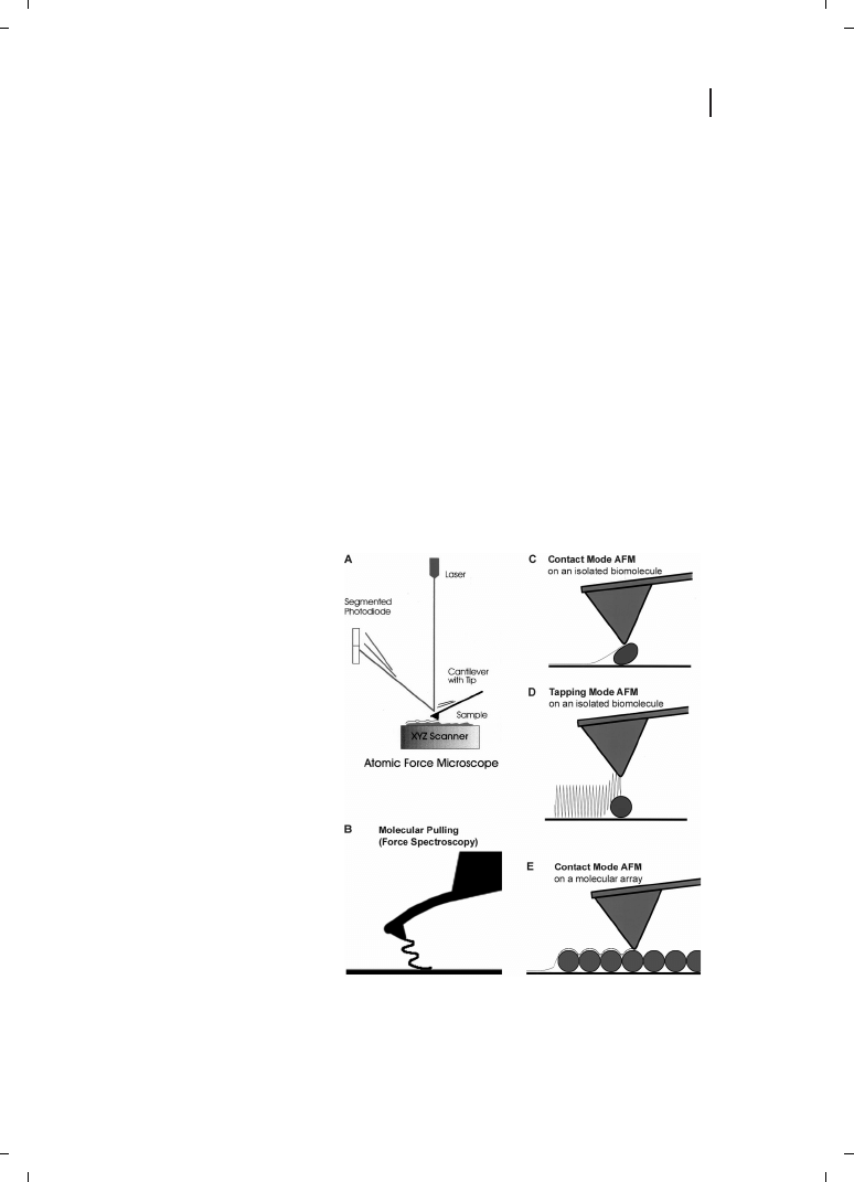
Figure 24.2
Atomic force microscopy
(AFM) and some of its uses for bio-
molecular imaging and probing. (A)
AFM schematic; see, for example, Ref.
[20] for details. (B) Molecular pulling,
or (Single Molecule) Force Spectro-
scopy is a method of obtaining infor-
mation about the mechanics and
folding of one or a few biomolecules.
It is also used to measure unbinding
forces and thermodynamic para-
meters of ligand–receptor interac-
tions. (C) Contact mode AFM tends
to deform, move or damage isolated
biomolecules. (D) Tapping mode AFM
reduces lateral forces on isolated
biomolecules. (E) Contact mode AFM
is often used for imaging biomolecu-
lar arrays, such as this “purple mem-
brane protein” array. Resolution is ty-
pically higher on closely packed arrays
of macromolecules than on isolated
macromolecules. There are at least
two explanations for this. First, the
packed macromolecules have less
freedom to move on the substrate.
Second, less of the tip interacts with the surface of the arrays; many tips have an asperity at the apex,
which can provide high-resolution imaging of relatively flat surfaces, such as those in protein arrays.
392
24 Surface Biology : Analysis of Biomolecular Structure by Atomic Force Microscopy and Molecular Pulling
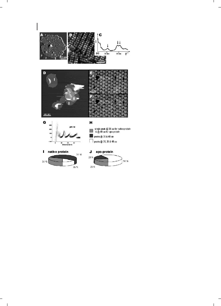
Figure 24.3
Membrane protein arrays
[40, 41]. (A–C) Native eye disc mem-
brane from the rod outer segment of a
vertebrate retina, adsorbed on mica
[40]. (A) Deflection image, showing that
three different surface types are evi-
dent : 1, the cytoplasmic side of the
disc membrane; 2, lipid; and 3, mica. To
avoid the formation of opsin, the chro-
mophore-depleted form of rhodopsin,
membrane samples were never ex-
posed to light. After adsorption of os-
motically shocked disc membranes
onto mica, their topography was mea-
sured in buffer solution (20 mM Tris–
HCl (pH 7.8), 150 mM KCl and 25 mM
MgCl
2
). (B) Height image of the cyto-
plasmic surface of the disc membrane,
showing rows of rhodopsin dimers, as
well as individual dimers (inside
dashed ellipse), presumably broken
away from one of the rows. The rho-
dopsin molecules protrude from the
lipid bilayer by 1.4
e 0.2 nm (n = 111).
This topograph is shown in relief, tilted
by 5
h. Vertical brightness range is
1.6 nm. (C) Angularly averaged powder-
diffraction pattern, showing peaks at
(8.4 nm)
–1
, (4.2 nm)
–1
and (3.8 nm)
–1
.
(D–J) Purple membranes from
Halo-
bacterium salinarum : imaging and ma-
nipulation of bacteriorhodopsin (BR)
molecules [41]. (D) Purple membrane patches (I) adsorbed onto freshly cleaved mica and imaged in
buffer solution (pH 7.8, 20 mM Tris-HCl, 300 mM KCl). In some areas, purple membrane patches overlap
with other membranes, forming double layers (II). (E) High-resolution image of the cytoplasmic purple
membrane surface showing BR trimers (outline) arranged into a hexagonal lattice. The topograph was
recorded at minimum force allowing the longest cytoplasmic loops of the individual BR molecules (loop
EF) to protrude fully from the membrane surface [88]. Individual defects show single or multiple BR
monomers missing. After imaging, the AFM tip was brought into contact to the membrane surface
(circle). This allowed the polypeptides of individual BR molecules to adsorb to the tip. During separation
of tip and sample, this molecular bridge was used to pull on the protein, and the force spectrum was
recorded. (F) Same purple membrane area imaged after the manipulation shows one individual BR
monomer missing (outline, now enclosing dimer instead of trimer). Vertical brightness range corresponds
to 50 nm (D) and 1.2 nm (E, F). (G) Force spectrum for unfolding of BR at pH 7.8 (n = 32). All molecules
were unfolded by grabbing the C-terminus at the cytoplasmic surface [46]. (H–J) Probability distribution of
pathways detected upon unfolding BR helices G and F. Probability of the unfolding pathways are shown
for native BR and for the apoprotein. Although 58 % of the unfolding events of apoprotein showed an
additional unfolding barrier at 26 aa, this barrier was observed in only 16 % of the unfolding traces of wild-
type BR.
24.2.2.2
Membrane Proteins
Protein arrays in membranes can typically be imaged at higher resolution than isolated
proteins, as shown diagrammatically for the “purple membrane” in Figure 24.2E and
the isolated “protein” in Figure 24.2C and D. This permits the visualization submolecular
structures and sometimes even substructural changes induced by such variables as pH
and imaging force. Gap-junction membranes, for example, show Ca(II)-dependent reduc-
tion in the diameter of the gap-junction pore at the extracellular surface and a force-depen-
dent collapse in pore structures at the cytoplasmic surface [5]. Figure 24.3 illustrates two
recent research accomplishments on membrane protein arrays. First, rhodopsin dimers in
a vertebrate retina were observed in a vertebrate retina (Figure 24.3A–C; see Ref. [40]). Sec-
ond, the structure of bacteriorhodopsin has been analyzed by pulling apart individual bac-
teriorhodopsin molecules (Figure 24.3D–J; see Ref., [41]). The basic principle of molecular
pulling is shown diagrammatically in Figure 24.2B.
393
24.2 Recent Results
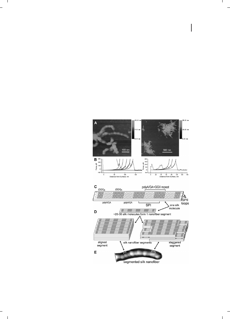
Figure 24.4
The molecular structure
of spider dragline silk nanofibers by
AFM and molecular pulling [42, 89].
(A) Nanofibers form from molecules
of a soluble bioengineered silk pro-
tein. These tapping-mode AFM
images in air show segmented sub-
structure of nanofibers and nanofi-
ber aggregates; similar images are
seen in aqueous fluid. (B) Two pull-
ing curves (single-molecule force
spectroscopy) on the silk molecules,
with WLC fits to rupture peaks. In
the first pull, much loosely adsorbed
protein fell off the tip before the ap-
pearance of sawtooth rupture peaks.
Rupture peaks are attributed to the
unfolding of 38-aa sequence repeats
from the silk protein. Peaks are typi-
cally 15 or 30 nm apart, which cor-
responds to the extended length of
one or two polyA/GA+GGX repeats
as shown in (C), below. (C) A model
for the folding of single silk mole-
cules. Length of the folded molecule
is based on the mean length of the
nanofiber segments (35
e 9 nm)
seen in (A) and (E). Amino-acid se-
quence is from [90]; zig-zags are
polyA/GA repeats, and spirals are
GGX repeats. D. Models for the
stacking of silk molecules into na-
nofiber segments. (E) AFM image of a segmented silk nanofiber, showing proposed relationship between
segments and nanofiber.

24.2.2.3
Spider Silk
Probe microscopy of a soluble bioengineered dragline silk protein provided results from
both AFM imaging and molecular pulling approaches (Figure 24.4). These results were
integrated into a molecular model for the folding pattern of individual silk molecules,
plus a multi-molecular model for the association of these molecules into segmented
silk nanofibers [42, 43].
This research involves the examination of mesostructures, which are more than single
molecules but much less than bulk materials. Pulling on native capture silk also produced
new information about spider silk at the mesostructure level [44].
24.2.3
Fossils
Today, even fossils are the subject of AFM research [45]. In the past, morphology has re-
presented the major approach to characterizing fossils, although the earliest fossils –
those of microorganisms – are too small for typical morphological observations to be
made. AFM has been used to detect 200 nm-sized angular platelets stacked in arrays in
the walls of petrified 650-million-year-old unicellular protists. In addition, Raman spectro-
scopy has been used to identify polycyclic aromatic kerogenous organic matter in the fos-
sils. These results support the conclusion that the microscopic structures are true fossils,
and not pseudo-fossil “look-alikes”.
24.2.4
Science and Nature
Reports judged worthy of inclusion into Nature and Science [46–63] are worth citing in
a review such as this, and news articles reported in both journals [64–68] are similarly
valuable to the reader. The reports described in more detail below are representative
of the breadth of topics covered in PubMed searches for AFM papers in Science and
Nature.
Titin has been a popular molecule for single-molecule force spectroscopy or molecular
pulling [69–71], and recent pulling research has focused on the relationship between the
mechanical pulling of single molecules and the physiological milieu within the muscle
[62, 72]. The tandem Ig domains of titin can withstand more force in vivo than was pre-
viously predicted, based on pulling experiments with mutant Ig domains [72]. In addition
to the tandem Ig domains, titin in muscle has other types domains along its elastic region.
Pulling experiments have been carried out on these other types of domains and, when
combined with the results from Ig domains, the single-molecule pulls produced results
which were similar to those observed with titin in situ in muscle sarcomere. In both
cases, titin was seen not to be a simple entropic spring but rather to have a complex struc-
ture-dependent elasticity [62].
Protein mimics formed from the polymerization of isocyanopeptides fold into beta-
helices that are different from the beta-helices found in proteins. The natural beta-helices
contain arrays of large beta-sheets stacked in a helical fashion, while the beta-helices of the
mimics have a central helical core with a beta-sheet-like arrangement of side arms [53].
394
24 Surface Biology : Analysis of Biomolecular Structure by Atomic Force Microscopy and Molecular Pulling

The number-averaged molecular mass and polydispersity index were calculated from AFM
images of the protein mimics.
Conductivity of DNA is an ongoing area of interest. DNA “combed” across a sub-micron
slit between two rhenium/carbon superconducting electrodes showed ohmic conductance
at temperatures between room temperature and 1 Kelvin, with a resistance per molecule
of ca. 100 kV. Below 1 Kelvin, a proximity-induced superconductivity was observed. AFM
was used in this research to characterize the topography of the samples, including DNA
density and surface structures; the carbon film exhibited a “forest structure” [54].
Dip-pen nanolithography (DPN) was developed using DNA as the “ink”, by dipping the
tip into DNA and then tracing patterns [73]. This technique is now being used with pro-
tein “ink” to form arrays for AFM-based observation of specific protein–antigen interac-
tions. There was virtually no nonspecific protein binding outside of the dots of the
array. The array and background were formed by the deposition of two different alkane
thiols on gold : a reactive (carboxy-terminated) alkane thiol in a patterned array, followed
by a passive (ethylene glycol-terminated) alkane thiol deposited as a drop onto the pat-
terned gold surface [60].
The nanoindentation of bulk amorphous metal induces crystallization at the indenta-
tions, at room temperature, as observed by both AFM and transmission electronic micro-
scopy (TEM). The observed crystallites are similar to those formed by annealing at 783
Kelvin [59].
The interactions between biological surfaces and mineral surfaces are a novel and ra-
pidly growing area of research. Genetically engineered bacterial viruses and ZnS quan-
tum-dot solutions spontaneously formed
Z72-mm ordered domains, arrays of which
formed a complex hybrid film that was continuous over a 1 cm-long distance [61].
Force measurements between an iron-reducing bacterium and an iron oxide showed a
several-fold increase in attraction when oxygen levels were reduced. In the absence of oxy-
gen, the bacteria (Shewanella) transfer electrons to the iron oxide (goethite), near which
they typically live. On the basis of Worm-Like Chain fits to the force curves, it appears
that there is a 150-kDa iron reductase near the outer membrane of the bacterium [55].
Magnetoreceptors in rainbow trout formed the focus of one investigation, in which
magnetic force microscopy was used to locate and characterize magnetic domains in re-
ceptor cells within olefactory lamellae. Magnetic crystals were arranged in a chain
Z1 mm
long in a single, multilobed cell [48].
24.3
Methodology
The basic design of an atomic force microscope and its main imaging modes are shown
diagrammatically in Figure 24.2, and a collection of scanning-probe-microscopy methodol-
ogies has been published recently in Methods in Cell Biology [74].
AFM imaging can be carried out both in air and in a liquid environment. Imaging in
liquid [75] is more demanding and more difficult to accomplish, but provides important
advantages. Specifically, the imaging environment in the liquid cell can, ideally, be pre-
cisely controlled by using a buffer in which biological macromolecules are most stable
and are held under native and optimal conditions for their functions. This also makes
395
24.3 Methodology

it possible to study the dynamics of behavior and function for many biological macromo-
lecules. Two major scanning modes can be employed for image-producing purposes [17,
76] (Figure 24.2C–E). In contact mode, the AFM probe is kept in contact with the sample
at all times during observation. In the second mode – the tapping or oscillating mode –
the AFM probe oscillates at or near its resonant frequency. In this mode, the probe comes
into contact with sample only at the lower end of each oscillation and, as a result, exerts
much smaller lateral forces on the sample. The amplitude and phase of the oscillation sig-
nal recorded from the probe are very sensitive to interaction forces between probe and the
sample. These changes are used to record sample topography (height imaging) plus am-
plitude and/or phase images.
24.3.1
The Probe
The resolving power of the AFM probe is one of the most critical issues, and silicon and
silicon nitride probes are currently used for all AFM applications. The AFM probe consists
of a cantilever (shaped usually like a diving board or as a “V”) and a pyramidal or conical
tip at the far end of the cantilever. The main characteristics of the cantilever are its soft-
ness (spring constant for cantilever) and resonant frequency. The softness and resonant
frequency of each cantilever are related to each other such that softer cantilevers have
lower resonant frequencies (provided that these cantilevers are of comparable size and
shape). Soft cantilevers are preferable in contact mode on biological objects because
they exert smaller forces and, thus, will not damage or dislocate the object from the sur-
face during imaging. They are also often used for tapping in liquid. In contrast, imaging
in tapping mode in air requires stiffer cantilevers that oscillate at higher resonant frequen-
cies. This helps them overcome repulsive and attractive forces between tip and sample as
well as meniscus forces at the water/air interface covering any surface in air [77]. To re-
duce meniscus forces, imaging in a constant flow of dry gas (e. g., nitrogen, helium)
can be employed [78].
The tip of the AFM probe is characterized by its aspect ratio (ratio between width and
height of the tip’s pyramid or cone) and tip sharpness (expressed as tip radius of curva-
ture). While the tip’s aspect ratio is not critical for most applications (standard tips
have cone or pyramid angles
Z20–30h), for some samples higher aspect ratio tips can
be crucial, as these will permit observation of features that otherwise are hidden by inter-
actions between the sample and sides of the tip (deep pockets, etc).
An image obtained by AFM is the result of interactions between the very end of the tip
and the biological macromolecules. The geometry of the tip end affects the final appear-
ance of the image, including the dimensions of the sample structures and any artifacts
and distortions of biological macromolecules [76]. For the majority of AFM samples, it
is impossible to obtain an image resolution that exceeds the tip radius. Current silicon
tips have radii of curvature of 2–10 nm, and silicon nitride tips of 20–60 nm. Silicon ni-
tride tips can be further oxide sharpened to improve their sharpness up to 5 10 nm. Oc-
casionally, resolution greater than tip radius is detected when imaging relatively hard sam-
ples or densely packed two-dimensional arrays of proteins. These unusually high-resolu-
tion images are attributed to the presence of small asperities on the end of the particular
396
24 Surface Biology : Analysis of Biomolecular Structure by Atomic Force Microscopy and Molecular Pulling
tip that effectively reduced the tip radius to a subnanometer range. In the case of imaging
well-oriented or at least highly packed 2-D arrays of proteins and DNA, occasional high-
resolution imaging can be attributed to the Fourier components from periodic nature of
these arrays [29].
Hydronamic drag can be a problem for imaging in fluids, and can be increased by both
large cantilevers and large movements. One promising improvement in AFM probe devel-
opment is the development of small cantilevers [71, 79] with lengths of only 10–20 mm,
unlike conventional cantilevers which have lengths of 100–400 mm (Figure 24.5). These
small cantilevers have significantly higher resonant frequencies without the concomitant
increase in spring constant, and this permits them to scan samples at much higher speeds
but without increasing the forces applied to the samples by stiff cantilevers.
A recently emerging commercial system is that of nanotube AFM probes, in which the
tip end is either a single- or multi-walled carbon nanotube with a tip radius as small as
0.5–2 nm and a 0
h cone angle. Although to date these tips have seen only limited use,
they show a significant improvement in spatial resolution and, due to their optimal aspect
ratio, they may also be preferred for imaging objects with deep crevices.
24.3.2
The Sample
One of the first requirements for any object undergoing analysis by AFM is to be attached
or adsorbed onto a hard (noncompressible) surface that is smooth and flat enough not to
obscure the sample’s topography. Freshly cleaved mica crystal (sometimes mounted on a
support) is the most common mounting surface for imaging biomolecules by AFM, al-
though some other materials (HOPG, glass, etc.) have also been used for this purpose.
Although mica has a negatively charged hydrophilic surface that is well-suited for
mounting many positively charged biological macromolecules, the researcher often has
397
24.3 Methodology
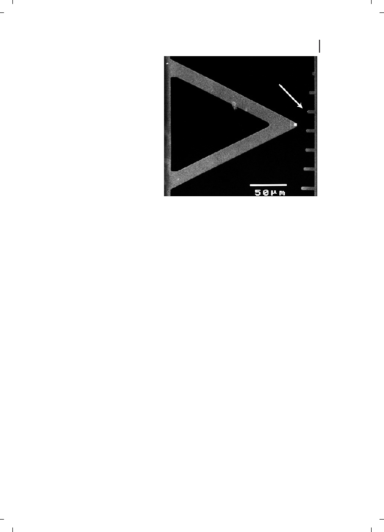
Figure 24.5
Small cantilevers (right)
give better signal-to-noise ratio and
permit higher imaging speeds than
standard cantilevers (left). At the same
spring constant, small cantilevers have
higher resonant frequencies than large
cantilevers. Arrow points to a cantilever
similar to the one used to image pro-
tein–protein interactions in real time in
Ref. [38]. Resonant frequency for small
cantilever (arrow) is 30 times larger
than that for large cantilever on left.

to modify the mica surface in order to facilitate binding of the biological macromolecules
to the surface. These modifications include treatment with ions of divalent metals (e. g.,
Ni(II) or Mg(II)) or long polymers carrying positive charges (e. g., poly-l-lysine, spermine,
spermidine). This treatment leads to the formation of positively charged clusters on the
mica surface and makes it possible for negatively charged molecules (such as DNA and
RNA) to bind to the mica surface.
Another commonly used means of modifying mica is to treat it with derivatives of si-
lane that can form monolayers on its surface. These monolayers are shown to be strongly
attached to mica and to be smooth enough for imaging even relatively small objects. The
outer ends of the silane molecules in the formed film can carry at their termini a number
of different chemical groups (e. g., amino-, carboxyl-, epoxy-, mercapto-, alkyl-) to create a
surface with the desired chemistry. For further details, the reader is referred to Refs. [34,
75]; for other articles, see Ref. [74].
24.4
The Future
Although some of the most exciting future developments in biological probe microscopy
remain dreams in the minds of their inventors, two possible future uses are outlined in
the following section.
24.4.1
Unity or Diversity?
Currently, single-molecule techniques are yielding information about the diversity seen in
the fine structures of the individual molecules within a single population of molecules.
One of the most exciting discoveries in single-molecule biophysics is that individual bio-
molecules of a particular type typically show qualitatively similar patterns of behavior, yet
the quantitative behavior of the individual molecules often differs significantly – perhaps
many-fold – between one molecule and the next. Xie observed this inter-molecular varia-
bility and defined static and dynamic forms of molecular variations [80]. Dynamic varia-
tions occur over time in a single molecule, while static variations occur between different
molecules. Static variations in molecular behavior have also been noted in the interactions
of the molecular chaperonins GroEL and GroES (Ref. [38] and P. Hansma, unpublished
results), in lactate dehydrogenase by Xue [81], in the RedBCD enzyme by S. Kowalczy-
kowski [82], and in a ribozyme by S. Chu [83], who named these variations “molecular
memory”.
These variations between one molecule and the next might be due to differences in the
interactions between the molecule and its environment, such as surface effects for mole-
cules attached to surfaces. While it is easy to label such variability as “surface artifacts”,
the cells in which these molecules reside are themselves filled with surfaces. In any
case, this question is now at the forefront of single-molecule biophysics : namely, “Why
are there such large differences between the individual molecules in a single so-called
‘homogeneous’ population?”
398
24 Surface Biology : Analysis of Biomolecular Structure by Atomic Force Microscopy and Molecular Pulling

24.4.2
World-wide Research
Among the AFM-related reports listed in PubMed during this millennium, the vast ma-
jority were from the United States and other developed countries. Almost one-third
were from the U. S., and one-third was from the United Kingdom, Germany, and
Japan. The developed countries [84] together account for over 91 % of the AFM-related de-
tailed papers in PubMed. One hope for the future is to see leading-edge research spread-
ing to other countries around the world, and a related hope was expressed by Kofi Annan,
Secretary-General of the United Nations, who extended such a challenge to the world’s
scientists [85].
References
399
References
[1]
D. P. Allison, P. Hinterdorfer, W. Han,
Biomolecular force measurements and the
atomic force microscope, Curr. Opin. Bio-
technol. 2002, 13, 47–51.
[2]
L. P. da Silva, Atomic force microscopy and
proteins, Protein Pept. Lett. 2002, 9, 117–
126.
[3]
Y. F. Dufrene, Atomic force microscopy, a
powerful tool in microbiology, J. Bacteriol.
2002, 184, 5205–5213.
[4]
A. Engel, H. Stahlberg, Aquaglyceropor-
ins : channel proteins with a conserved
core, multiple functions, and variable sur-
faces, Int. Rev. Cytol. 2002, 215, 75–104.
[5]
D. Fotiadis, S. Scheuring, S. A. Muller, A.
Engel, D. J. Muller, Imaging and manipu-
lation of biological structures with the
AFM, Micron 2002, 33, 385–397.
[6]
M. K. Higgins, H. T. McMahon, Snap-shots
of clathrin-mediated endocytosis, Trends
Biochem. Sci. 2002, 27, 257–263.
[7]
C. S. Hodges, Measuring forces with the
AFM : polymeric surfaces in liquids, Adv.
Colloid Interface Sci. 2002, 99, 13–75.
[8]
R. E. Marchant, I. Kang, P. S. Sit, Y. Zhou,
B. A. Todd, S. J. Eppell, I. Lee, Molecular
views and measurements of hemostatic
processes using atomic force microscopy,
Curr. Protein Pept. Sci. 2002, 3, 249–274.
[9]
D. J. Muller, H. Janovjak, T. Lehto, L.
Kuerschner, K. Anderson, Observing
structure, function and assembly of single
proteins by AFM, Prog. Biophys. Mol. Biol.
2002, 79, 1–43.
[10]
D. J. Muller, K. Anderson, Biomolecular
imaging using atomic force microscopy,
Trends Biotechnol. 2002, 20, S45–S49.
[11]
P. J. Werten, H. W. Remigy, B. L. de Groot,
D. Fotiadis, A. Philippsen, H. Stahlberg, H.
Grubmuller, A. Engel, Progress in the
analysis of membrane protein structure
and function, FEBS Lett. 2002, 529, 65–72.
[12]
J. Zlatanova, S. M. Lindsay, S. H. Leuba,
Single molecule force spectroscopy in
biology using the atomic force microscope,
Prog. Biophys. Mol. Biol. 2000, 74, 37–61.
[13]
H. G. Hansma, L. I. Pietrasanta, I. D.
Auerbach, C. Sorenson, R. Golan, P. A.
Holden, Probing biopolymers with the
atomic force microscope : a review, J. Bio-
mater. Sci. Polymer Edition 2000, 11, 675–
683.
[14]
T. E. Fisher, M. Carrion-Vazquez, A. F.
Oberhauser, H. Li, P. E. Marszalek, J. M.
Fernandez, Single molecular force spec-
troscopy of modular proteins in the ner-
vous system, Neuron 2000, 27, 435–446.
[15]
T. E. Fisher, P. E. Marszalek, J. M. Fernan-
dez, Stretching single molecules into novel
conformations using the atomic force mi-
croscope, Nature Struct. Biol. 2000, 7, 719–
724.
[16]
A. Engel, D. J. Muller, Observing single
biomolecules at work with the atomic force
microscope, Nature Struct. Biol. 2000, 7,
715–718.
[17]
W. R. Bowen, R. W. Lovitt, C. J. Wright,
Application of atomic force microscopy to

400
24 Surface Biology : Analysis of Biomolecular Structure by Atomic Force Microscopy and Molecular Pulling
the study of micromechanical properties of
biological materials [Review], Biotechnol.
Lett. 2000, 22, 893–903.
[18]
H. G. Hansma, Surface Biology of DNA by
Atomic Force Microscopy, Annu. Rev. Phys.
Chem. 2001, 52, 71–92.
[19]
H. G. Hansma, L. I. Pietrasanta, R. Golan,
J. C. Sitko, M. Viani, G. Paloczi, B. L.
Smith, D. Thrower, P. K. Hansma, Recent
Highlights from Atomic Force Microscopy
of DNA, Biol. Struct. Dynamics, Conversa-
tion 2000, 11, 271–276.
[20]
H. G. Hansma, D. O. Clegg, E. Kokkoli, E.
Oroudjev, M. Tirrell, Analysis of matrix
dynamics by atomic force microscopy, in :
J. Adams (ed.), Methods in Cell Biology, vol.
69, San Diego, Academic Press, 2002, pp.
163–193.
[21]
H. G. Hansma, L. Pietrasanta, Atomic force
microscopy and other scanning probe mi-
croscopies, Curr. Opin. Chem. Biol. 1998, 2,
579–584.
[22]
N. Kaji, M. Ueda, Y. Baba, Direct
measurement of conformational changes
on DNA molecule intercalating with a
fluorescence dye in an electrophoretic buf-
fer solution by means of atomic force
microscopy, Electrophoresis 2001, 22,
3357–3364.
[23]
D. V. Klinov, I. V. Lagutina, V. V. Prokhorov,
T. Neretina, P. P. Khil, Y. B. Lebedev, D. I.
Cherny, V. V. Demin, E. D. Sverdlov, High
resolution mapping DNAs by R-loop
atomic force microscopy, Nucleic Acids Res.
1998, 26, 4603–4610.
[24]
M. Koping-Hoggard, I. Tubulekas,
H. Guan, K. Edwards, M. Nilsson,
K. M. Varum, P. Artursson, Chitosan as a
nonviral gene delivery system. Structure–
property relationships and characteristics
compared with polyethylenimine in
vitro and after lung administration in vivo,
Gene Ther. 2001, 8, 1108–1121.
[25]
M. Rief, H. Clausen-Schaumann, H. E.
Gaub, Sequence-dependent mechanics of
single DNA molecules, Nature Struct. Biol.
1999, 6, 346–349.
[26]
X. E. Cai, J. Yang, Molecular forces for
the binding and condensation of DNA
molecules, Biophys. J. 2002, 82,
357–365.
[27]
B. Sampaolese, A. Bergia, A. Scipioni,
G. Zuccheri, M. Savino, B. Samori,
P. De Santis, Recognition of the DNA
sequence by an inorganic crystal surface,
Proc. Natl. Acad. Sci. USA 2002, 99,
13566–13570.
[28]
R. Krautbauer, L. H. Pope, T. E. Schrader, S.
Allen, H. E. Gaub, Discriminating small
molecule DNA binding modes by single
molecule force spectroscopy, FEBS Lett.
2002, 510, 154–158.
[29]
J. H. Hafner, C. L. Cheung, A. T. Woolley,
C. M. Lieber, Structural and functional
imaging with carbon nanotube AFM
probes, Prog. Biophys. Mol. Biol. 2001, 77,
73–110.
[30]
H. G. Hansma, Varieties of imaging with
scanning probe microscopes, Proc. Natl.
Acad. Sci. USA 1999, 96, 14678–14680.
[31]
Z. Shao, Y. Zhang, Biological cryo atomic
force microscopy: a brief review, Ultrami-
croscopy 1996, 66, 141–152.
[32]
D. M. Czajkowsky, Z. Shao, Submolecular
resolution of single macromolecules with
atomic force microscopy, FEBS Lett. 1998,
430, 51–54.
[33]
D. J. Muller, A. Engel, Conformations,
flexibility, and interactions observed on in-
dividual membrane proteins by atomic
force microscopy, Methods Cell Biol. 2002,
68, 257–299.
[34]
D. M. Czajkowsky, Z. Shao, Supported lipid
bilayers as effective substrates for atomic
force microscopy, Methods Cell Biol. 2002,
68, 231–241.
[35]
M. Guthold, X. Zhu, C. Rivetti, G. Yang,
N. H. Thomson, S. Kasas, H. G. Hansma,
B. Smith, P. K. Hansma, C. Bustamante,
Direct observation of one-dimensional dif-
fusion and transcription by Escherichia coli
RNA polymerase, Biophys. J. 1999, 77,
2284–2294.
[36]
S. Kasas, N. H. Thomson, B. L. Smith,
H. G. Hansma, X. Zhu, M. Guthold,
C. Bustamante, E. T. Kool, M. Kashlev,
P. K. Hansma, E. coli RNA polymerase
activity observed using atomic force
microscopy, Biochemistry 1997, 36,
461–468.
[37]
C. Bustamante, S. B. Smith, J. Liphardt, D.
Smith, Single-molecule studies of DNA
mechanics, Curr. Opin. Struct. Biol. 2000,
10, 279–285.
[38]
M. B. Viani, L. I. Pietrasanta, J. B. Thomp-
son, A. Chand, I. C. Gebeshuber,
J. H. Kindt, M. Richter, H. G. Hansma,
P. K. Hansma, Probing protein–protein

401
References
interactions in real time, Nature Struct.
Biol. 2000, 7, 644–647.
[39]
L. Bousset, N. H. Thomson, S. E. Radford,
R. Melki, The yeast prion Ure2p retains its
native alpha-helical conformation upon
assembly into protein fibrils in vitro,
EMBO J. 2002, 21, 2903–2911.
[40]
D. Fotiadis, Y. Liang, S. Filipek, D. A. Sa-
perstein, A. Engel, K. Palczewski, Atomic-
force microscopy: Rhodopsin dimers in
native disc membranes, Nature 2003, 421,
127–128.
[41]
D. J. Muller, M. Kessler, F. Oesterhelt,
C. Moller, D. Oesterhelt, H. Gaub, Stability
of bacteriorhodopsin alpha-helices and
loops analyzed by single-molecule force
spectroscopy, Biophys. J. 2002, 83, 3578–
3588.
[42]
E. Oroudjev, J. Soares, S. Arcidiacono,
J. B. Thompson, S. A. Fossey, H. G.
Hansma, Segmented nanofibers of spider
dragline silk : atomic force microscopy and
single-molecule force spectroscopy, Proc.
Natl. Acad. Sci. USA 2002, 99, 6460–
6465.
[43]
E. Oroudjev, C. Y. Hayashi, J. Soares,
S. Arcidiacono, S. A. Fossey, H. G.
Hansma, Nanofiber Formation in Spider
Dragline-Silk as Probed by Atomic Force
Microscopy and Molecular Pulling, in :
D. A. Bonnell, J. Piqueras, A. P. Shreve,
F. Zypman (eds), Spatially Resolved Char-
acterization of Local Phenomena in Materials
and Nanostructures, vol. 738, Boston, MA,
2003, in press.
[44]
N. Becker, E. Oroudjev, S. Mutz, J. P. Cle-
veland, P. K. Hansma, C. Y. Hayashi, D. E.
Makarov, H. G. Hansma, Molecular nano-
springs in spider capture silk threads, Na-
ture Mater. 2003, 2, 278–283.
[45]
A. Kempe, J. W. Schopf, W. Altermann,
A. B. Kudryavtsev, W. M. Heckl, Atomic
force microscopy of Precambrian micro-
scopic fossils, Proc. Natl. Acad. Sci. USA
2002, 99, 9117–9120.
[46]
F. Oesterhelt, D. Oesterhelt, M. Pfeiffer, A.
Engel, H. E. Gaub, D. J. Muller, Unfolding
pathways of individual bacteriorhodopsins,
Science 2000, 288, 143–146.
[47]
T. R. Serio, A. G. Cashikar, A. S. Kowal,
G. J. Sawicki, J. J. Moslehi, L. Serpell,
M. F. Arnsdorf, S. L. Lindquist, Nucleated
conformational conversion and the
replication of conformational information
by a prion determinant, Science 2000, 289,
1317–1321.
[48]
C. E. Diebel, R. Proksch, C. R. Green, P.
Neilson, M. M. Walker, Magnetite defines a
vertebrate magnetoreceptor, Nature 2000,
406, 299–302.
[49]
L. K. Nielsen, T. Bjornholm, O. G. Mourit-
sen, Fluctuations caught in the act, Nature
2000, 404, 352.
[50]
S. R. Whaley, D. S. English, E. L. Hu,
P. F. Barbara, A. M. Belcher, Selection of
peptides with semiconductor binding
specificity for directed nanocrystal assem-
bly, Nature 2000, 405, 665–668.
[51]
S. T. Yau, P. G. Vekilov, Quasi-planar nu-
cleus structure in apoferritin crystalliza-
tion, Nature 2000, 406, 494–497.
[52]
H. Seelert, A. Poetsch, N. A. Dencher,
A. Engel, H. Stahlberg, D. J. Muller,
Structural biology. Proton-powered turbine
of a plant motor, Nature 2000, 405,
418–419.
[53]
J. J. Cornelissen, J. J. Donners, R. de
Gelder, W. S. Graswinckel, G. A. Metselaar,
A. E. Rowan, N. A. Sommerdijk, R. J. Nolte,
Beta-helical polymers from isocyanopep-
tides, Science 2001, 293, 676–680.
[54]
A. Y. Kasumov, M. Kociak, S. Gueron,
B. Reulet, V. T. Volkov, D. V. Klinov,
H. Bouchiat, Proximity-induced supercon-
ductivity in DNA, Science 2001, 291,
280–282.
[55]
S. K. Lower, M. F. Hochella, Jr., T. J. Bever-
idge, Bacterial recognition of mineral sur-
faces : nanoscale interactions between She-
wanella and alpha-FeOOH, Science 2001,
292, 1360–1363.
[56]
D. Y. Takamoto, E. Aydil, J. A. Zasadzinski,
A. T. Ivanova, D. K. Schwartz, T. Yang, P. S.
Cremer, Stable ordering in Langmuir-
Blodgett films, Science 2001, 293, 1292–
1295.
[57]
C. A. Orme, A. Noy, A. Wierzbicki, M. T.
McBride, M. Grantham, H. H. Teng, P. M.
Dove, J. J. DeYoreo, Formation of chiral
morphologies through selective binding of
amino acids to calcite surface steps, Nature
2001, 411, 775–779.
[58]
J. B. Thompson, J. H. Kindt, B. Drake,
H. G. Hansma, D. E. Morse, P. K. Hansma,
Bone indentation recovery time correlates
with bond reforming time, Nature 2001,
414, 773–776.

402
24 Surface Biology : Analysis of Biomolecular Structure by Atomic Force Microscopy and Molecular Pulling
[59]
J. J. Kim, Y. Choi, S. Suresh, A. S. Argon,
Nanocrystallization during nanoindenta-
tion of a bulk amorphous metal alloy at
room temperature, Science 2002, 295,
654–657.
[60]
K. B. Lee, S. J. Park, C. A. Mirkin, J. C.
Smith, M. Mrksich, Protein nanoarrays
generated by dip-pen nanolithography,
Science 2002, 295, 1702–1705.
[61]
S. W. Lee, C. Mao, C. E. Flynn, A. M.
Belcher, Ordering of quantum dots using
genetically engineered viruses, Science
2002, 296, 892–895.
[62]
H. Li, W. A. Linke, A. F. Oberhauser, M.
Carrion-Vazquez, J. G. Kerkvliet, H. Lu,
P. E. Marszalek, J. M. Fernandez, Reverse
engineering of the giant muscle protein
titin, Nature 2002, 418, 998–1002.
[63]
H. Yan, X. Zhang, Z. Shen, N. C. Seeman,
A robust DNA mechanical device con-
trolled by hybridization topology, Nature
2002, 415, 62–65.
[64]
J. G. Forbes, G. H. Lorimer, Structural
biology. Unraveling a membrane protein,
Science 2000, 288, 63–64.
[65]
R. F. Service, DNA imaging. Getting a feel
for genetic variations, Science 2000, 289,
27–28.
[66]
D. R. Meldrum, TechSight. Sequencing
genomes and beyond, Science 2001, 292,
515–517.
[67]
D. K. Newman, Microbiology. How bacteria
respire minerals, Science 2001, 292, 1312–
1313.
[68]
S. Bunk, Better microscopes will be in-
strumental in nanotechnology develop-
ment, Nature 2001, 410, 127–129.
[69]
M. Rief, M. Gautel, F. Oesterhelt, J. M.
Fernandez, H. E. Gaub, Reversible unfold-
ing of individual titin immunoglobulin
domains by AFM, Science 1997, 276, 1109–
1112.
[70]
P. E. Marszalek, H. Lu, H. B. Li, M. Car-
rion-Vazquez, A. F. Oberhauser, K. Schul-
ten, J. M. Fernandez, Mechanical unfolding
intermediates in titin modules, Nature
1999, 402, 100–103.
[71]
M. B. Viani, T. E. Schaeffer, G. T. Paloczi,
L. I. Pietrasanta, B. L. Smith, J. B. Thomp-
son, M. Richter, M. Rief, H. E. Gaub,
K. W. Plaxco, A. N. Cleland, H. G. Hansma,
P. K. Hansma, Fast imaging and fast force
spectroscopy of single biopolymers with a
new atomic force microscope designed for
small cantilevers, Rev. Sci. Instruments
1999, 70, 4300–4303.
[72]
P. M. Williams, S. B. Fowler, R. B. Best,
J. L. Toca-Herrera, K. A. Scott, A. Steward,
J. Clarke, Hidden complexity in the me-
chanical properties of titin, Nature 2003,
422, 446–449.
[73]
L. M. Demers, D. S. Ginger, S. J. Park,
Z. Li, S. W. Chung, C. A. Mirkin, Direct
patterning of modified oligonucleotides on
metals and insulators by dip-pen nano-
lithography, Science 2002, 296, 1836–1838.
[74]
B. P. Jena, J. K. H. Horber, Atomic Force
Microscopy in Cell Biology, vol. 68. Amster-
dam : Academic Press, 2002.
[75]
J. H. Kindt, J. C. Sitko, L. I. Pietrasanta, E.
Oroudjev, N. Becker, M. B. Viani, H. G.
Hansma, Methods for biological probe
microscopy in aqueous fluids, Methods Cell
Biol. 2002, 68, 213–229.
[76]
C. A. Siedlecki, R. E. Marchant, Atomic
force microscopy for characterization of the
biomaterial interface, Biomaterials 1998, 19,
441–454.
[77]
B. Drake, C. B. Prater, A. L. Weisenhorn,
S. A. Gould, T. R. Albrecht, C. F. Quate,
D. S. Cannell, H. G. Hansma, P. K.
Hansma, Imaging crystals, polymers, and
processes in water with the atomic force
microscope, Science 1989, 243, 1586–1589.
[78]
H. G. Hansma, I. Revenko, K. Kim, D. E.
Laney, Atomic force microscopy of long
and short double-stranded, single-stranded
and triple-stranded nucleic acids, Nucleic
Acids Res. 1996, 24, 713–720.
[79]
M. B. Viani, T. E. Schaffer, A. Chand, M.
Rief, H. E. Gaub, P. K. Hansma, Small
cantilevers for force spectroscopy of single
molecules, J. Appl. Physics 1999, 86, 2258–
2262.
[80]
X. S. Xie, H. P. Lu, Single-molecule enzy-
mology, J. Biol. Chem. 1999, 274, 15967–
15970.
[81]
Q. F. Xue, E. S. Yeung, Differences in the
chemical reactivity of individual molecules
of an enzyme, Nature 1995, 373, 681–683.
[82]
P. R. Bianco, L. R. Brewer, M. Corzett, R.
Balhorn, Y. Yeh, S. C. Kowalczykowski, R. J.
Baskin, Processive translocation and DNA
unwinding by individual RecBCD enzyme
molecules, Nature 2001, 409, 374–378.
[83]
X. Zhuang, H. Kim, M. J. Pereira, H. P.
Babcock, N. G. Walter, S. Chu, Correlating
structural dynamics and function in single

403
References
ribozyme molecules, Science 2002, 296,
1473–1476.
[84]
http ://millenniumindicators.un.org/unsd/
mi/mi_dict_xrxx.asp?def_code=491.
[85]
K. Annan, A challenge to the world
ls
scientists, Science 2003, 299, 1485.
[86]
J. C. Sitko, E. M. Mateescu, H. G. Hansma,
Sequence-dependent DNA condensation
and the electrostatic zipper, Biophys. J.
2003, 84, 419–431.
[87]
A. A. Kornyshev, S. Leikin, Electrostatic
zipper motif for DNA aggregation, Physical
Rev. Lett. 1999, 82, 4138–4141.
[88]
D. J. Mueller, H.-J. Sass, S. Mueller, G.
Bueldt, A. Engel, Surface structures of na-
tive bacteriorhodopsin depend on the mo-
lecular packing arrangement in the mem-
brane, J. Mol. Biol. 1999, 285, 1903–1909.
[89]
E. Oroudjev, C. Y. Hayashi, J. Soares, S.
Arcidiacono, S. A. Fossey, H. G. Hansma,
Nanofiber Formation in Spider Dragline-
Silk as Probed by Atomic Force Microscopy
and Molecular Pulling, presented at MRS
Fall Meeting, Boston, MA, 2002.
[90]
J. T. Prince, K. P. McGrath, C. M. Digio-
lamo, D. L. Kaplan, Construction, cloning,
and expression of synthetic genes encoding
spider dragline silk, Biochemistry 1995, 34,
10879–10885.
Wyszukiwarka
Podobne podstrony:
24.Kroukovci, Biologia Geografia
NLP How to Live on 24 Hours a Day by Arnold Bennett
23 24 JM by AZ
bio spr 24.04.07, biologia liceum ściągi
24. Omów działanie mięśni, studia-biologia, Opracowane pytania do licencjatu
opracowania zestawow (2010-2011) by fabs, Medycyna, I rok, Biologia, Biologia
Biologia medyczna 24.10.08r, fizjoterapia, biologia medyczna
Zegar biologiczny. 24 godziny z życia organizmu, Zdrowie,zioła,wegetarianizm,zdrowe odżywianie,oczys
5 Biologia molekularna 24.10.2011, Biotechnologia UTP, Biologia molekularna
Biologia wykład 24 10 2006
moleki, Biologia molekularana by anuha, Biologia molekularana::::
In vitro biological effects of titanium rough surface obtain
Improved biological performance of Ti implants due to surfac
Essentials of Biology 1e c 24
By GuiTop Nicolo Paganini Caprice 24 ( XXIV Tr john Williams) Sheet Score Classical Spanish Gui
24 piątek
1Ochr srod Wyklad 1 BIOLOGIA dla studid 19101 ppt
więcej podobnych podstron