
A
PPLIED AND
E
NVIRONMENTAL
M
ICROBIOLOGY
, Jan. 2003, p. 97–101
Vol. 69, No. 1
0099-2240/03/$08.00
⫹0 DOI: 10.1128/AEM.69.1.97–101.2003
Copyright © 2003, American Society for Microbiology. All Rights Reserved.
Improved Understanding of the Bacterial Vaginal Microbiota of
Women before and after Probiotic Instillation
Jeremy P. Burton,
1,2
* Peter A. Cadieux,
2
and Gregor Reid
1,2
Canadian Research and Development Centre for Probiotics, The Lawson Health Research Institute,
1
and
Department of Microbiology and Immunology, University of Western Ontario,
2
London, Ontario, Canada
Received 26 June 2002/Accepted 24 September 2002
The vaginal bacterial microbiota of 19 premenopausal women was examined by PCR-denaturing gradient gel
electrophoresis (DGGE) and sequencing of the V2-V3 region of the 16S rRNA gene. Ten of the women were
studied further to investigate the effect and persistence of vaginally inserted capsules containing viable
lactobacilli. PCR-DGGE indicated that most subjects had a microbiota represented by one to three dominant
DNA fragments. Analysis of these fragments revealed that 79% of the women possessed sequences with high
levels of similarity to Lactobacillus species sequences. Sequences homologous to Lactobacillus iners sequences
were the most common and were detected in 42% of the women tested. Alteration of the vaginal microbiota
could be detected by PCR-DGGE in several women after the instillation of lactobacilli. Additionally, randomly
amplified polymorphic DNA analysis of lactobacilli isolated from selective media demonstrated that the
exogenous strains could be detected for up to 21 days in some subjects. This study demonstrates that
non-culture-based techniques, such as PCR-DGGE, are useful adjuncts for studies of the vaginal microbiota.
The microbes that inhabit the vagina play a major role in
illnesses of the host, including bacterial vaginosis, yeast vagi-
nitis, cancer, and sexually transmitted diseases, such as human
immunodeficiency virus infection, as well as in the mainte-
nance of a healthy tract. Our understanding of the nature and
functionality of these organisms has progressed in recent years,
but it is still far from optimal. For some time the microbiota of
so-called normal women of child-bearing age was believed to
be dominated by Lactobacillus acidophilus and Lactobacillus
fermentum, followed by Lactobacillus brevis, Lactobacillus
jensenii, Lactobacillus casei, and other species (12). More re-
cently, molecular methods have shown that Lactobacillus
crispatus and Lactobacillus jensenii are the most common iso-
lates (2, 12), and in one study a previously undescribed Lac-
tobacillus species was found in 15% of women (2). The devel-
opment of denaturing gradient gel electrophoresis (DGGE)
has provided an exciting tool to analyze a given population of
organisms within a host. To date, this method has been used
successfully to examine the intestinal microbiotas of adults and
children (5, 18).
Continuous application of certain Lactobacillus strains vag-
inally and orally has been shown to alter the microbiota from
a microbiota indicative of bacterial vaginosis to a microbiota
that is dominated by lactobacilli and regarded as normal (10).
Instillation of probiotic lactobacilli has the potential to make a
significant impact on the health of women, and therefore, it is
important to understand how the vaginal microbiota changes
and adapts to the presence of these strains. Therefore, the first
goal of the present study was to utilize PCR-DGGE and to
sequence different 16S DNA fragments to determine which
bacterial species were most common among the vaginal sam-
ples of premenopausal women. The second goal was to use
DGGE to examine the impact of probiotic strains on the vag-
inal bacterial microbiota and determine the persistence of ex-
ogenous lactobacillus strains by using selective medium and
randomly amplified polymorphic DNA (RAPD) profiling (7).
MATERIALS AND METHODS
Subjects, probiotic instillation, and sample collection.
Nineteen premeno-
pausal Caucasian women who had no symptoms or signs of vaginal or urinary
tract infection and were otherwise healthy were recruited. Each woman signed an
informed consent under a protocol approved by the human ethics review board
at the University of Western Ontario. None of the recruits was receiving anti-
microbial prescribed therapy or using spermicidal products. Deep vaginal sam-
ples were collected by rotating swabs throughout the vagina of each of the
subjects prior to the start of the study at zero time and at 6 months. For the 10
subjects in whom lactobacilli were vaginally instilled (subjects 260 to 269), one
capsule containing 1
⫻ 10
9
total CFU of Lactobacillus fermentum RC-14 and
Lactobacillus rhamnosus GR-1 was inserted daily into the vagina following the
initial swabbing for 3 days. Additional swabs were collected on days 3, 7, 14, and
21 from the subjects who received probiotics. Two swabs were collected per
subject at each sampling point, one for the culture of lactobacilli for RAPD
analysis and the other for direct bacterial DNA extraction for PCR-DGGE.
Once taken, the swabs were immediately placed in transport medium (NCS
Diagnostics Inc., Etobicoke, Ontario, Canada) and taken to the lab for process-
ing within 3 h.
Culturing and DNA fingerprinting of Lactobacillus strains by RAPD analysis.
Vaginal swabs were agitated in 1 ml of sterile phosphate-buffered saline (PBS)
(pH 7.5) and serially diluted. To determine the persistence of L. rhamnosus
GR-1 and L. fermentum RC-14 within the vagina, aliquots of each dilution were
plated onto MRS plates (BBL, Becton Dickinson, Cockeysville, Md.) containing
selective agents for each strain (7) (fusidic acid [32
g/ml; Sigma Chemical Co.,
St. Louis, Mo.] and tetracycline [8
g/ml; Sigma], respectively) and incubated
anaerobically by using the BBL GasPack system at 37°C for 48 h. Ten colonies
from each subject were selected for testing by RAPD analysis by the method of
Gardiner et al. (7).
Extraction of bacterial DNA from swabs for PCR.
Swabs were vigorously
agitated in 1 ml of PBS to dislodge the cells. The cells were pelleted by centrif-
ugation (10,000
⫻ g, 5 min) and washed once in PBS, and total DNA was
extracted by using Instagene matrix (Bio-Rad Laboratories, Hercules, Calif.)
according to the manufacturer’s instructions. PCRs were carried out in 0.2-ml
* Corresponding author. Mailing address: Lawson Health Research
Institute, St. Joseph’s Hospital, 268 Grosvenor St., London, Ontario
N6A 4V2, Canada. Phone: (519) 646-6100, ext. 65120. Fax: (519)
646-6110. E-mail: jburton@lri.sjhc.london.on.ca.
97
tubes with a thermocycler (Mastercycler; Eppendorf, Wesseling-Berzdorf, Ger-
many). The HDA eubacterial PCR primers and amplification conditions of
Walter et al. were utilized (18).
DGGE, DNA fragment excision from gels, reamplification, and sequencing.
Preparation of DGGE gel gradients and electrophoresis were carried out by
using the manufacturer’s guidelines for the D-code universal detection system of
Bio-Rad. A 100% solution was defined as a mixture of 7 M urea and 40%
formamide. The concentrations of polyacrylamide, denaturant, and Tris-acetate
buffer (40 mM Tris, 20 mM glacial acetic acid, 1 mM EDTA [pH 8.0]) were 8%,
30 to 50%, and 1
ⴛ, respectively. Other parameters have been described previ-
ously (18). Fragments of interest were excised from DGGE gels with a sterile
scalpel, washed once in 1
ⴛ PCR buffer, and incubated in 20 l of the same buffer
overnight at 4°C. Five microliters of the buffer solution was used as the template
for PCR. Reamplification was conducted by using the primers described previ-
ously but without the GC clamp (18). Sequences of the reamplified fragments
were determined by the dideoxy chain termination method (Sequencing Facility,
John P. Robarts Research Institute, London, Ontario, Canada). Analysis of the
partial 16S rRNA sequences was conducted by using the GenBank database and
the BLAST algorithm (1). Identities of isolates were determined on the basis of
the highest score.
RESULTS
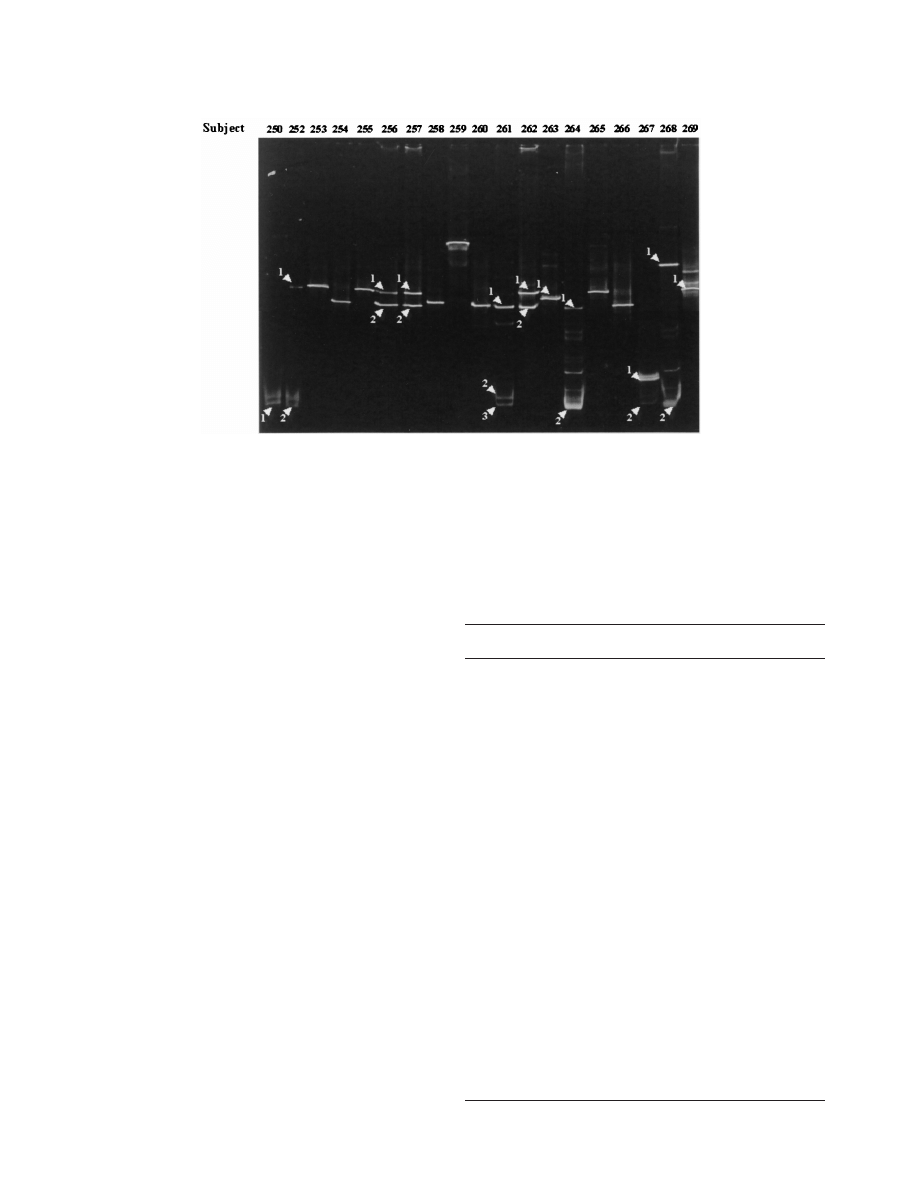
DGGE and sequencing of DNA fragments before probiotic
use indicated that most of the vaginal samples from the 19
women studied had one to three dominant fragments, as ob-
served within a lane of a DGGE gel (Fig. 1). For subjects 261,
264, and 268 5 to 10 fragments were detected (Fig. 1). When
the dominant fragments from every sample were sequenced,
the majority of women tested (15 of 19 women) had at least
one sequence homologous to a sequence of a species of Lac-
tobacillus (Table 1). A significant discovery was that an organ-
ism that was recently found in the vagina (4), Lactobacillus
iners, was the most commonly recovered species and was de-
tected in 42% of the women.
Sequence analysis indicated that Gardnerella vaginalis was
present in six of the study participants at zero time; three of
these women (subjects 250, 267, and 268) would have been
characterized as having asymptomatic bacterial vaginosis by
the Nugent criteria (9). In three of the subjects with G. vagi-
nalis, other microorganisms not commonly found in the vagina,
including Arthrobacter sp., Caulobacter sp., and Butyrivibrio
fibrisolvens, were detected. G. vaginalis and Lactobacillus spe-
cies were simultaneously detected in three subjects at the first
sampling time (Table 1).
FIG. 1. DGGE of the V2-V3 16S rRNA gene amplicons from vaginal samples: profiles for 19 subjects (zero time, prestudy samples). The
arrowheads indicate the DNA fragments sequenced from specific lanes, while in unmarked lanes only the dominant fragment was sequenced.
BLAST sequence homologies are shown in Table 1.
TABLE 1. BLAST analysis of vaginal bacterial V2-V3 16S rRNA
sequences of excised fragments from DGGE gels (zero time)
Subject
Fragment
in gel
Most closely related
bacterial sequence
% Identity
Accession no.
250
1
Gardnerella vaginalis
98
M58744
252
1
Lactobacillus crispatus
100
AF257097
2
Gardnerella vaginalis
98
M58744
253
1
Lactobacillus crispatus
98
AF257097
254
1
Lactobacillus iners
100
Y16329
255
1
Lactobacillus crispatus
97
AF257097
256
1
Lactobacillus crispatus
100
AF257097
2
Lactobacillus iners
99
Y16329
257
1
Lactobacillus crispatus
98
AF257097
2
Lactobacillus iners
100
Y16329
258
1
Streptococcus
agalactiae
100
AF015927
259
1
Lactobacillus gasseri
100
AF243165
260
1
Lactobacillus iners
100
Y16329
261
1
Lactobacillus iners
99
Y16329
2
Arthrobacter sp.
100
AJ243423
3
Gardnerella vaginalis
99
M58744
262
1
Lactobacillus
acidophilus
97
AF375937
2
Lactobacillus iners
96
Y16329
263
1
Lactobacillus
delbrueckii
97
AF375917
264
1
Lactobacillus iners
92
Y16329
2
Gardnerella vaginalis
98
M58744
265
1
Lactobacillus crispatus
98
AF257097
266
1
Lactobacillus iners
96
Y16329
267
1
Caulobacter sp.
98
M83799
2
Gardnerella vaginalis
97
M58744
268
1
Butyrivibrio
fibrisolvens
95
AF125217
2
Gardnerella vaginalis
97
M58744
269
1
Lactobacillus crispatus
99
AF257097
98
BURTON ET AL.
A
PPL
. E
NVIRON
. M
ICROBIOL
.
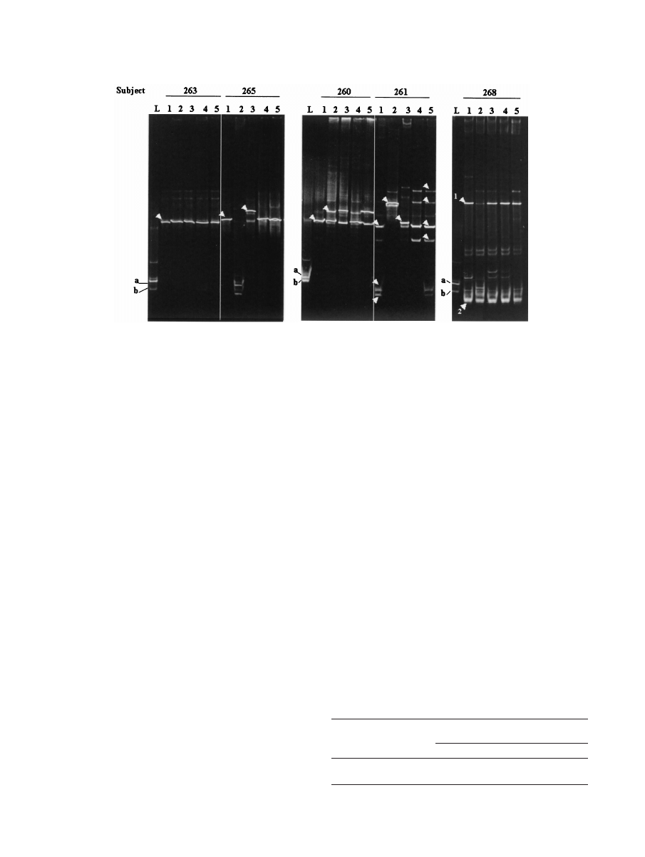
After probiotic instillation, DGGE and sequencing results
showed that in five patients there was no apparent major al-
teration in the existing vaginal microbiota, regardless of
whether one fragment (subject 263) (Fig. 2) or more DNA
fragments (subjects 262, 264, 267, and 269) (data not shown)
were initially detected. No changes were observed in the
DGGE profile of subject 266, other than detection of the
exogenous lactobacilli in the first sample after instillation. Sub-
ject 260 acquired an L. crispatus strain (100% homology with
accession no. AF257097 sequence) in addition to the original
L. iners strain 3 days after probiotic instillation was begun (Fig.
2).
The G. vaginalis DNA fragment present in subject 261 dis-
appeared immediately following lactobacillus treatment and
was detected again only at day 21. This subject and subject 265
retained their indigenous lactobacilli (excluding day 3 data for
subject 261) but also acquired a Pseudomonas strain (on days
3 and 7, respectively); subject 261 acquired a Streptococcus
agalactiae strain on day 7. When other DNA fragments ob-
served in the last two samples in the DGGE gel from subject
261 were sequenced, they were found to be homologous to L.
iners and were likely to be spurious PCR artifacts (17). There-
fore, if the spurious DNA fragments in subject 261 were ig-
nored, the day 21 microbiota was the same as the microbiota
prior to treatment in both subjects. In subject 268 a DNA
fragment of B. fibrisolvens was present at zero time, and al-
though the intensity of the fragment significantly decreased at
day 3, the intensity was similar to the intensity in the zero-time
microbiota in subsequent day 7, 14, and 21 samples tested (Fig.
2). The follow-up samples obtained from the women after 6
months showed that most women (10 of 18 women, with one
woman noncompliant) had altered DGGE profiles, indicating
that their bacterial microbiota had changed compared to the
microbiota in the prestudy samples.
The presence of the instilled exogenous Lactobacillus spe-
cies could not always be detected within the vaginal samples by
PCR-DGGE. However, RAPD profiling (Table 2) detected
the exogenous lactobacillus strains in 80% of the women after
1 week and in 20% of the women after 3 weeks (L. rhamnosus
GR-1 only). The detection of instilled Lactobacillus strains by
RAPD analysis inversely correlated with detection of G. vagi-
nalis by DGGE and sequence analysis in samples from subject
261 (data not shown).
DISCUSSION
A number of interesting findings emerged from this study. L.
iners, which was not detected in other studies of the vaginal
microbiota (2, 12), is clearly a common constituent of the
women sampled in this study. This species does not grow on
the major selective media used for isolation of Lactobacillus,
including MRS and Rogosa-Sharp medium (4). This might
FIG. 2. DGGE profiles of the vaginal microbiota from five women during the study. Lanes L contained known isolates L. fermentum RC-14
(a) and L. rhamnosus GR-1 (b). Lanes 1 to 5 contained amplicons from samples taken at zero time (prestudy) and at 3, 7, 14, and 21 days after
instillation of capsules containing lactobacilli, respectively. The arrowheads indicate DNA fragments that were sequenced. Presumptive identities
based on closest BLAST homologies are as follows: for subject 263, lane 1, L. delbrueckii; for subject 265, lane 1, L. crispatus; for subject 265, lane
3, Pseudomonas sp.; for subject 260, lane 1, L. iners; for subject 260, lane 2, L. crispatus; for subject 261, lane 1 (from top to bottom), L. iners,
Arthrobacter sp., and G. vaginalis; for subject 261, lane 2, Pseudomonas sp.; for subject 261, lane 3, S. agalactiae; for subject 261, lane 5, L. iners
(all fragments); for subject 268, lane 1, B. fibrisolvens and G. vaginalis (fragments 1 and 2, respectively).
TABLE 2. Detection of Lactobacillus strains by selective culturing
and subsequent RAPD analysis in a group of 10 women
Lactobacillus strain
No. of women positive on the following days
after instillation:
3
7
14
21
GR-1
10
8
6
2
RC-14
5
4
2
0
V
OL
. 69, 2003
TRACKING VAGINAL BACTERIAL MICROBIOTA BY DGGE AND RAPD
99

explain the failure to detect this organism, or the organism may
have been confused with members of the L. acidophilus com-
plex (4). The potential importance of L. iners in protecting the
vagina from disease and its possible use as a probiotic remain
to be determined. Since most of the urogenital bacterial mi-
crobiota originates from the gastrointestinal tract (14) and
while species of vaginal lactobacilli have also been detected in
feces (14, 15, 18), we can only assume that this is the origin of
L. iners. However, because there has been no selective medium
or species-specific primers described for L. iners, this cannot be
confirmed at present.
The discovery of three strains not commonly detected in the
vagina is also intriguing. Arthrobacter spp. are gram-positive
organisms typically isolated from soil, although some are now
regarded as opportunistic pathogens, having been recovered
from blood and urine (6). Caulobacter spp. are freshwater
organisms, and B. fibrisolvens is a fecal organism. Although we
cannot be certain of the precise origin of these organisms in
the three subjects in which they were found, the findings sug-
gest that the vaginal microbiota may also be influenced by
environmental organisms, perhaps acquired through bathing
and exposure to the soil.
The correlation between a healthy vaginal tract, as defined
by lack of symptoms and signs of disease, and dominance of
lactobacilli (9) supports the belief that these commensals play
a major role in preventing certain types of vaginal infections. In
the zero-time samples of three of six subjects G. vaginalis was
detected in conjunction with a species of Lactobacillus. Thus,
the presence of lactobacilli does not necessarily exclude poten-
tial pathogens from the vagina. The question becomes, what
virulent properties or other factors result in an infection? The
balance between an infectious state and a healthy state is likely
a constant battle, and we speculate that this battle involves
interactions between bacteria and interactions between bacte-
ria and host defenses (11).
The immediate detection of changes in the DGGE profiles
of four subjects (subjects 260, 261, 265, and 268) following
lactobacillus instillation and the subsequent reversion of the
profiles to the profiles of the prestudy state in three of the
subjects over the course of the study suggest that these changes
were probably not attributable to temporal variation of the
microbiota. Pseudomonas species can be a cause of urinary
tract infections (3, 13). The detection of Pseudomonas sp. in
samples from subjects 261 and 265 following instillation of the
probiotic might have been due to emergence of endogenous
and potentially opportunistic microorganisms within the vagina
at levels below the detection limit of PCR-DGGE (8, 17). Such
microorganisms may become increasingly prevalent upon mi-
nor alteration of the vaginal microenvironment. Persistence of
microorganisms at levels below the detection threshold of
PCR-DGGE was demonstrated by culturing vaginal swabs on
selective antibiotic media preferential for the supplanted Lac-
tobacillus strains and typing isolates by RAPD analysis. For up
to 21 days after the initial instillation, the exogenous strains
could be detected in the samples from some women by RAPD
analysis but not by PCR-DGGE. Whether probiotic microor-
ganisms create a slight perturbation of the microbiota follow-
ing which other persistent endogenous microorganisms, in-
cluding lactobacilli (such as L. crispatus in subject 260), take
advantage to replenish their populations has yet to be deter-
mined. However, the instillation of two probiotic strains
showed that non-hydrogen-peroxide-producing L. rhamnosus
GR-1 persisted longer than the L. fermentum RC-14 strain, a
known H
2
O
2
producer, emphasizing that expression of this
factor alone is probably insufficient for restoration of a lacto-
bacillus-dominant microbiota, as previously proposed (16).
The detection of instilled lactobacillus strains by RAPD
analysis of cultured organisms at low levels but not by DGGE
in certain samples may have been the result of the ability to
plate out the entire contents of a vaginal sample on agar. PCRs
for DGGE, however, rely on efficient DNA extraction and
multiple cells to be present to ensure that a representative
DNA molecule from each bacterial type is present in each
aliquot used for a reaction. Other factors that may also influ-
ence amplification strength may include dominant DNA tem-
plates outcompeting lesser species, PCR primer bias, and
rRNA operon copy numbers that are different in different
microorganisms (8, 17). However, previous culture studies
have failed to identify the presence of certain species, including
L. iners. Our data suggest that PCR-DGGE may be a superior
technique for detecting the dominant microbiota that may not
be detectable by standard culture techniques. Furthermore,
PCR-DGGE was a useful tool for detecting changes in the
vaginal microbiota after the addition of lactobacillus strains.
We suggest that the DGGE technique is a very useful adjunct
for clinical studies of the vaginal tract.
ACKNOWLEDGMENTS
We thank Dee Beuerman and Ivo Braunstein for recruiting subjects
and performing bacterial culturing and DNA extraction.
REFERENCES
1. Altschul, S. F., W. Gish, W. Miller, E. W. Myers, and D. J. Lipman. 1990.
Basic local alignment search tool. J. Mol. Biol. 215:403–410.
2. Antonio, M. A., S. E. Hawes, and S. L. Hillier. 1999. The identification of
vaginal Lactobacillus species and the demographic and microbiologic char-
acteristics of women colonized by these species. J. Infect. Dis. 180:1950–
1956.
3. Bonadio, M., M. Meini, P. Spitaleri, and C. Gigli. 2001. Current microbio-
logical and clinical aspects of urinary tract infections. Eur. Urol. 40:439–444.
(Discussion, 40:445.)
4. Falsen, E., C. Pascual, B. Sjoden, M. Ohlen, and M. D. Collins. 1999.
Phenotypic and phylogenetic characterization of a novel Lactobacillus spe-
cies from human sources: description of Lactobacillus iners sp. nov. Int. J.
Syst. Bacteriol. 49:217–221.
5. Favier, C. F., E. E. Vaughan, W. M. De Vos, and A. D. Akkermans. 2002.
Molecular monitoring of succession of bacterial communities in human ne-
onates. Appl. Environ. Microbiol. 68:219–226.
6. Funke, G., R. A. Hutson, K. A. Bernard, G. E. Pfyffer, G. Wauters, and M. D.
Collins.
1996. Isolation of Arthrobacter spp. from clinical specimens and
description of Arthrobacter cumminsii sp. nov. and Arthrobacter woluwensis
sp. nov. J. Clin. Microbiol. 34:2356–2363.
7. Gardiner, G., C. Heinemann, M. Baroja, A. W. Bruce, D. Beuerman, J.
Madrenas, and G. Reid.
2002. Oral administration of the probiotic combi-
nation Lactobacillus rhamnosus GR-1 and L. fermentum RC-14 for human
intestinal applications. Int. Dairy J. 12:191–196.
8. Muyzer, G., and K. Smalla. 1998. Application of denaturing gradient gel
electrophoresis (DGGE) and temperature gradient gel electrophoresis
(TGGE) in microbial ecology. Antonie Leeuwenhoek 73:127–141.
9. Nugent, R. P., M. A. Krohn, and S. L. Hillier. 1991. Reliability of diagnosing
bacterial vaginosis is improved by a standardized method of Gram stain
interpretation. J. Clin. Microbiol. 29:297–301.
10. Reid, G., D. Beuerman, C. Heinemann, and A. W. Bruce. 2001. Probiotic
Lactobacillus dose required to restore and maintain a normal vaginal flora.
FEMS Immunol. Med. Microbiol. 1351:1–5.
11. Reid, G., J. Howard, and B. S. Gan. 2001. Can bacterial interference prevent
infection? Trends Microbiol. 9:424–428.
12. Sobel, J. D. 1999. Biotherapeutic agents as therapy for vaginitis, p. 221–244.
In G. Elmer, L. V. McFarland, and C. Surawicz (ed.), Biotherapeutic agents
100
BURTON ET AL.
A
PPL
. E
NVIRON
. M
ICROBIOL
.

and infectious diseases. Humana Press, Totowa, N.J.
13. Takeyama, K., Y. Kunishima, M. Matsukawa, S. Takahashi, T. Hirose, N.
Kobayashi, I. Kobayashi, and T. Tsukamoto.
2002. Multidrug-resistant
Pseudomonas aeruginosa isolated from the urine of patients with urinary tract
infection. J. Infect. Chemother. 8:59–63.
14. Tannock, G. W. 1999. The bowel microflora: an important source of urinary
tract pathogens. World J. Urol. 17:339–344.
15. Tannock, G. W. 1995. Normal microflora: an introduction to microbes inhabit-
ing the human body, 1st ed. Chapman & Hall, London, United Kingdom.
16. Vallor, A. C., M. A. Antonio, S. E. Hawes, and S. L. Hillier. 2001. Factors
associated with acquisition of, or persistent colonization by, vaginal lactoba-
cilli: role of hydrogen peroxide production. J. Infect. Dis. 184:1431–1436.
17. von Wintzingerode, F., U. B. Gobel, and E. Stackebrandt. 1997. Determina-
tion of microbial diversity in environmental samples: pitfalls of PCR-based
rRNA analysis. FEMS Microbiol. Rev. 21:213–229.
18. Walter, J., G. W. Tannock, A. Tilsala-Timisjarvi, S. Rodtong, D. M. Loach,
K. Munro, and T. Alatossava.
2000. Detection and identification of gastro-
intestinal Lactobacillus species by using denaturing gradient gel electro-
phoresis and species-specific PCR primers. Appl. Environ. Microbiol. 66:
297–303.
V
OL
. 69, 2003
TRACKING VAGINAL BACTERIAL MICROBIOTA BY DGGE AND RAPD
101
Wyszukiwarka
Podobne podstrony:
Drilling Fluid Yield Stress Measurement Techniques for Improved understanding of critical fluid p
Drilling Fluid Yield Stress Measurement Techniques for Improved understanding of critical fluid p
Eric Racine Pragmatic Neuroethics Improving Treatment and Understanding of the Mind Brain Basic Bioe
Improved Characterization of Nitromethane, Nitromethane Mixtures, and Shaped Charge Jet
A chemical analog of curcumin as an improved inhibitor of amyloid abetaoligomerization
Towards an understanding of the distinctive nature of translation studies
(Trading) Paul Counsel Towards An Understanding Of The Psychology Of Risk And Succes
Kuijpers Towards a deeper understanding of metalworking technology
Salvation and Creativity Two Understandings of Christianity
perkins feminist understanding of productivity
keith reid a whiter shade of pale
Improved Performance of SPME Fibers and Applications
American Psychological Association Anwsers to Your Questions for a Better Understanding of Sexual O
British Patent 8,575 Improved Methods of and Apparatus for Generating and Utilizing Electric Energy
Charles Tart On Emergent Interactionist Understanding of Human Consciousness
Baruch Spinoza On the Improvement of the Understanding
Improvised Explosive Devices Booklet of Related Readings
więcej podobnych podstron