Antibody conjugated magnetic PLGA nanoparticles for diagnosis and
treatment of breast cancer
Jaemoon Yang,
a
Choong-Hwan Lee,
b
Joseph Park,
a
Sungbaek Seo,
a
Eun-Kyung Lim,
a
Yong Jin Song,
c
Jin-Suck Suh,
d
Ho-Geun Yoon,
e
Yong-Min Huh*
d
and Seungjoo Haam*
a
Received 28th February 2007, Accepted 17th April 2007
First published as an Advance Article on the web 27th April 2007
DOI: 10.1039/b702538f
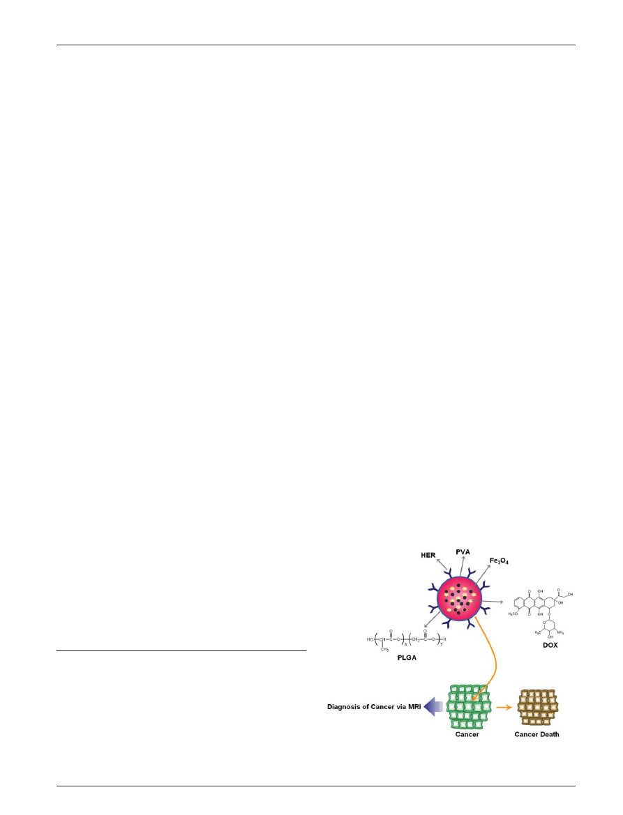
DOX–magnetic PLGA nanoparticles conjugated with well-tailored antibodies were synthesized
for the detection and therapy of breast cancer. Magnetic nanocrystals embedded in polymeric
nanoparticles did not inhibit the nanoparticle formulation or drug release kinetics. The
multimodal nanoparticles demonstrated remarkable cancer cell affinity and ultrasensitivity via
magnetic resonance imaging. Furthermore, the loaded anticancer drugs were released and
sustained for three weeks.
1. Introduction
Inorganic/organic nanoparticles hold significant potential for
biomedical applications due to their molecular size.
1
Recently,
highly crystalline and monodisperse magnetic nanocrystals
(MNCs) have demonstrated excellent properties that can be
applied to areas such as drug delivery, magnetic resonance
(MR) imaging, cell separation and hyperthermia.
2–5
In general,
MNCs are combined with organic compounds such as small
ligands or biocompatible polymers, for various applications.
6
For example, organic ligands on the MNC surface were
exchanged with hydrophilic, functionalized ligands to mediate
conjugation with a peptide or antibody.
3
Furthermore, the
MNCs were used as seeds to polymerize the monomers.
7
Unfortunately, these approaches demand sophisticated proce-
dures or preparatory stages for preparation of organo-
magnetic hybrids.
In the present study, we synthesized a multimodal nano-
composite using inorganic/organic materials for detection and
treatment of cancer (see Scheme 1). The nano-emulsion
method was used to incorporate the anticancer drug doxoru-
bicin (DOX) into magnetic poly(
D
,
L
-lactide-co-glycolide)
(PLGA) nanoparticles (DMPNP). The physico-chemical
properties of the nanoparticles were evaluated for nano-
structure and characteristic features. The antibody Herceptin
1
(HER) used for targeting breast cancer was conjugated to
DMPNP using bioconjugation chemistry (HER-DMPNP).
The breast cancer cell binding affinity of HER-DMPNP and
its potential as an MR probe were estimated. To assess the
therapeutic potential of HER-DMPNP, the drug loading
content, entrapment efficiency and release behavior were also
investigated.
2. Experimental
2.1. Materials
Polyoly(
D
,
L
-lactide-co-glycolide) (PLGA, M
w
: 5 000) was
obtained from Wako Chemicals. Iron(
III
) acetylacetonate, 1,2-
hexadecanediol, dodecanoic acid, dodecylamine, benzyl ether,
1-ethyl-3-(3-dimethylaminopropyl)carbodiimide,
N-hydroxy-
succinimide and polyvinyl alcohol (PVA, M
w
: 15 000–20 000)
were purchased from Sigma-Aldrich. Doxorubicin (DOX) was
obtained from Fluka. Phosphate buffered saline (PBS; 10 mM,
pH 7.4) was purchased from Gibco. All other chemicals and
reagents were of analytical grade.
2.2. Synthesis of magnetic nanocrystals (MNCs)
For the synthesis of the MNCs, 2 mmol iron(
III
) acetylaceto-
nate, 10 mmol 1,2-hexadecanediol, 6 mmol dodecanoic acid,
6 mmol dodecylamine, and benzyl ether (20 mL) were mixed
a
Department of Chemical Engineering, Yonsei University, Seoul 120-
749, South Korea. E-mail: haam@yonsei.ac.kr; Fax: +82-2-312-6401;
Tel: +82-2-2123-3554
b
ATGen, Advanced Technology Research Center, 68 Yatap-dong,
Bundang-gu, Seongnam-si, Gyeonggi-do, 463-816, South Korea
c
Department of Physics, College of Natural Science, Ajou University,
Suwon 433-749, South Korea
d
Department of Radiology, College of Medicine, Yonsei University,
Seoul 120-752, South Korea
e
Department of Biochemistry and Molecular Biology, Center for Chronic
Metabolic Disease Research, College of Medicine, Yonsei University,
Seoul 120-752, South Korea
Scheme 1
Schematic illustration of magnetic PLGA nanoparticles for
diagnosis and treatment of cancer.
PAPER
www.rsc.org/materials
| Journal of Materials Chemistry
This journal is ß The Royal Society of Chemistry 2007
J. Mater. Chem.
, 2007, 17, 2695–2699 | 2695

under a nitrogen atmosphere. The mixture was preheated to
150
uC for 30 min and refluxed at 300 uC for 30 min. After the
reactants were cooled to room temperature, the products were
purified with an excess of pure ethanol. Approximately 12 nm
of MNCs were synthesized after the seed-mediated growth.
3,8
2.3. Preparation of DOX–magnetic PLGA nanoparticles
(DMPNP)
PLGA (100 mg), MNCs (20 mg), DOX (5 mg) and triethyl-
amine (50 mL) were dissolved in 10 mL of dichloromethane.
The organic phase was added to 20 mL of an aqueous phase
containing 3% PVA as a stabilizer. After mutual saturation of
the organic and aqueous phases, the mixture was emulsified
for 10 min with an ultrasonicator (ULH700S, Ulssohitech)
operated at 450 W.
9
After solvent evaporation, the products
were purified with three cycles of centrifugation at 20 000 rpm.
The precipitated nanoparticles were redispersed in 10 mM
sodium phosphate buffer (2 mL, pH 7.4). Due to the presence
of amphiphilic PVA, the prepared nanoparticles were well
suspended in the aqueous phase. The PLGA nanoparticles
(PNPs) and DOX-PLGA nanoparticles (DPNPs) were pre-
pared in the same manner as previously mentioned.
2.4. Characterization of DOX–magnetic PLGA nanoparticles
(DMPNP)
The size distribution of the nanoparticles was analyzed by laser
scattering (ELS-Z, Otsuka electronics). The morphology and
presence of MNCs were evaluated using a transmission
electron microscope (TEM, JEM-1100, JEOL Toko). DPNP
and DMPNP were negatively stained with phosphotungstic
acid.
10
Fourier transform infrared spectroscopy (FT-IR,
ExcaliburTM series, Varian Inc.) was used to confirm the
characteristic bands of the DMPNP. The saturation magne-
tizations of the MNC and DMPNP were evaluated using
a vibrating-sample magnetometer (VSM, MODEL-7300,
Lakeshore). The quantity of MNCs encapsulated in the
DMPNP was analyzed with a thermogravimetric analyzer
(SDT-Q600, TA instrument). The surface compositions were
evaluated
by
X-ray
photoelectron
spectroscopy
(XPS,
ESCALAB MK II, V.G. Scientific Ltd.).
2.5. Drug release test
The drug loading content, entrapment efficiency and drug
release profile were determined using a UV spectrophotometer
(Optizen 2120UV, MECASYS Co). To obtain the drug release
profiles, 20 mg of DPNP and DMPNP were suspended in 5 mL
of PBS sealed in dialysis tubing and immersed in 20 mL of
buffer solution at 37.5
uC. The system was shaken at a
moderate speed and the amount of DOX released was
monitored at l
max
(480 nm) over regular time intervals. In
addition, the drug loading content and entrapment efficiency
were also measured in the same manner.
2.6. Antibody conjugation with DMPNP (HER-DMPNP)
In order to conjugate the antibody with the prepared DMPNP,
1 mg of HER (Herceptin
1
, Roche Pharma Ltd.) was dissolved
in 400 mL of PBS and mixed with 100 mL of the DMPNP
solution (10 mg mL
2
1
). N-Hydroxysuccinimide (2.0 mM)
and 1-ethyl-3-(3-dimethylaminopropyl)carbodiimide (2.0 mM)
were added to the previous solution. After 4 h, HER-DMPNP
were purified with a Sephacryl S-300 column (Amersham
Biosciences). A BCA kit was used to measure the amount of
HER conjugated to the DMPNP surface.
3
2.7. Cell affinity test
The cancer cell affinity of HER-DMPNP was investigated
using flow cytometry and epifluorescence microscopy. Target
cancer cells (NIH3T6.7, SK-BR3 and MDA-MB-231 cells,
10
6
cells mL
2
1
) were incubated and treated with HER-
DMPNP for 30 min. The solution was washed three times
with 0.2% fetal bovine serum (FBS) and 0.02% NaN
3
in PBS.
The samples were resuspended in 400 mL 4% paraformalde-
hyde and FACScalibur (Beckton-Dickinson, Mansfield, MA)
was used to monitor the cell-associated fluorescence. The
DOX was excited with an argon laser (488 nm) and
fluorescence was detected at 560 nm. Data were collected
and analyzed from 10 000 gated events.
2.8. In vitro MR imaging procedure
All MR imaging experiments were performed with a 1.5 T
clinical MRI instrument with a micro-47 surface coil (Intera;
Philips Medical Systems, Best). For T2-weighted MR imaging
of in vitro cells at 1.5 T, the following parameters were
adopted: point resolution: 156 6 156 mm, section thickness
of 0.6 mm, TE = 60 ms, TR = 4000 ms and number of
acquisitions = 1. For T2 mapping of in vitro cells, the following
parameters were adopted: point resolution of 156 6 156 mm,
section thickness of 0.6 mm, TE = 20, 40, 60, 80, 100, 120, 140,
160 ms, TR = 4000 ms and number of acquisitions = 2. R2 was
defined as 1/T2 s
2
1
.
3. Results and disscusion
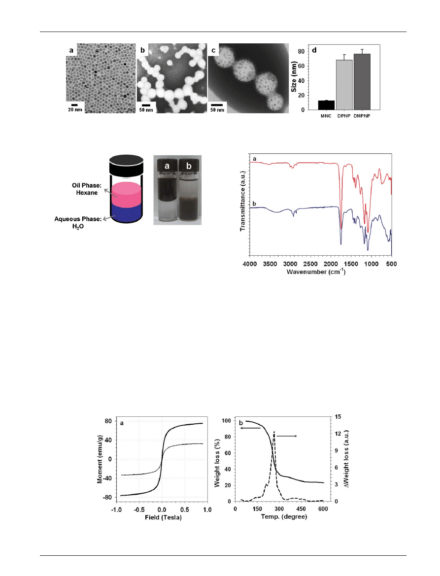
MNCs were synthesized using the high temperature seed-
mediated growth method for magnetic components of MR
probes for cancer detection. The MNC morphology was
examined by TEM, as shown in Fig. 1(a). The MNC size was
determined by laser scattering to be 12.6 ¡ 0.5 nm.
DMPNP for MR probes and drug carriers were prepared by
the nano-emulsion method in the presence of DOX, MNC and
PLGA. Similarly, PNP was formulated without DOX or MNC
for reference. TEM images of PNPs and DMPNPs are
presented in Fig. 1(b) and (c). MNCs dispersed in organic
PVA solvent were well encapsulated in the polymeric
nanoparticles due to the hydrophobic interaction of MNCs
and PLGA. Using laser scattering, the sizes of PNP and
DMPNP were 68.5 ¡ 7.2 nm and 76.8 ¡ 6.2 nm, respectively.
Compared to PNP, the DMPNP size slightly increased due to
incorporation of MNC.
The solubility of MNC and DMPNP is shown in Fig. 2. The
oil phase of hexane exists above water due to its lower density.
The MNC was soluble only in hexane and the DMPNP was
dispersed only in water due to the surface PVA. The nonionic
surfactant, PVA, successfully increased the water solubility of
DMPNP. Furthermore, after several days, the DMPNP was
2696 |
J. Mater. Chem.
, 2007, 17, 2695–2699
This journal is ß The Royal Society of Chemistry 2007
well dispersed in the aqueous phase. To this end, DMPNP
demonstrated excellent colloidal stability in an aqueous phase
(Fig. 2).
For the assessment of the potential of DMPNP as an MR
probe, the magnetic sensitivity was estimated. The hysteresis
loops of MNC and DMPNP were investigated using a
vibrating sample magnetometer at 300 K (Fig. 3(a)). The
MNC and DMPNP exhibited superparamagnetic behavior
without magnetic hysteresis. The saturation magnetizations
at 0.9 T of MNC and DMPNP were 74.7 emu g
2
1
and
32.5 emu g
2
1
, respectively. Due to the presence of organic
components such as DOX, PLGA and PVA, the saturation
magnetization of DMPNP was lower than that of MNC. The
quantity of magnetic nanoparticles in DMPNP was measured
using a thermogravimetric analyzer and the results are
shown in Fig. 3(b). All organic compounds were removed at
210–270
uC. The quantity of MNC encapsulated in DMPNP
was 23.1 wt%.
FT-IR was used to evaluate the chemical structure of
PLGA (Fig. 4(a)) and DMPNP (Fig. 4(b)). The characteristic
peak of PLGA at 1749 cm
2
1
is due to the ester group
(Fig. 4(a)). During preparation of DMPNP, the characteristic
peak of PLGA was not altered (Fig. 4(b)). The hydroxy
group of PVA was confirmed at 3500–3100 cm
2
1
(Fig. 4(b)).
Furthermore, the MNC characteristic peak of the Fe–O bond
was observed at 585 cm
2
1
.
11
These results suggest that the
MNC of DMPNP successfully coexisted with PLGA due to
the presence of PVA.
Fig. 1
TEM images of (a) MNC in hexane, (b) DOX-PLGA nanoparticles (DPNP), (c) DMPNP in an aqueous solution and (d) laser scattering
size distributions of MNC, DPNP and DMPNP.
Fig. 2
Solubility test of (a) MNC in hexane and (b) DMPNP in an
aqueous phase.
Fig. 3
(a) Magnetic hysteresis loops of MNC (black solid line) and DMPNP (dotted line). (b) Thermogravimetry analysis of DMPNP: weight loss
vs. temperature (black solid line), D weight loss vs. temperature (dashed line).
Fig. 4
FT-IR spectra of (a) PLGA and (b) DMPNP.
This journal is ß The Royal Society of Chemistry 2007
J. Mater. Chem.
, 2007, 17, 2695–2699 | 2697
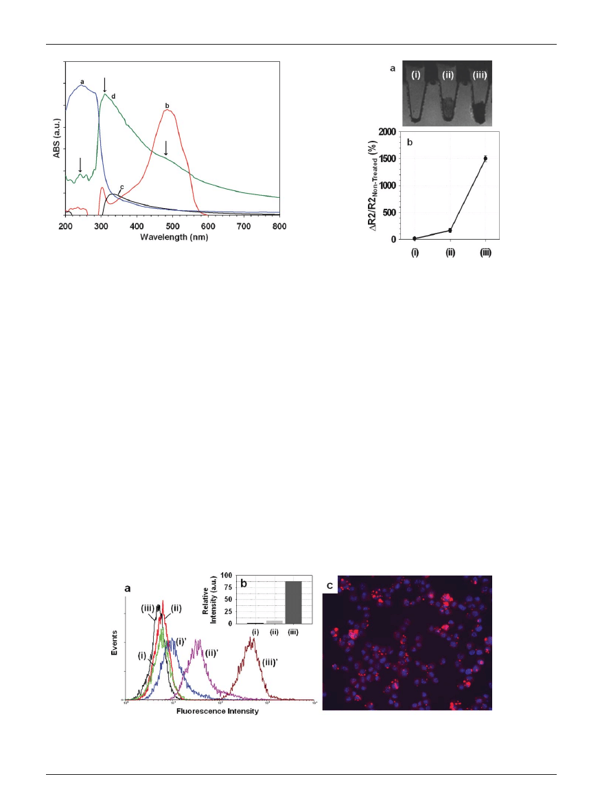
To further confirm the DMPNP multi-structure, UV-vis
absorption spectroscopy was performed (Fig. 5). The charac-
teristic bands of MNC, DOX and PLGA were observed at
245 nm, 485 nm and 335 nm, respectively. The characteristic
bands of DMPNP are shown in Fig. 5(d).
The breast cancer cell targeting efficacy of HER-DMPNP
was investigated. The human epidermal growth factor
receptor-2 (HER2) was used as a tumor targeting marker for
the treatment of patients with metastatic breast cancer.
12
Fibroblast NIH3T6.7 cells, which express high levels of the
HER2/neu cancer markers, were compared with SK-BR3 and
MDA-MB-231 cells, which express low levels of the cancer
markers.
3
In Fig. 6(a), the NIH3T6.7 cells incubated with
HER-DMPNP demonstrated a greater fluorescence intensity
by FACS analysis than other cells, and the relative intensity
was 87.4 times higher than that of non-treated cells. Compared
to non-treated control cells, the relative fluorescence intensities
of SK-BR3 and MDA-MB-231 cells were 6.5 and 1.5,
respectively. In addition, the cellular binding efficiency of
HER-DMPNP to the NIH3T6.7 cells was visualized using an
epifluorescence microscope (Fig. 6(c)). The red is due to DOX
of the HER-DMPNPs in the target cells (the blue of the
nucleus is due to 49,6-diamidino-2-phenylindole (DAPI)
staining), indicating acceptable cellular binding efficiency.
FACS analysis and fluorescence microscopy demonstrated
that HER-MFND successfully bound target cancer cells.
The T2-weighted MR images and the change of relaxivity
figure for three types of breast cancer cell lines indicated the
potential for cancer detection (Fig. 7). The MR image of
NIH3T6.7 cells incubated with HER-DMPNP exhibited a
black color. Other cells presented gray due to low levels of
the cancer markers. The changes of DR2/R2
Non-treated
in the
HER-DMPNP treated cells compared to the non-treated cells
were
y1 500% (NIH3T6.7), y166.7% (SK-BR3) and y14.3%
(MDA-MB-231), as shown in Fig. 7(b). These results
demonstrated
the
efficient
targeted
delivery
of
HER-
DMPNP for the HER2/neu receptor of cancer cells.
To test the influence of MNCs in polymeric nanoparticles
on the drug release profile, a release test was performed in
triplicate to calculate a mean value and standard deviation.
The quantities of encapsulated DOX in DPNP and DMPNP
were 17.4% and 5.8%, respectively. However, the drug
entrapment efficiencies for DPNP and DMPNP were 62.7%
Fig. 5
UV-vis adsorption spectra of (a) MNC in hexane, (b) DOX,
(c) PNP and (d) DMPNP in an aqueous solution.
Fig. 6
(a) FACS analysis of HER-DMPNP against (i)9 MDA-MB-231, (ii)9 SK-BR3 and (iii)9 NIH3T6.7 cells. The non-treated control cells are
also presented: (i) MDA-MB-231, (ii) SK-BR3 and (iii) NIH3T6.7 cells. (b) Relative intensity via FACS analysis. (c) Fluorescence microscopy
image of NIH3T6.7 cells incubated with HER-DMPNP; red: DOX and blue: DAPI.
Fig. 7
(a) T2-weighted MR images and (b) DR2/R2
Non-treated
graph
for (i) MDA-MB-231, (ii) SK-BR3 and (iii) NIH3T6.7 cells.
2698 |
J. Mater. Chem.
, 2007, 17, 2695–2699
This journal is ß The Royal Society of Chemistry 2007
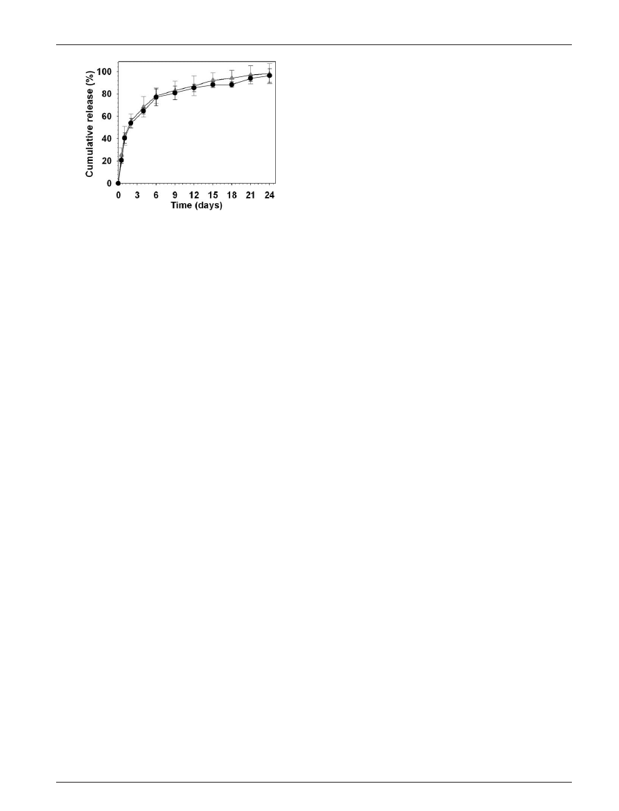
and 67.9%, respectively. The amount of encapsulated drug in
DMPNP was lower than that in DPNP due to the presence of
the MNC. The drug entrapment efficiencies were analogous
for both cases. Similarly, the drug release profiles of DPNP
and DMPNP were comparable (Fig. 8). After 3 and 12 days,
60% and 80% of the encapsulated DOX was released from
DPNP and DMPNP due to polymer degradation. To this end,
DOX loaded DMPNP was released from drug carriers without
any inhibition due to MNC.
Conclusions
We successfully synthesized antibody conjugated DOX–
magnetic PLGA nanoparticles (HER-DMPNP) for detection
and treatment of cancer. The well-tailored DMPNP was
prepared using a surfactant through a nano-emulsion
method. MNC was embedded in polymeric nanoparticles
through hydrophobic interaction. The DMPNP demonstrated
excellent sensitivity as MR probes for detection of cancer
cells. Moreover, affinity of the HER-DMPNP to cancer
cells was predominant. In addition, DOX encapsulated in
polymeric nanoparticles released sustainably without any
inhibition due to the presence of MNC. These multifunctional
nanocomposites can be applied to various biomedical
fields such as targeted drug delivery, MRI probes and cell
separation.
Acknowledgements
This work was supported by KOSEF through National Core
Research Center for Nanomedical Technology (R15-2004-024-
00000-0 and R01-2006-000-10023-0), the National R&D
Program for Cancer Control, Ministry of Health & Welfare,
Republic of Korea (0620190-1), and Yonsei University
Research Fund of 2006.
References
1 P. Alivisatos, Science, 1996, 271, 933; W. Li, C. Gao, H. Qian,
J. Ren and D. Yan, J. Mater. Chem., 2006, 16, 1852; Y.-W. Jun,
Y.-M. Huh, J.-S. Choi, J.-H. Lee, H.-T. Song, S.-J. Kim, S. Yoon,
K.-S. Kim, J.-S. Shin, J.-S. Suh and J. Cheon, J. Am. Chem. Soc.,
2005, 127, 5732; Y. Li, M. Afzaal and P. O’Brien, J. Mater. Chem.,
2006, 16, 2175.
2 S. Rana, A. Gallo, R. S. Srivastava and R. D. K. Misra, Acta
Biomater., 2007, 16, 233.
3 Y.-M. Huh, Y. Jun, H. Song, S. Kim, J. Choi, J. Lee, S. Yoon,
K. Kim, J. Shin, J. Suh and J. Cheon, J. Am. Chem. Soc., 2005,
127, 12387; J. Lee, Y.-M. Huh, Y. Jun, J. Seo, J. Jang, H. Song,
S. Kim, E. Cho, H. Yoon, J. Suh and J. Cheon, Nat. Med., 2006,
13, 95.
4 T. Schneider, L. R. Moore, Y. Jing, S. Haam, P. S. Williams,
A. J. Fleischman, S. Roy, J. J. Chalmers and M. Zborowski,
J. Biochem. Biophys. Methods, 2006, 68, 1.
5 S. I. Park, Y. H. Hwang, J. H. Lim, J. H. Kim, H. I. Yun and
C. O. Kim, J. Magn. Magn. Mater., 2006, 304, 403.
6 X. Michalet, F. F. Pinaud, L. A. Bentolila, J. M. Tsay, S. Doose,
J. J. Li, G. Sundaresan, A. M. Wu, S. S. Gambhir and S. Weiss,
Science, 2005, 307, 538.
7 Y. Sun, X. Ding, Z. Zheng, X. Cheng, X. Hua and Y. Peng, Chem.
Commun., 2006, 2765.
8 S. Sun, H. Zeng, D. B. Robinson, S. Raoux, P. M. Rice, S. X. Wang
and G. Li, J. Am. Chem. Soc., 2004, 126, 273.
9 J. Yang, S. B. Park, H. G. Yoon, Y.-M. Huh and S. Haam, Int. J.
Pharm., 2006, 324, 185.
10 S. Sengupta, D. Eavarone, I. Capila, G. Zhao, N. Watson,
T. Kiziltepe and R. Sasisekharan, Nature, 2005, 28, 568.
11 C. Rocchiccioli-Deltcheff, R. Franck, V. Cabuil and R. Massart,
J. Chem. Res., 1987, 5, 126.
12 R. M. Hudziak, G. D. Lewis, M. Winget, B. M. Fendly,
H. M. Shepard and A. Ullrich, Mol. Cell. Biol., 1989, 9, 1165.
Fig. 8
The drug release profiles for (m) DPNP and ($) DMPNP.
This journal is ß The Royal Society of Chemistry 2007
J. Mater. Chem.
, 2007, 17, 2695–2699 | 2699
Wyszukiwarka
Podobne podstrony:
Diagnosis and Treatment of Autoimmune Hepatitis
In vivo MR spectroscopy in diagnosis and research of
Diagnosis and Management of Hemochromatosis
Best Available Techniques for the Surface Treatment of metals and plastics
Diagnosis and Management of hepatitis
Osteoporosis ľ diagnosis and treatment
In vivo MR spectroscopy in diagnosis and research of
Mathematica package for anal and ctl of chaos in nonlin systems [jnl article] (1998) WW
PSYCHIC METHODS OF DIAGNOSIS AND TREATMENT IN ACUPUNCTURE …
Young Internet Addiction diagnosis and treatment considerations
Step by step instructions activation of all brands of machines for EasyDiag and completion of the sc
EASE Guidelines for Authors and Translators of Scientific Articles to be Published in English june20
Diagnosis and Management of the Painful Shoulder Part 1 Clinical Anatomy and Pathomechanics
więcej podobnych podstron