
Human heat shock protein 70 (Hsp70) as a peripheral membrane protein
Ajay K. Mahalka
, Thomas Kirkegaard
, Laura T.I. Jukola
, Marja Jäättelä
, Paavo K.J. Kinnunen
a
Helsinki Biophysics and Biomembrane Group, Department of Biomedical Engineering and Computational Science, Aalto University, Espoo, Finland
b
Cell Death and Metabolism, Danish Cancer Society Research Center, Copenhagen, Denmark
c
Orphazyme ApS, Copenhagen, Denmark
a b s t r a c t
a r t i c l e i n f o
Article history:
Received 1 October 2013
Received in revised form 13 January 2014
Accepted 17 January 2014
Available online 28 January 2014
Keywords:
Hsp70
Liposomes
Tryptophan
Fluorescence
Extended lipid conformation
Langmuir-
films
While a signi
ficant fraction of heat shock protein 70 (Hsp70) is membrane associated in lysosomes, mitochon-
dria, and the outer surface of cancer cells, the mechanisms of interaction have remained elusive, with no conclu-
sive demonstration of a protein receptor. Hsp70 contains two Trps, W90 and W580, in its N-terminal nucleotide
binding domain (NBD), and the C-terminal substrate binding domain (SBD), respectively. Our
fluorescence
spectroscopy study using Hsp70 and its W90F and W580F mutants, and Hsp70-
ΔSBD and Hsp70-ΔNBD
constructs, revealed that binding to liposomes depends on their lipid composition and involves both NBD and
SBD. Association of Hsp70 with phosphatidylcholine (PC) liposomes is weak, with insertion of its Trps into the
bilayer hydrocarbon region. In the presence of cardiolipin (CL), bis-monoacylglycero phosphate (BMP), or
phosphatidylserine (PS) Hsp70 attaches to membranes peripherally, without penetration. Our data suggest
that the organelle distribution of Hsp70 is determined by their speci
fic lipid compositions, with Hsp70 associating
with the above lipids in mitochondria, lysosomes, and the surface of cancer cells, respectively. NBD and SBD attach
to lipids by extended phospholipid anchorage, with speci
fic acidic phospholipids associating with Hsp70 in the ex-
tended conformation with acyl chains inserting into hydrophobic crevices within Hsp70, and other chains remaining
in the bilayer. This anchorage is expected to cause a stringent orientation of Hsp70 on the surface. Our data further
suggest that acidic phospholipids induce a transition of SBD into the molten globule state, which may be essential
to allow SBD
–substrate interaction also within the hydrophobic bilayer interior acyl chain region.
© 2014 Elsevier B.V. All rights reserved.
1. Introduction
Heat shock protein 70 (Hsp70) constitutes a highly conserved family
of protein chaperones, which under physiological conditions regulate
protein homeostasis and promote cell survival
. Some Hsps are con-
stitutively expressed, whereas others are strictly stress-inducible
.
The major stress-induced human Hsp70 (also referred to as Hsp72) is
expressed when the cell is exposed to stress such as heat shock or UV
radiation. Escherichia coli Hsp70 chaperone is DnaK, which is regulated
by two protein modulators, DnaJ and GrpE
. Saccharomyces cerevisiae
has several Hsp family members, the most studied of these being the cy-
tosolic Ssa1p
. Eight different and unique Hsp70 have been reported
to be present in eukaryote cells, distributed in different subcellular com-
partments, including cytosol, nucleus, endoplasmic reticulum, and mi-
tochondria
. The main function of these ubiquitous chaperones is to
bind to denatured proteins and to assist in their refolding, in order to
prevent their aggregation, and to guide them to their native conforma-
tions, in a manner requiring ATP
, thus preventing cellular damage
and apoptosis induced by unfolded aggregated proteins
. Hsp70s
consist of two domains: NBD (residues 1
–386) and the C-terminal sub-
strate binding domain (SBD, residues 386
–640,
,
, panel A).
Three distinct conformations: nucleotide free, ADP-dependent, and
ATP-dependent, have been demonstrated for E. coli DnaK
. The
Biochimica et Biophysica Acta 1838 (2014) 1344
Abbreviations: AcrA, acrylamide; aSM, acid sphingomyelinase; a.u., arbitrary unit; BMP,
bis(monoacylglycero)phosphate; Br
2
PC, brominated phosphatidylcholine; 6,7Br
2
-PC, 1-
palmitoyl-2-(6,7-dibromo)stearoyl-sn-glycero-3-phosphocholine; 9,10Br
2
-PC, 1-palmitoyl-
2-(9,10-dibromo)stearoyl-sn-glycero-3-phosphocholine; 11,12Br
2
-PC, 1-palmitoyl-2-(11,12-
dibromo)stearoyl-sn-glycero-3-phosphocholine; Br
4
BMP, bis[mono(9,10)-dibromostearoyl]
glycerophosphate; 9,10Br
2
-PS, 1-palmitoyl-2-(9,10-dibromo)stearoyl-sn-glycero-3-
phospho-
L
-serine; Br
8
CL, tetra(9,10-dibromo stearoyl)cardiolipin; Br
2
PS, brominated
phosphatidylserine; CD, circular dichroism; Chol, cholesterol; CL, cardiolipin; DnaK, E. coli
heat shock protein 70; DTT, dithiothreitol; EDTA, ethylenediamine-N,N,N
′,N′-tetraacetic
acid; F,
fluorescence intensity; F
0
, initial
fluorescence intensity; FA, fatty acid; Grp78, endo-
plasmic reticulum heat shock protein 70; HD, Huntington disease; HDP, host defense pep-
tides; Hepes, 4-(2-Hydroxyethyl)-1-piperazineethanesulfonic acid; Hsc70, constitutively
expressed heat shock protein 70; Hsp70, Heat shock protein of
≈70kDa; Hsp70-ΔNBD, re-
combinant Hsp70 lacking the nucleotide binding domain; Hsp70-
ΔSBD, recombinant
Hsp70 lacking the substrate binding domain; Hsp70-W90F, recombinant Hsp70 with substi-
tution W90F; Hsp70-W580F, recombinant Hsp70 with substitution W580F; KCL, potassium
chloride; K
sv
, Stern
–Volmer quenching constants; L/P, lipid/protein molar ratio; LUV, large
unilamellar vesicles; MES, 2-(N-morpholino)ethanesulfonic acid; NBD, nucleotide binding
domain; NPD, Niemann
–Pick disease; PEG, polyethylene glycol; POPC, 1-palmitoyl-2-
oleoyl-sn-glycero-3-phosphocholine; POPS, 1-palmitoyl-2-oleoyl-sn-glycero-3-phospho-
L
-
serine; PS, phosphatidylserine; RFI, relative
fluorescence intensity; Spm, sphingomyelin;
SBD, substrate binding domain; Ssa1p, S. cerevisiae heat shock protein 70; tOCL, 1,1
′,2,2′-
tetraoleoyl cardiolipin; wtHsp70, wild type Hsp70;
π, surface pressure; π
0
, initial surface pres-
sure;
Δπ, increment in surface pressure; π
c
, critical packing pressure;
λ, wavelength; Δλ,
spectral center of mass
⁎ Corresponding author at: Helsinki Biophysics & Biomembrane Group, Department of
Biomedical Engineering and Computational Science, P.O. Box 12200 (Rakentajanaukio
3), FIN-00076, Aalto, Finland. Tel.: +358 50 540 4600; fax: +358 9 470 23182.
E-mail address:
(P.K.J. Kinnunen).
0005-2736/$
– see front matter © 2014 Elsevier B.V. All rights reserved.
http://dx.doi.org/10.1016/j.bbamem.2014.01.022
Contents lists available at
Biochimica et Biophysica Acta
j o u r n a l h o m e p a g e : w w w . e l s e v i e r . c o m / l o c a t e / b b a m e m
conformations of NBD and SBD have been shown to be coupled for
Hsp70
, DnaK
, and Grp78
. The presence of ATP accelerates
the binding and release of polypeptides, but it still remains unclear
whether it is the binding or hydrolysis of ATP that causes the release
of peptides from SBD
. Formation of dimers and higher-order oligo-
mers of Hsp70 has been suggested, with the monomeric protein
representing the functionally active chaperone
.
Hsp70 is additionally involved in the control of cell signaling for
growth, differentiation, and apoptosis
and its overexpression is
required for the growth and survival of human tumors
, with
elevated expression of Hsp70 correlating with poor prognosis in
human breast cancer and endometrial tumors
. Hsp70 is highly
expressed in the cytosol, outer surface of the plasma membrane and
the membranes of the endo-lysosomal compartment in primary tumors
of different origins, whereas its expression in unstressed normal cells is
low and restricted to the cytosol
Hsp70 may also provide a recognition structure for natural killer
cells
. Multiple reports have demonstrated the association of
Hsp70 family members with biomembranes in normal and tumor
cells, and tumor-derived cell lines
. Bovine Hsc70 binds to
cell surface sulfogalactolipids through its N-terminal nucleotide binding
domain (NBD,
). Electron microscopy shows Hsp70 on the cell sur-
face, in clathrin coated pits, and within endosome/lysosome-related
vesicles
. There is also evidence that Hsp70 is associated with the
so-called detergent resistant microdomains in the plasma membrane
.
Direct interaction of Hsp70 with bis-monoacylglycero phos-
phate (BMP), an acidic phospholipid enriched in late endosomes and
lysosomes
appears to be required for the activation of lysosomal
acid sphingomyelinase (aSM), whose activity is essential for the down-
stream cytoprotective effect of lysosomal Hsp70. Our previous studies
revealed that NBD contains a speci
fic and pH-dependent binding site
for BMP. Trp90 of NBD is required for this interaction and its mutation
to Phe results in an Hsp70 with compromised BMP-binding, rendering
Hsp70 unable to prevent lysosomal membrane permeabilization
This interaction can also be blocked by an antibody against BMP
Despite the accumulating information on the membrane association
of Hsp70 and its potential signi
ficance to the functions of Hsp70, lipid–
Hsp70 interactions have not been assessed in detail. Accordingly, the
exact molecular mechanisms and the mode of attachment of Hsp70 to
membrane bilayers remain to be elucidated. Intriguingly, Hsp70 has
been demonstrated to contain two fatty acid binding sites. Membrane
association of Hsp70 and its lipid-interactions demonstrated so far al-
ready suggest that Hsp70 could be a peripheral membrane protein. In
this study, we exploited the intrinsic Trp
fluorescence of human
Hsp70 to evaluate possible lipid speci
ficity in the membrane binding
of Hsp70. Two mutants, Hsp70-W90F and Hsp70-W580F as well as
NBD and SBD constructs Hsp70-
ΔSBD and Hsp70-ΔNBD were addition-
ally compared with wtHsp70 for their interactions with 1-palmitoyl-2-
oleoyl-sn-glycero-3-phosphocholine (POPC), as well as cardiolipin (CL),
BMP, and 1-palmitoyl-2-oleoyl-sn-glycero-3-phospho-
L
-serine (POPS)
containing PC liposomes, complementing our previous studies on BMP
, and those by Arispe et al.
on PS.
The aim of the present study was to explore in more detail the inter-
actions of Hsp70 and NBD and SBD with different lipids, providing at
this stage a qualitative understanding of the interactions and how
they in
fluence the orientation and conformation of Hsp70 in mem-
branes. Our present results demonstrate a complex array of interactions
of Hsp70 with phospholipid membranes. These interactions are highly
sensitive to the membrane lipid composition, with Hsp70 selectively
binding to membranes containing negatively charged phospholipids
such as CL, BMP, and PS. Accordingly, the distribution of Hsp70 in lyso-
somes, mitochondria, and on the outer surface of cancer cells, respec-
tively, could re
flect the enrichment of these lipids in the above
organelles and their interactions with Hsp70. Using single Trp Hsp70
mutants W90F and W580F we showed that both NBD and SBD contrib-
ute to the attachment of Hsp70 to lipid surfaces. In NBD the phospholip-
id binding site involves W90, which is also involved in the cationic site
responsible for the binding of ATP
. Our data derived from Langmuir
balance and Trp
fluorescence spectroscopy experiments using collision-
al quenching by brominated phospholipids (6,7-Br
2
PC, Br
2
PS, Br
4
BMP,
and Br
8
CL), and acrylamide as well as wtHsp70, its W90F and W580F
mutants, and the NBD and SBD constructs allow us to conclude that
(i) Hsp70 binds to CL, BMP, and PS containing membrane surfaces
peripherally, most likely by extended phospholipid anchorage
.
(ii) Both NBD and SBD appear to interact with lipids.
(iii) Our data further suggest that under these conditions SBD is likely
to adopt the molten globule conformation.
2. Experimental procedures
2.1. Materials
1-Palmitoyl-2-oleoyl -sn -glycero -3-phosphocholine (POPC), 1-
palmitoyl- 2-(6,7-dibromo)stearoyl -sn -glycero-3- phosphocholine
(6,7Br
2
-PC), 1-palmitoyl-2-(9,10 -dibromo)stearoyl- sn-glycero-3-
phosphocholine (9,10Br
2
-PC), 1-palmitoyl-2-(11,12-dibromo)stearoyl-
sn-glycero-3-phosphocholine (11,12Br
2
-PC), 1-palmitoyl-2-oleoyl-sn-
glycero-3-phospho-
L
-serine (POPS), 1,1
′,2,2′-tetraoleoyl cardiolipin
(toCL), bis-monoacylglycero phosphate (BMP), cholesterol, N-acyl-
phosphatidylethanolamine, and sphingomyelin were from Avanti
Polar-Lipids Inc. (Alabaster, AL, USA). PEG 400 was from ABCR GmbH
& Co.KG (Karlsruhe, Germany). Lipids were dissolved in chloroform
and their concentrations were determined gravimetrically using
a high precision electrobalance (Cahn, Cerritos, CA) as described
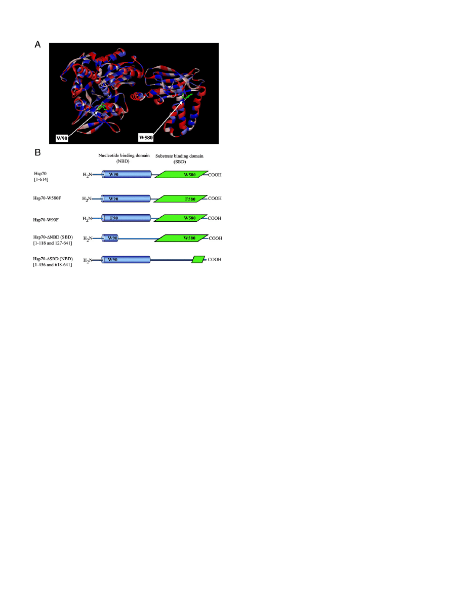
Fig. 1. Panel A: Tentative 3D ribbon structure of Hsp70 based on the crystal structures of
bovine Hsc70 (PDB ID: 1YUW) and SBD of Hsp70 (PDB ID: 2P32), homology modeled by
the Discovery studio. Surface coloring illustrates hydrophobicity (blue) and hydrophilicity
(red). W90 and W580 are shown as ball and stick models (green). Panel B: Hsp70 con-
structs used in the present study.
1345
A.K. Mahalka et al. / Biochimica et Biophysica Acta 1838 (2014) 1344
–1361

previously
. Acrylamide (AcrA, 99% purity) was from Eastman-
Kodak (Rochester, NY, US). All other chemicals were of analytical
grade from standard sources.
2.2. Expression and puri
fication of Hsp70
wtHsp70, Hsp70-W580F, Hsp70-W580F, Hsp70-
ΔNBD, and Hsp70-
ΔSBD constructs (
, panel B) were generated using the pET-16b
vector system and Ni
2+
-af
finity-purification system (Novagen, Merck
KGaA, Darmstadt, Germany). The recombinant Hsp70 constructs con-
taining His
6
-tag were separated using a Ni
2+
-column, with the bound
protein eluted with 0.5 mM imidazole at pH 7.4, subsequently medium
exchanged for Dulbecco's phosphate buffered saline (D-PBS) using
chromatography over a PD10 column (Amersham). The factor Xa recog-
nition site located between the His
6
-tag and the Hsp70 domains was
then cleaved by factor Xa and the His
6
-tag and factor Xa removed by
filtering through Amicon Ultra 50MWCA (Amersham). The purified
Hsp70 constructs were stored at
−20 °C in 25 mM Hepes, 0.1 mM
EDTA, 10% glycerol, 50 mM KCL, 1 mM DTT, and pH 7.6. Charges of
Hsp70 and its constructs at pH 7.4 and 6.0 were estimated with the
web-based calculator (
http://www.scripps.edu/~cdputnam/protcalc.
Importantly, our attempts to utilize His-tag containing proteins
showed, that this addition has a signi
ficant influence on the lipid bind-
ing properties of Hsp70 (Mahalka et al., to be published), yielding con-
structs, which deviated drastically in their lipid binding properties
from the wild-type protein. This behavior appears to result from the
binding of His-tag to phospholipids.
2.3. Preparation of liposomes
Lipids were dissolved and mixed in chloroform to obtain the indicat-
ed compositions, where after this solvent was removed under a stream
of nitrogen. The lipid residues were subsequently maintained under
reduced pressure for at least 2 h and subsequently hydrated for
60 min at 60 °C in 20 mM Hepes, 0.1 mM EDTA to yield a lipid concen-
tration of 2 mM. pH of the buffers was adjusted with HCl to pH 7.4 or
6.0 as indicated. In order to obtain large unilamellar vesicles (LUV),
the hydrated lipid mixtures were extruded through 100 nm pore size
polycarbonate membranes (Nucleapore Inc., Pleasanton, CA, USA)
with a LiposoFast small-volume homogenizer (Avestin, Ottawa,
Canada,
), yielding vesicles with mean diameters of 120
–140 nm
measured by dynamic light scattering.
2.4. Tryptophan
fluorescence spectroscopy
All
fluorescence measurements were conducted with a Perkin-
Elmer LS50B spectro
fluorometer. Hsp70 and buffer (20 mM Hepes,
0.1 mM EDTA, pH 7.4 or 6.0, total volume 2 ml) were mixed in a mag-
netically stirred PEG-treated
quartz cuvettes, with 10 mm optical
path length in a holder thermostated at 37 °C with a circulating
waterbath. Coating of the quartz glass cuvettes by PEG was used to min-
imize the binding of the Hsp70 to the cuvettes
. The initial concen-
tration of Hsp70 in the cuvette was 0.43
μM in the indicated buffers.
Trp emission was recorded between 308 and 450 nm with excitation
and emission bandwidths of 10 nm and with excitation at 295 nm.
When indicated liposomes were added in ten subsequent 20 nmol
aliquots (in 10
μl) into the cuvette and spectra recorded after a
20 min stabilization period for each addition. From these data the
emission peak positions, peak intensities, and spectral centers of mass
were determined.
Brominated lipids were used to monitor possible contacts of the
Trp residues of Hsp70 with the lipid acyl chains
. BMP, POPS,
and tOCL were brominated as described by East and Lee
to
yield bis[mono(9,10)-dibromostearoyl]glycerol-phosphate (Br
4
BMP),
1-palmitoyl-2-(9,10-dibromo)-stearoyl-sn-glycero-3-phospho-
L
-serine
(Br
2
PS), and tetra(9,10-dibromostearoyl) cardiolipin (Br
8
CL). Lipo-
somes containing brominated phospholipids (6,7-Br
2
PC, 9,10Br
2
-, and
11,12Br
2
PC, Br
2
PS, Br
4
BMP, or Br
8
CL) were added into a solution of
Hsp70 as above, and the ratios (F
0
/F) of the Trp
fluorescence peak inten-
sities upon adding control liposomes and liposomes containing the indi-
cated brominated lipids, respectively were calculated from the emission
spectra. Differences in the quenching of Trp
fluorescence by (6,7)-,
(9,10)-, and (11,12)-Br
2
-PC were used to estimate by the parallax
method the apparent depth of penetration of Hsp70 Trps into the lipid
bilayers
In order to determine the exposure of Trps to the aqueous phase,
AcrA, a water-soluble collisional quencher
, was added as six subse-
quent micromolar aliquots. Spectra were recorded using excitation at
295 nm to minimize inner
filter effect due to AcrA. All data have been
subtracted for background and light scattering due to liposomes, nor-
malized, and corrected for volume changes and the inner
filter effect.
2.5. Binding of Hsp70 to lipid monolayers
Appropriate amounts of lipid stock solutions were mixed in chloro-
form to obtain the desired compositions. The indicated lipid mixtures
were subsequently spread onto an air/buffer interface in magnetically
stirred circular Te
flon coated wells (diameter of 17.8 mm and a sub-
phase volume of 1.2 ml) drilled in aluminum. Dynamic surface pressure
(
π) was monitored by the Wilhelmy technique using miniature cylindri-
cal probes (Dyneprobe, Kibron Inc., Espoo, Finland) attached to the sen-
sors of a four channel Langmuir tensiometer (DeltaPi-4, Kibron Inc.).
Data were recorded using the embedded features of the instrument
control software allowing for simultaneous monitoring of four reac-
tions. After stabilization of the applied monolayers to a range of initial
surface pressure values
π
0
the indicated proteins were injected into
the subphase (0.1
μM final concentration), where after the increment
in
π (Δπ) due to their intercalation into the lipid film was recorded.
These data are represented as
Δπ vs π
0
, yielding upon least-squares lin-
ear
fitting straight lines with negative slopes. The x-axis intercepts rep-
resent critical lateral packing densities of lipids corresponding to surface
pressure
π
c
, above which the protein no longer can intercalate into the
monolayer
. All measurements were performed at ambient temper-
ature of ~23 °C.
2.6. Circular dichroism (CD)
CD spectra were recorded with Olis RSF 1000F (On-line Instrument
Systems Inc., Bogart, GA) CD spectrophotometer using a 0.1 mm optical
path length quartz cuvette thermostated at 37 °C with a circulating
waterbath. The instrument was calibrated with d-(+)-camphorsulfonic
acid. Near and far UV-CD spectra were recorded from 260 to 198 nm,
and 310 to 145 nm, respectively, at increment of 1 nm with 1 s integra-
tion time. To remove chloride and DTT aliquots of Hsp70 stock solution
were dialyzed against MES buffer. The concentration of Hsp70 used in
far and near UV-CD were 2 and 8.5
μM, respectively in a final volume
of 2 ml of 20 mM MES, 0.1 mM EDTA at pH 6.0. All spectra have been
corrected for circular differential scattering due to buffer and liposomes.
The spectra shown represent the averages of four scans, and were ana-
lyzed with the computer program K2d2
.
3. Results
3.1. Lipid
–protein interactions of Hsp70
We investigated the interactions of wtHsp70 with liposomes com-
posed of POPC together with a range of different acidic phospholipids.
To begin with, we
first assessed the association of Hsp70 with the liquid
expanded (
fluid) state zwitterionic POPC, and then proceeded to com-
pare this with its binding to POPC liposomes containing cardiolipin,
BMP, and phosphatidylserine.
1346
A.K. Mahalka et al. / Biochimica et Biophysica Acta 1838 (2014) 1344
–1361
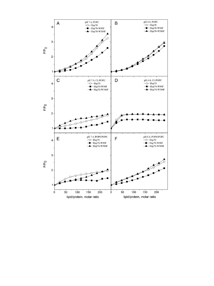
3.1.1. Binding of Hsp70 to phosphatidylcholine liposomes
Fluorescence spectroscopy of Trp provides a sensitive tool to obtain
information on changes in protein conformation and interactions with
e.g. lipids
. Human Hsp70 contains two Trps, W90 and W580, in its
NBD and SBD, respectively (
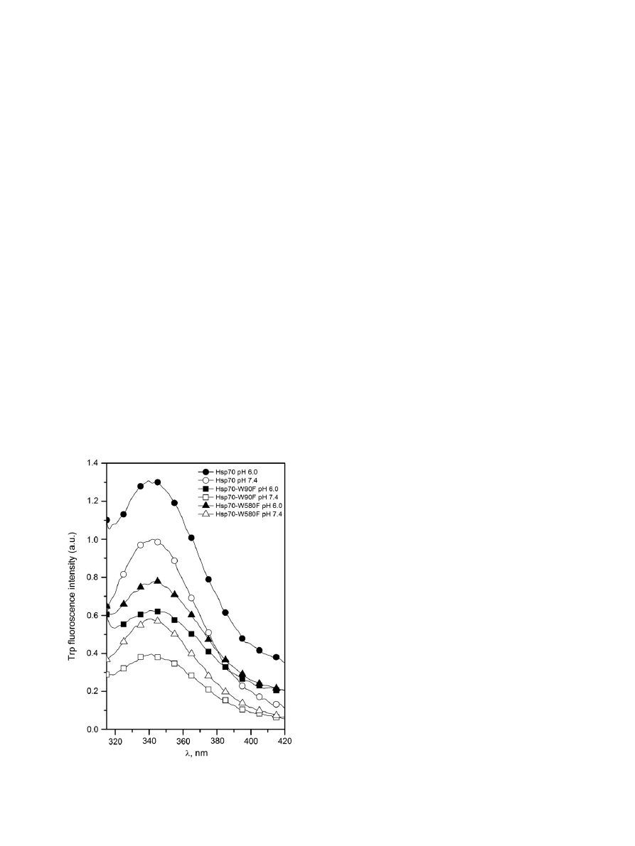
, panel B). Steady state Trp
fluores-
cence emission depends on solvent polarity, and re
flects in part the ex-
posure of this residue to water
. Emission of Hsp70 in buffer was
sensitive to pH with nearly identical spectral centers of mass observed
at both pH 7.4 and 6.0, yet with approximately 30% higher overall emis-
sion evident at more acidic pH (
). Augmented quantum yields at
pH 6.0 were also seen for both Hsp70-W90F and Hsp70-W580F, thus
suggesting that at pH 6.0 NBD and SBD both adopt conformations
in which the two Trps become embedded in more hydrophobic
environments.
We
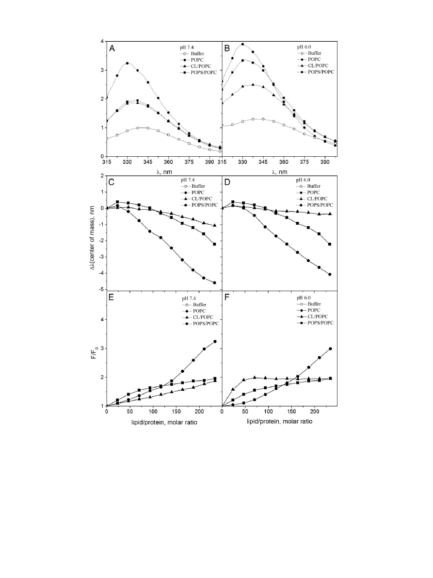
first explored by Trp fluorescence the binding of wtHsp70 to
POPC liposomes. POPC LUV induced profound changes in the Trp
fluorescence of Hsp70 (
), with a large increase in the relative
fluo-
rescence intensity (RFI), together with a decrease (blue shift) in the
wavelength of maximal emission (
λ
max
), thus indicating an increase in
the hydrophobicity of the environments accommodating either W90
or W580, or both (
, panels C and D). Together with the above
shift in
λ
max
POPC LUV also caused a narrowing of the major peak at
λ
max
≈ 340 nm seen in the absence of liposomes (
). The RFI vs
L/P curve, for POPC was biphasic at pH 7.4 (
, panel E), with an
initial linear component followed by a second linear component at
L/P
N150 (i.e. corresponding to a higher surface dilution of Hsp70).
Especially at higher L/P ratios the RFI values equilibrated slowly, requir-
ing up to 15
–20 min. The curves reveal no sign of saturation thus indicat-
ing a low af
finity interaction, most likely arising from weak, non-specific
hydrophobic partitioning of Hsp70 to the POPC bilayer. These changes
suggest that compared to Hsp70 in buffer, binding of Hsp70 to POPC
liposomes causes at least one of its Trp residues to become accommodat-
ed in a more hydrophobic environment. Complementary
fluorescence
experiments and spectral characteristics of W90 and W580 (
see below) suggest that the Trp primarily contacting the POPC bilayer
acyl chain region is W90 of NBD, which consistently emits at slightly
shorter value for
λ
max
. Compared to the spectra measured in the pres-
ence of POPC LUV at pH 7.4 the values for RFI recorded at pH 6.0 are
somewhat lower and the initial linear component seen at pH 7.4 spans
only a narrow L/P range (
, panels E & F).
The above data complies with an intercalation of Hsp70 into POPC
bilayers. In order to check for this possibility we investigated if
phospholipids with brominated stearoyl chains, i.e. 6,7-, 9,10-, and
11,12Br
2
-PC quench Trp
fluorescence of Hsp70 bound to POPC lipo-
somes. Bromine is a collisional quencher of Trp and in a bilayer in the
absence of chain reversal the above phospholipid acyl chains should re-
main in the hydrophobic region of a bilayer. Trp
fluorescence quenching
by these three brominated PCs in POPC liposomes reveals contacts of
the bromines with at least one of the Trp residues (
, panel A). In
a neutral bilayer and at pH 7.4 the Trp residue(s) in question seem(s)
to reside in the vicinity of acyl chain carbon atoms 6,7 and 9,10. At
pH 6 there is slightly less quenching, thus suggesting attenuated pene-
tration of the Trp(s) into POPC bilayers (
, panel B). This pH depen-
dence could re
flect an increase in the net charge of Hsp70 at pH 6.0,
results in less penetration into the bilayer. Interestingly, quenching by
6,7-, and 9,10Br
2
-PCs diminishes the blue shift in the spectral center of
mass (Table SI). Since collisional quenching by Br should not alter the
shape of the spectra, these data suggest that one of the Trp residues
preferentially contacts the brominated acyl chains and becomes
quenched, with less quenching of the
fluorescence from Trp residing
in a less hydrophobic environment and emitting at a longer
λ
max
. The
latter Trps are likely to include the fraction of Hsp70 in solution (not at-
tached to liposomes) and W580. Comparison of
λ
max
values for W90
and W580 (
) suggests that the brominated PCs in POPC lipo-
somes preferentially interact with NBD, quenching W90. From X-ray
diffraction, the average distances of the bromines of 6,7-, 9,10-, and
11,12Br
2
-PC, from the bilayer center are 10.8, 8.3, and 6.3 Å, respective-
ly
. Parallax analysis
of our data suggests, that the distance of
the Trp-residue (W90) quenched is approximately 11.5 Å from the
bilayer center, thus indicating only a shallow insertion of W90 into the
bilayer, in keeping with partitioning of Trp into the interfacial region
of phospholipid bilayers
In order to resolve the contributions of W90 and W580 to the above
Trp
fluorescence signals and the involvement of NBD and SBD in lipid
interactions, we repeated the above experiments using Hsp70 mutants
Hsp70-W90F and Hsp70-W580F. We previously showed that the W90F
mutant fails to activate acid sphingomyelinase in cells, thus resulting in
a lack of stabilization of lysosomal membranes when endocytosed.
Interactions of Hsp70-W90F with liposomes containing BMP are im-
paired
. While F instead of W is generally considered to represent
a minimally perturbing mutation, the activity of Hsp70 in cells is lost, re-
vealing a crucial functional role of W90. The extent of perturbation of
the overall conformation of Hsp70 and the lipid interactions of Hsp70
by the W90F mutation remain to elucidated. Yet, the results obtained
from this construct allow us to decipher molecular level understanding
of the phospholipid
–Hsp70 interactions. Comparison of the spectra of
Hsp70 and the above constructs (
) reveals that the combined emis-
sion from W90 and W580 closely parallel the RFI of wtHsp70, thus sug-
gesting that the overall conformations of the Trp-containing domains in
these mutants are similar to those when present in the wtHsp70.
Similar to wtHsp70 a signi
ficant enhancement in Trp fluorescence is
seen for the Hsp70-W580F as well as Hsp70-W90F in the presence of PC
LUV (
, panels A & B). Accordingly, the non-speci
fic, low affinity in-
teraction of Hsp70 with POPC liposomes seems to involve contributions
from both NBD and SBD, most likely driven by hydrophobicity, and with
intercalation of Trp-containing sequences into the lipid hydrocarbon re-
gion. 6,7-Br
2
PC quenching data reveal contacts of both NBD and SBD
Trps with the POPC bilayer region at both pH 7.4 and 6.0 (
, panels
A & B), with more pronounced quenching of W90. The above conclusion
is supported by large
fluorescence enhancement of Hsp70-W580F
Fig. 2. Trp emission spectra in 20 mM Hepes, 0.1 mM EDTA for Hsp70 (
○/●), Hsp70-
W90F (W580,
□/■), and Hsp70-W580F (W90, Δ/▲). Open symbols pH 7.4 and closed
symbols pH 6.0. The concentrations of Hsp70 and the two mutants were 0.43
μM.
1347
A.K. Mahalka et al. / Biochimica et Biophysica Acta 1838 (2014) 1344
–1361
compared to Hsp70-W90F (
, panels A & B) and diminished extent
of quenching of Hsp70-W90F by AcrA compared to Hsp70-W580F
(
, panels A & B). Moreover, comparison of
λ
max
values also suggests
that the brominated PCs in POPC liposomes preferentially interact with
NBD, quenching W90 (
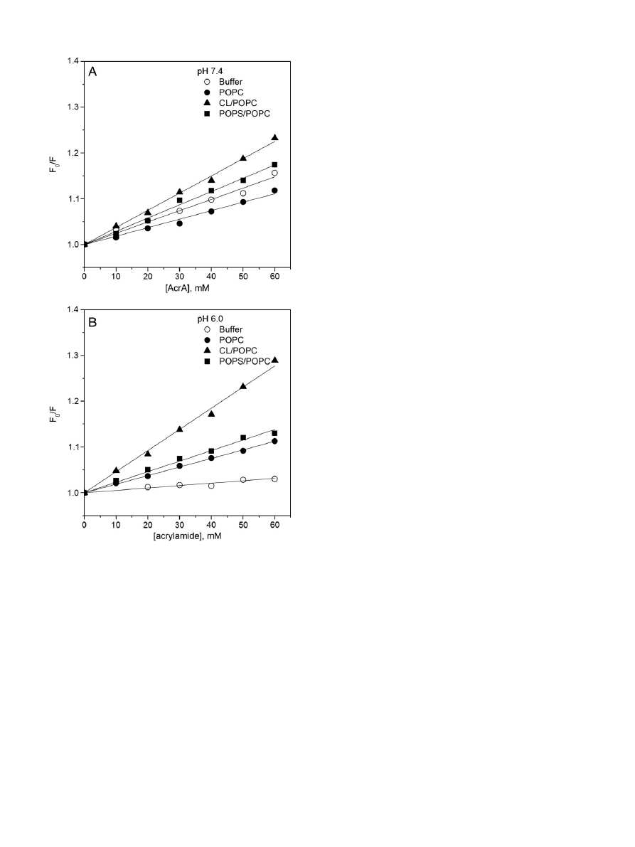
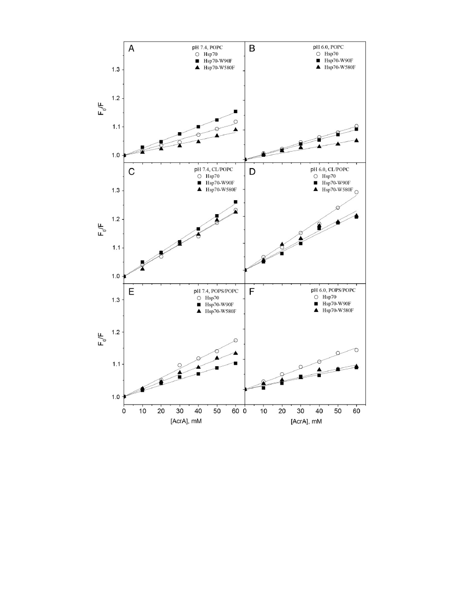
In order to verify the intercalation of Hsp70 into POPC bilayers we
assessed the ef
ficiency of quenching of Trp emission by the water-
soluble collisional quencher acrylamide [AcrA,
]. In keeping with the
immersion of Hsp70 Trps into the POPC bilayer at pH 7.4, their
quenching by AcrA was attenuated in the presence of POPC LUV
(
, panel A), suggesting shielding of the Trps from contacts with
the water soluble AcrA upon the POPC bilayer
–Hsp70 interaction. At
pH 6.0, however more ef
ficient quenching by AcrA becomes evident,
showing an increased exposure to AcrA (
, panel B), in keeping
with a more super
ficial location of Hsp70 on the POPC bilayer surface
at pH 6.0, as concluded from Trp
fluorescence and quenching by bromi-
nated PCs (
, panel B,
, panel B). These data aligns with a
shielding from AcrA of W90 in Hsp70-W580F in the presence of PC
Fig. 3. Tryptophan
fluorescence spectra for wtHsp70 (○) in buffer (20 mM Hepes, 0.1 mM EDTA) and in the same buffer in the presence of 95 μM (total lipid) LUV at pH 7.4 (panel A) and
6.0 (panel B). The effects of lipid binding on the decrement in the spectral center of mass (
Δλ) for Hsp70 Trp fluorescence at pH 7.4 (panel C) and 6.0 (panel D) and the relative fluores-
cence intensity (F/F
0
) at pH 7.4 (panel E) and 6.0 (panel F). LUV were composed of POPC (
●), CL/POPC (X
CL
= 0.2,
▲), and POPS/POPC (X
POPS
= 0.2,
■). Initial protein concentration was
0.43
μM and the total concentration of lipids were increased in 10 μM increments up to 100 μM.
1348
A.K. Mahalka et al. / Biochimica et Biophysica Acta 1838 (2014) 1344
–1361
Table 1
Values for the spectral center of mass (
Δλ) for Hsp70, Hsp70-W90F, Hsp70-W580F, Hsp70-ΔNBD, and Hsp70-ΔSBD in buffer and with the indicated LUV at L/P ≈ 234 and at pH 7.4 and
6.0.
Hsp70
Hsp70-W90F
(W580)
Hsp70-W580F
(W90)
Hsp70-
ΔNBD
(SBD)
Hsp70-
ΔSBD
(NBD)
Buffer
pH 7.4
341.9
341.9
341.9
342.8
341.2
pH 6.0
341.8
341.9
342
342.2
341.2
POPC
pH 7.4
337.3
339.4
337.7
338.3
339.2
pH 6.0
337.7
337.4
338.5
338.2
339.2
CL/PC (X
CL
= 0.2)
pH 7.4
341.0
343.2
342.8
343.0
342.2
pH 6.0
340.8
342.4
343.0
343.2
342.2
BMP/PC (X
BMP
= 0.2)
pH 6.0
341.5
341.9
342.5
342.9
341.2
PS/PC (X
PS
= 0.2)
pH 7.4
339.6
341.6
340.5
341.3
340.2
pH 6.0
339.5
339.3
340.3
340.2
339.2
Fig. 4. Quenching of Hsp70 Trp
fluorescence by 6,7-(■), 9,10-(●), or 11,12Br
2
-PC (
▲) containing (X = 0.3) LUV. The latter was composed of POPC (panels A & B), CL/POPC (X
CL
= 0.2,
panels C & D), and POPS/POPC (X
POPS
= 0.2, panels E & F) LUV. The quenching ef
ficiencies are depicted as the ratio of relative fluorescence intensities with LUV as such (F
0
) and LUV
with the indicated Br
2
PCs (F), data measured at pH 7.4 (lefthand panels), or pH 6.0 (righthand panels).
1349
A.K. Mahalka et al. / Biochimica et Biophysica Acta 1838 (2014) 1344
–1361
LUV at pH 6.0 (
, panel B), supporting the conclusion that W90
in NBD contacts bilayer hydrocarbon region, while W580 in SBD still
remains accessible also to the bulk aqueous phase (
, panel B).
Lipid monolayers (Langmuir-
films) residing on a gas/water interface
provide an excellent model to study protein
–lipid interactions and pro-
tein penetration into membranes under highly controlled conditions.
Lipid lateral packing density in monolayers can be precisely adjusted,
which allows the extent of the insertion of Hsp70 into the
film to be in-
vestigated as a function of the initial value
π
0
. As an independent check
for a possible intercalation of Hsp70 into POPC membranes suggested
by our Trp
fluorescence data (e.g. experiments demonstrating signifi-
cant quenching by brominated PCs included in POPC LUV), we mea-
sured the penetration of Hsp70 into POPC monolayers residing on an
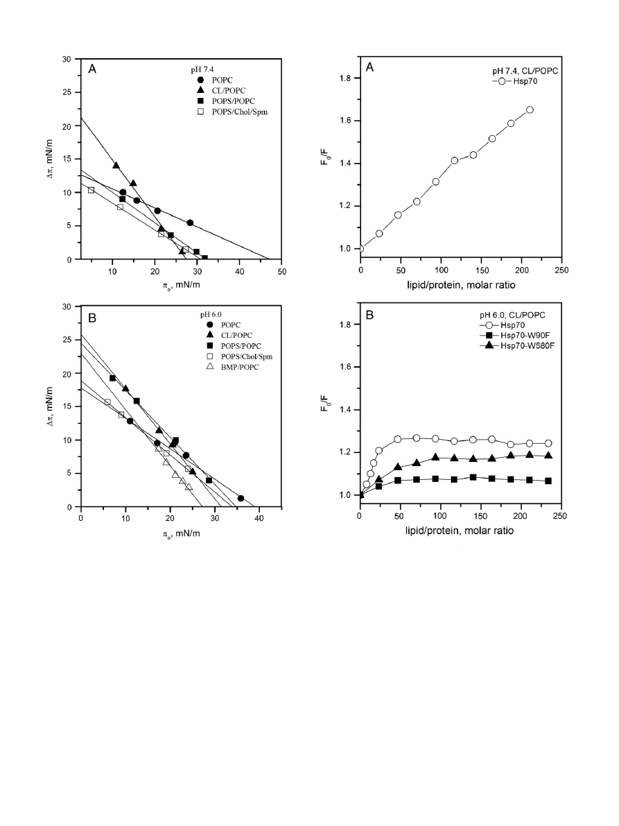
air/buffer interface, at a range of initial lateral pressures (
, panels
A & B). The equilibrium lateral pressures estimated for biomembranes
are approximately 33
–35 mN/m
. The exclusion pressures
π
c
of 47
and 38 mN/m, measured for Hsp70 and POPC monolayers at pH 7.4
and 6.0, respectively, demonstrate that Hsp70 can ef
ficiently incorpo-
rate into PC
films, at neutral pH, in particular. The intercalation of
Hsp70 into POPC monolayers is reduced at pH 6.0, again in keeping
with results from Trp
fluorescence.
3.1.2. Binding of Hsp70 to liposomes containing cardiolipin
Judged from Trp
fluorescence the binding of Hsp70 to CL/PC
(X
CL
= 0.2) LUV (
, panel F) resembles that described previously
by us for BMP/PC bilayers
. Lower RFI in the spectra for CL containing
Fig. 5. Relative
fluorescence intensities for Hsp70 (○), Hsp70-W90F (W580, ■), and Hsp70-W580F (W90, ▲) in the presence of POPC (panels A & B), CL/POPC (X
CL
= 0.2 panels C & D),
and POPC/POPS (X
POPS
= 0.2, panels E & F) LUV. Initial protein concentration was 0.43
μM and the concentration of lipid was increased in 10 μM increments. The data are depicted as the
ratio (F/F
0
) of the emission measured in the presence of the indicated LUV (F) and the emission intensity in 20 mM Hepes, 0.1 mM EDTA (F
0
) at pH 7.4 (lefthand panels), or pH 6.0
(righthand panels).
1350
A.K. Mahalka et al. / Biochimica et Biophysica Acta 1838 (2014) 1344
–1361
LUV compared to POPC could re
flect the vicinity of the Trps to the
surface charges of CL/PC liposomes (
). No signi
ficant differences
in the values of
λ
max
were seen for spectra recorded between pH 7.4
and 6.0 with CL/PC (X
CL
= 0.2) liposomes (
, panels C & D),
although at pH 6.0 the overall values for RFI were approx. 26% higher
(
, panels A & B).
The signi
ficant quenching of Hsp70 by 11,12-Br
2
PC in the presence
of cardiolipin at pH 7.0 reveals that the Trps are in contact with the
sn-2 acyl chain of PC (
, panel C). Binding of Hsp70 to CL-
containing liposomes with brominated PCs (
, panel D) at pH 6.0 re-
veals ef
ficient quenching with similar dependence on L/P as seen for the
Trp emission of Hsp70 in the presence of CL/PC LUV, with saturation
observed at L/P
≈ 50 (
, panel F). Accordingly, at pH 6.0 the af
finity
of Hsp70 to CL/POPC LUV seems to be high (
, panel D). This behav-
ior was observed neither for POPC (
, panels A & B) nor for POPS/
POPC LUV (see below). Of the brominated PCs, the 6,7-Br
2
-PC caused
the most ef
ficient quenching and was thus selected for subsequent
experiments.
Compared to wtHsp70 the af
finities for CL of the W90F and W580F
mutants seem to be different, saturating at approx. L/P
≈ 50, and 75, re-
spectively (
, panel D). Accordingly, both NBD and SBD contribute
to the attachment of Hsp70 with CL containing bilayer. The membrane
af
finity of the NBD domain appears to be slightly reduced by the
W580F mutation in SBD, in keeping with a conformational coupling
Fig. 6. Quenching by 6,7Br
2
-PC (X = 0.3) of Hsp70 (
○), Hsp70-W90F (W580, ■), and Hsp70-W580F (W90, ▲) Trp fluorescence in POPC (panels A & B), CL/POPC (X
CL
= 0.2, panels C & D),
or in POPS/POPC (X
POPS
= 0.2, panels E & F) LUV. The quenching ef
ficiencies are depicted as the ratio of relative fluorescence intensities with liposomes as such (F
0
) and LUV with
6,7-Br
2
PC (F), measured at pH 7.4 (lefthand panels), or pH 6.0 (righthand panels).
1351
A.K. Mahalka et al. / Biochimica et Biophysica Acta 1838 (2014) 1344
–1361
between the two domains. Ef
ficient quenching of W580 in the Hsp70-
W90F mutant by 6,7-Br
2
PC in CL/POPC LUV revealing a high af
finity in-
teraction of the SBD domain with CL/PC LUV (
, panel D), suggests
that SBD possesses a high af
finity binding site for CL.
Interestingly, at both pH 7.4 and 6.0, the binding of Hsp70 to CL/PC
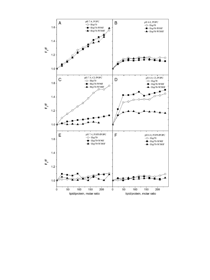
bilayers results in an augmented quenching by AcrA (
), at acidic
pH in particular, indicating a conformational change which renders at
least one of the Trp residues in Hsp70 more exposed to water. This is
in keeping with a red shift which is evident in the
fluorescence spec-
trum of the Trp residue, which is not contacting the brominated acyl
chains of PCs (
), suggesting that at least one of the Trp residues
remains accommodated in a more hydrophilic environment in the pres-
ence of CL/PC LUV. Interestingly, compared to Hsp70, there is attenuated
quenching of Hsp70-W90F and Hsp70-W580F Trp
fluorescence by AcrA
(
, panel D), suggesting that both domains are required for the
opening of the Hsp70 structure when bound to CL.
Importantly, the association of Hsp70 with CL/PC monolayers is dis-
tinctly different from that with POPC, with
π
0
vs
Δπ data revealing low
exclusion pressures
π
c
of 27.5 and 31 mN/m, respectively, at pH 7.4
and 6.0 (
). Lack of increment in the surface pressure of CL/PC
monolayers at the above initial surface pressures following the injection
of the Hsp70 into the subphase suggests a lack of intercalation of Hsp70
into CL/PC bilayer, estimated to have lateral pressure of approximately
33
–35 mN/m
.
To further explore the interactions of CL with the Hsp70 domains we
used brominated toCL (Br
8
CL). The ef
ficient quenching of Hsp70 Trps by
Br
8
CL at pH 7.4 reveals that the Trps are in direct contact with the CL
acyl chain bromides (
, panel A). Quenching of wtHsp70 at
pH 6.0 reveals a high af
finity interaction, saturating at L/P ≈ 30
(
, panel B). For X
CL
= 0.2 this corresponds to CL/P
≈ 6. Assuming
uniform distribution of CL in the two lea
flets of the LUV bilayers the
number of CL reacting with Hsp70 is close to 3:1. The ef
ficient
quenching for both Hsp70-W580F and Hsp70-W90F by Br
8
CL at
pH 6.0, saturating at approx. L/P
≈ 50 and 100, respectively, shows
that both NBD and SBD contain CL binding sites (
, panel B).
These interactions with CL further result in direct contacts of the bromi-
nated acyl chains of CL and W90 and W580. Yet, the af
finity of Hsp70 to
the above CL/PC bilayers is signi
ficantly reduced by both W90F and
W580F mutations, again in keeping with a conformational coupling of
the two domains.
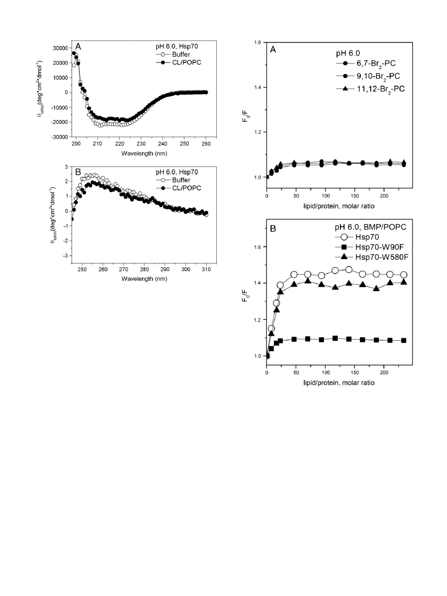
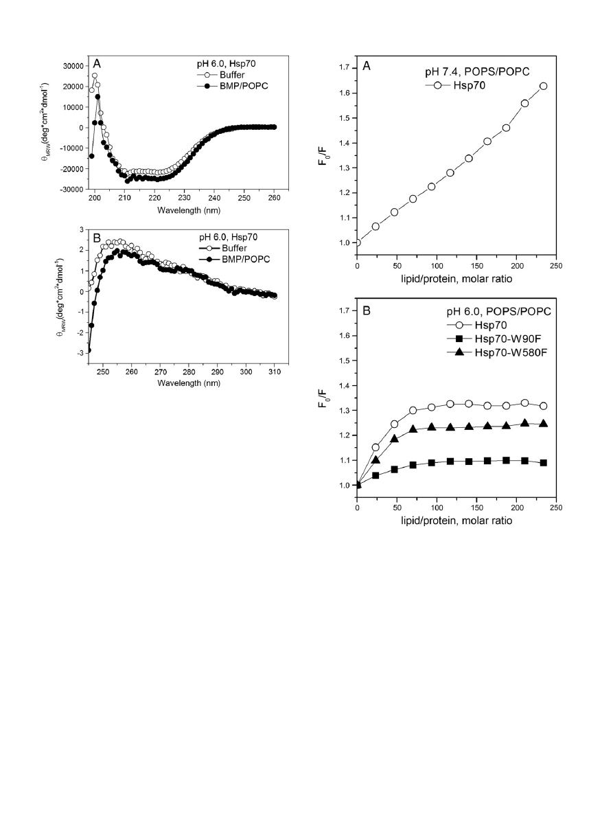
The far-UV CD spectra of 2
μM Hsp70 in 20 mM MES, 0.1 EDTA at
pH 6.0 show double minimum at 222 and 210 nm (
, panel A)
characteristic to a protein containing predominantly
α-helical structure.
The calculated helical content decreased slightly, from 84 to 83% by the
presence of CL/POPC (X
CL
= 0.2) LUV, suggesting CL-containing mem-
brane induce a minor change in the overall secondary structure of
Hsp70. Yet, near UV-CD of Hsp70 in the presence CL/POPC (X
CL
= 0.2)
LUV indicates loss of asymmetry in the aromatic environment (
,
panel B), in keeping with molten globule state.
3.1.3. Binding of Hsp70 to liposomes containing bis-monoacylglycero
phosphate
In order to relate the above data to our earlier results for the interac-
tion of Hsp70 with BMP, we additionally investigated the binding at
pH 6.0 of Hsp70 to PC LUV containing BMP, or brominated BMP
(Br
4
BMP), or Br
2
PCs (
). For BMP/POPC (X = 0.2) quenching by
Br
2
PCs is seen, saturating at L/P
≈ 30, similarly to CL/PC LUV (
,
panel A). Likewise, quenching by Br
4
BMP (X = 0.2) saturates at this
stoichiometry (
, panel B). Most ef
ficient quenching by Br
4
BMP
is seen for W90, in keeping with the suggested binding site for this
lipid in NBD
Binding to BMP/PC LUV and quenching of Hsp70 by Br
2
PCs (
,
panel A) at pH 6.0 reveal only weak quenching. In keeping with the
weak quenching by Br
2
PCs and the observed red shift in the
fluores-
cence spectrum (Table SI), Hsp70 does not seem to penetrate into
BMP/PC bilayers and at least one of the Trp residues becomes accommo-
dated in a more hydrophilic environment in the presence of BMP/PC
LUV. To further explore the BMP
–Hsp70 interactions we used Br
4
BMP.
Quenching of wtHsp70 at pH 6.0 reveals a high af
finity binding, saturat-
ing at L/P
≈ 30 (
, panel B). The ef
ficient quenching of both
Hsp70-W580F and Hsp70-W90F by Br
4
BMP contained in BMP/PC LUV
and at pH 6.0, both saturating at approx. L/P
≈ 30, suggests that there
are binding sites for BMP in both NBD as well as SBD (
), similar
to the observations on the binding of Hsp70 to CL/PC LUV. Langmuir
films of BMP/PC gave an exclusion pressure of 27 mN/m at pH 6.0, indi-
cating no intercalation into BMP/PC membranes (
, panel B), esti-
mated to have lateral pressure of approximately 33
–35 mN/m
.
Compared with buffer, the helical content calculated from CD spec-
tra increased slightly, from 84 to 85% by the presence of BMP/POPC
Fig. 7. Quenching by AcrA of Trp
fluorescence of membrane bound Hsp70. The concentra-
tions of lipids and Hsp70 were 95 and 0.4
μM, corresponding to L/P ≈ 234. The data are
represented as the ratio of initial
fluorescence intensity (F
0
) and the intensity measured
in the presence of AcrA (F), at pH 7.4 (panel A) and 6.0 (panel B). The liposomes were
composed of POPC (
●), CL/POPC (X
CL
= 0.2,
▲), and POPS/POPC (X
POPS
= 0.2,
■). Also
shown are data for Hsp70 in buffer (
○). Although the depicted results were from a single
experiment, these data were readily reproducible and consistent in all similar experiments
(data not shown).
1352
A.K. Mahalka et al. / Biochimica et Biophysica Acta 1838 (2014) 1344
–1361
LUV (X
BMP
= 0.2,
, panel A). Similar to CL/PC LUV near UV-CD of
Hsp70 in the presence BMP/POPC LUV (X
BMP
= 0.2) indicates loss of
asymmetry in the aromatic environment (
, panel B), suggesting
the molten globule state.
3.1.4. Binding of Hsp70 to liposomes containing phosphatidylserine
We then proceeded to study the interaction of Hsp70 with mem-
branes containing the acidic phospholipid PS. Compared to neat POPC
LUV, PS/PC LUV (X
PS
= 0.2) also cause an increase in RFI, yet with a
smaller decrement in
λ
max
). This could re
flect vicinity of W90
and W580 to the surface charges of the bilayers, similar to CL/PC LUV
with almost identical spectra seen at pH 7.4 for the PS and CL containing
membranes (
). No signi
ficant differences in the values of λ
max
were seen for the spectra recorded with PS/PC LUV at pH 7.4 and 6.0
(
, panels C & D). At pH 6.0 the values for the emission band RFI
increased and the peak was observed at a somewhat shorter
λ
max
.
Compared to POPC LUV the changes in
fluorescence induced by PS/PC
LUV were more rapid and apparent equilibria were reached faster.
In contrast to POPC LUV, judged from the lack of signi
ficant
quenching by the brominated PCs Hsp70 does not insert into PS con-
taining membranes (
, panels E & F).
Judged from relative
fluorescence intensities both Hsp70-W90F and
Hsp70-W580F seem to be involved in interactions with PS containing
membranes (
, panels E & F). The lack of quenching by 6,7-Br
2
PC re-
veal that neither NBD nor SBD insert into PS containing membranes
(
, panels E & F).
Fig. 8. AcrA quenching for Hsp70 (
○), Hsp70-W90F (W580, ■), and Hsp70-W580F (W90, ▲) in the presence of POPC (panels A & B), CL/POPC (X
CL
= 0.2, panels C & D), and POPS/POPC
(X
POPS
= 0.2, panels E & F) LUV. The concentrations of lipids and proteins were 95 and 0.4
μM, corresponding to L/P ≈ 234. The data are represented as the ratio of initial fluorescence
intensity (F
0
) and the intensity measured in the presence of increasing concentrations of AcrA (F), data measured at pH 7.4 (lefthand panels), or pH 6.0 (righthand panels).
1353
A.K. Mahalka et al. / Biochimica et Biophysica Acta 1838 (2014) 1344
–1361
The above aligns with the ef
ficient quenching of Hsp70 Trps seen for
AcrA in the presence of PS/PC LUV, revealing that the Trps remain acces-
sible to the bulk aqueous phase (
, panels A & B). However, this con-
tradicts the reduced quenching of W90F and W580F by AcrA in the
presence of PS/PC LUV, suggesting shielding of the Trps from access to
AcrA in the bulk aqueous phase for these mutants (
, panels E & F).
PS/PC Langmuir
films gave for Hsp70 exclusion pressures of 33 and
35 mN/m at pH 7.4 and 6.0, respectively, suggesting only weak interca-
lation into PS/PC membranes to be possible (
). For sphingomyelin/
cholesterol (1/1, molar ratio)
films with 10 mol% PS exclusion pressures
of 31 and 34 mN/m at pH 7.4 and 6.0, respectively, were recorded, thus
suggesting a lack of signi
ficant protein intercalation into lipid films cor-
responding to those found in the plasma membrane of cancer cells.
The lack of quenching of Trps by Br
2
PCs contained in PS/PC LUV
could indicate that PS is displacing the brominated PCs from their bind-
ing sites in Hsp70 (
, panels E & F). In keeping with binding of PS to
Hsp70 is an ef
ficient quenching of Trp emission by Br
2
PS was observed
(
, panels A & B). Further, the quenching of both Hsp70-W90F and
Hsp70-W580F Trps by Br
2
PS at pH 6.0, both saturating at approx.
L/P
≈ 100 and 50, respectively, suggests that both NBD as well as SBD
contained binding sites for PS (
, panel B).
3.2. Domain-speci
fic lipid interactions of Hsp70
In the second part of this study we aimed at the identi
fication of pos-
sible different interactions of the two domains (NBD & SBD) of Hsp70
Fig. 9. Penetration of Hsp70 into lipid monolayers residing on 20 mM Hepes, 0.1 mM
EDTA at pH 7.4 (panel A) or 6.0 (panel B), illustrated as a function of the initial surface
pressure
π
0
and the increment in surface pressure (
Δπ) following the injection of Hsp70
into the subphase (0.1
μM final concentration). Lipid monolayers were POPC (●), CL/POPC
(X
CL
= 0.2,
▲), POPS/POPC (X
POPS
= 0.2,
■), POPS/Chol/Spm (X
POPS
= 0.10, X
Chol
=
X
Spm
= 0.45,
□), and BMP/POPC (X
BMP
= 0.2,
Δ).
Fig. 10. Quenching of Hsp70 Trps (
○) by Br
8
CL (X = 0.2, in CL/POPC LUV) at pH 7.4 (panel
A) as well as Hsp70 (
○), Hsp70-W90F (W580, ■), and Hsp70-W580F (W90, ▲) at pH 6.0
(panel B). Quenching ef
ficiencies are depicted as the ratio of relative fluorescence intensi-
ties with LUV without (F
0
) and with Br
8
CL (F). Initial concentration of the proteins was
0.43
μM while the concentrations of lipids were increased in 10 μM (total lipid)
increments.
1354
A.K. Mahalka et al. / Biochimica et Biophysica Acta 1838 (2014) 1344
–1361
with the above three different phospholipids, employing the constructs
Hsp70-
ΔNBD, and Hsp70-ΔSBD, lacking NBD and SBD, respectively
, panel B). Unfortunately, the former construct also contains part
of the NBD (residues 1 to 118, including W90) as attempts to delete
this domain completely resulted in an expression product prone to ex-
tensive aggregation. The latter property of SBD has been observed pre-
viously in other laboratories
. Comparison of the spectra of Hsp70
and the above constructs reveals that the emission from the single Trp
containing NBD construct is insensitive to buffer pH (Fig. S1). Changes
in
fluorescence recorded in the presence of LUV composed of POPC, as
well as CL in POPC, or PS in POPC (as indicated) reveal that both con-
structs retain the ability to interact with phospholipid bilayers.
Comparison of the quantum yields of Hsp70 and the above
Trp
→ Phe mutants reveals conformational coupling between the
NBD and SBD. For example, the combined emission of Hsp70-
ΔSBD
and Hsp70-
ΔNBD at L/P ≈ 230 exceeds the intensity of wtHsp70
fluorescence (Fig. S2).
POPC LUV induced, similar to Hsp70, profound changes in Trp
fluo-
rescence of both Hsp70-
ΔSBD, and Hsp70-ΔNBD (Fig. S3, panels A &
B), with a large increase in the relative
fluorescence intensity values
(RFI) indicating a major change (increase) in the hydrophobicity in
the surroundings of their Trp residues. Interactions of NBD and SBD
with PC seem to be non-speci
fic and of low affinity. The fluorescence en-
hancement induced in the presence of PC LUV is larger for NBD than for
SBD, which contains two Trps, and thus supports our earlier conclusion
that in wtHsp70 it is the NBD and its W90 which insert into PC bilayers.
Compared to wtHsp70 the af
finity of the NBD construct for CL ap-
pears to be reduced, whereas for SBD and CL a high af
finity interaction
is evident, saturating at approx. L/P
≈ 50 (Fig. S3, panel D). Judged
from relative
fluorescence intensity values both NBD and SBD seem to
be involved in interactions with membranes containing PS (Fig. S3,
panels E & F). Interestingly, both NBD and SBD contribute to the bilayer
attachment of Hsp70.
The quenching of Hsp70-
ΔNBD and Hsp70-ΔSBD Trps by Br
2
-6,7PC
contained in POPC LUV suggests that Hsp70 penetrates into POPC LUV
(Fig. S4, panels A & B). Similar to the above, ef
ficient quenching of
Hsp70-
ΔNBD and Hsp70-ΔSBD Trps by Br
2
-6,7PC is observed with
CL/PC LUV (X
Br2-6,7PC
= 0.2, Fig. S4, panels C & D). The lack of quenching
of Hsp70-
ΔNBD and Hsp70-ΔSBD Trps by Br
2
PCs contained in PS/PC
LUV suggests that PS is displacing the brominated PCs from their
binding sites in Hsp70 (Fig. S4, panels E & F).
Fig. 11. Panel A: Far-UV CD spectra of 2
μM Hsp70 (○) in buffer (20 mM MES, 0.1 EDTA)
and in the same buffer in the presence of 200
μM (total lipid) CL/PC (X
CL
= 0.2,
●) LUV at
pH 6.0. Panel B: Near-UV CD spectra of 8.5
μM Hsp70 (○) in buffer (20 mM MES,
0.1 EDTA) and in the same buffer in the presence of 700
μM (total lipid) CL/PC
(X
CL
= 0.2,
●) LUV at pH 6.0.
Fig. 12. Quenching of Hsp70 Trp
fluorescence by 6,7-(■), 9,10-(●), or 11,12Br
2
-PC
(
▲, X = 0.3) contained in BMP/POPC (X
BMP
= 0.2) LUV at pH 6.0 (panel A). Quenching
of Hsp70 (
○), Hsp70-W90F (W580, ■), and Hsp70-W580F (W90, ▲) Trp fluorescence
by Br
4
BMP (X = 0.2, in POPC LUV) at pH 6.0 (panel B). The quenching ef
ficiencies are
depicted as the ratio of relative
fluorescence intensities with LUV without (F
0
) and with
the brominated lipid (F), as indicated.
1355
A.K. Mahalka et al. / Biochimica et Biophysica Acta 1838 (2014) 1344
–1361
At both pH 7.4 and 6.0, augmented quenching of Hsp70-
ΔNBD Trp
emission by AcrA is seen upon binding to CL/PC LUV, suggesting a con-
formational change, which opens the structure of SBD, enhancing the
exposure and access of AcrA to W580 (Fig. S5, panels C & D). In contrast,
less ef
ficient quenching of Hsp70-ΔSBD W90 by AcrA was evident at
both pH, suggesting that W90 was buried inside NBD in a more compact
structure when bound to CL/PC membranes (Fig. S5, panel D). At both
pH 7.4 and 6.0, augmented quenching of Hsp70-
ΔNBD Trp in the pres-
ence of PS/PC LUV by AcrA is evident, suggesting a conformational
change, which opens the structure of SBD making W580 more exposed
to water (Fig. S5, panels E & F). In contrast, less ef
ficient quenching of
Hsp70-
ΔSBD W90 in the presence of PS/PC LUV by AcrA was seen at
both pH 7.4 and 6.0, suggesting that W90 becomes buried inside NBD
folded into a compact structure when bound to PS/PC membranes
(Fig. S5, panel F), similar to the data measured in the presence of
CL/PC LUV.
4. Discussion
Studies on the interaction of Hsp70 with lipid membranes are sparse
and have been mainly limited to qualitative observations. We describe
here a somewhat more detailed assessment of the binding of Hsp70 to
speci
fic lipids. Our previous results demonstrated that BMP could repre-
sent the membrane ligand responsible for the localization of Hsp70 in
the lysosomal/endosomal membranes, required for the cytoprotective
effect of Hsp70, resulting from the activation of acidic sphingomyelinase
and subsequent formation of ceramide. In this study we investigated if
Hsp70 would exhibit similar speci
fic interactions with other acidic
phospholipids. Importantly, due to the limited availability of the
Hsp70 protein and the constructs, we focused at this stage for
first
obtaining a qualitative view on the mechanisms on Hsp70
–lipid interac-
tions, in order to provide a good starting point for possible later quanti-
tative analyses.
Regarding the association of Hsp70 with different lipids, the follow-
ing conclusions could be made,
(i) Hsp70 associates with POPC bilayers, with shallow penetration
into the membrane. W90 in NBD appears to signal this penetra-
tion, and resides at a distance of approximately 11 Å from the bi-
layer center, in the interfacial region of the bilayer and still also
accessible to weak collisional quenching by the water soluble
AcrA. At pH 6.0 the penetration of Hsp70 into POPC is less than
at pH 7.4, evidenced by both Trp
fluorescence as well as
Langmuir-
film penetration experiments.
Fig. 13. Panel A: Far-UV CD spectra of 2
μM Hsp70 (○) in buffer (20 mM MES, 0.1 EDTA)
and in the same buffer in the presence of 200
μM (total lipid) BMP/PC (X
BMP
= 0.2,
●)
LUV at pH 6.0. Panel B: Near-UV CD spectra of 8.5
μM Hsp70 (○) in buffer (20 mM MES,
0.1 EDTA) and in the same buffer in the presence of 700
μM (total lipid) BMP/PC
(X
BMP
= 0.2,
●) LUV at pH 6.0.
Fig. 14. Panel A, Quenching of Hsp70 Trps (
○) by Br
2
PS (X = 0.2, in PS/POPC LUV) at
pH 7.4, and panel B at pH 6.0 Hsp70 (
○), Hsp70-W90F (W580, ■), and Hsp70-W580F
(W90,
▲). Quenching efficiencies are depicted as the ratio of relative fluorescence intensi-
ties with LUV with PS (F
0
) and Br
2
PS (F). Initial concentration of the proteins was 0.43
μM
while the concentration of lipids was increased in 10
μM increments.
1356
A.K. Mahalka et al. / Biochimica et Biophysica Acta 1838 (2014) 1344
–1361
(ii) The presence of CL has a profound impact on the membrane as-
sociation of Hsp70. The interaction between CL and Hsp70 at
pH 6.0 seems to be of high af
finity and abolishes the penetration
of Hsp70 into CL/PC Langmuir
films. Taken together our data on
the binding of Hsp70 to CL/PC membranes suggest that CL adopts
the extended conformation, thus anchoring Hsp70 to the lipid
surface
.
In keeping with earlier observations, Hsp70 binds avidly to lipo-
somes containing negatively charged lipids
. The effects of LUV
with different lipid compositions correlate with the structure of the
acidic phospholipid headgroup. Insigni
ficant changes in the net charges
of POPC, POPS, and CL, viz. 0,
−1, and −2, respectively, are anticipated
upon decreasing pH from 7.4 to 6. In contrast, for Hsp70 net charges of
−10.9 and −3.5 at pH 7.4 and 6.0, respectively, can be estimated. The
corresponding values for NBD are
−4.8 and +1.0, and for SBD −9.1
and
−5.5 (at pH 7.4 and 6, respectively). These charges of Hsp70 and
its two domains should, however, be taken as tentative only as they
were calculated based on the amino acid sequence, neglecting any ef-
fects of protein three-dimensional structure, schematically illustrated
in (
, panel A). From the point of view of electrostatic interactions
with anionic lipids, it is of interest that both NBD and SBD expose on
their surface positively charged residues. At pH 7.4 Hsp70 binds to neg-
atively charged liposomes in a rather similar manner irrespective of the
acidic phospholipid species present (
). This may be ex-
plained by the net negative charge of the protein and its both domains
at pH 7.4 causing repulsion between Hsp70 and the negatively charged
membranes. Lowering pH to 6.0 changes the estimated net charge of
NBD from negative to positive (
−4.8 to +1.0), which could explain
the altered membrane binding (
). With respect to the pH depen-
dence of the lipid binding of Hsp70 as re
flected in Trp fluorescence,
the roles of His89 and 594 are of interest, being vicinal to W90 and
W580, respectively
.
We assessed possible speci
fic lipid interactions of Hsp70 by using
W90 and W580 as intrinsic
fluorescent probes. At this point it is impor-
tant to bear in mind the caveats involved. Accordingly, interpretation of
the Trp
fluorescence data for wtHsp70 is complicated by the presence of
the two Trps (in NBD and SBD), in particular with the conformations of
these two domains being coupled
. Moreover, both W90F and W580F
mutations are readily re
flected in the structural dynamics of native SBD
and NBD, respectively (
), in keeping with their conforma-
tional coupling. Likewise, the fact that the mutation W90F renders
Hsp70 inactive
may also mean that the perturbation that caused
this mutation in NBD is also re
flected in the conformational dynamics
of SBD, resulting in its altered interactions with lipids. Of the phospho-
lipids investigated POPC induced the most pronounced changes in Trp
fluorescence. The slow stabilization of the changes in Trp emission
upon binding to POPC requiring approx. 15
–20 min indicates that
what happens is more than simple non-speci
fic association. The expo-
nential increase in RFI upon surface dilution of Hsp70 could indicate
dimer/oligomer dissociation in POPC bilayers, resulting in a more ef
fi-
cient membrane intercalation (
, panels E and F). The shoulder
seen in the spectra in the presence of POPC liposomes suggests that
one of the Trps becomes accommodated in a more hydrophobic micro-
environment while the environment of the other Trp remains more or
less unaltered (
). The linear component observed at pH 7.4
, panel E) may be interpreted as binding, with the subsequent
component at L/P
N 150 representing a process associated with a reor-
ganization of Hsp70 in the membrane surface upon its surface dilution.
The exponential increase is accompanied with a blue shift of about
10 nm in the peak position and about 5 nm in the spectral center of
mass, in keeping with at least one of the two Trps entering a more hy-
drophobic environment. AcrA quenching shows shielding of the two
Trps in the presence of POPC LUV at pH 7.4 (
, panel A). Overall,
our results suggest that the Trp becoming immersed into POPC bilayers
is W90. The above data together with the demonstration of Trp
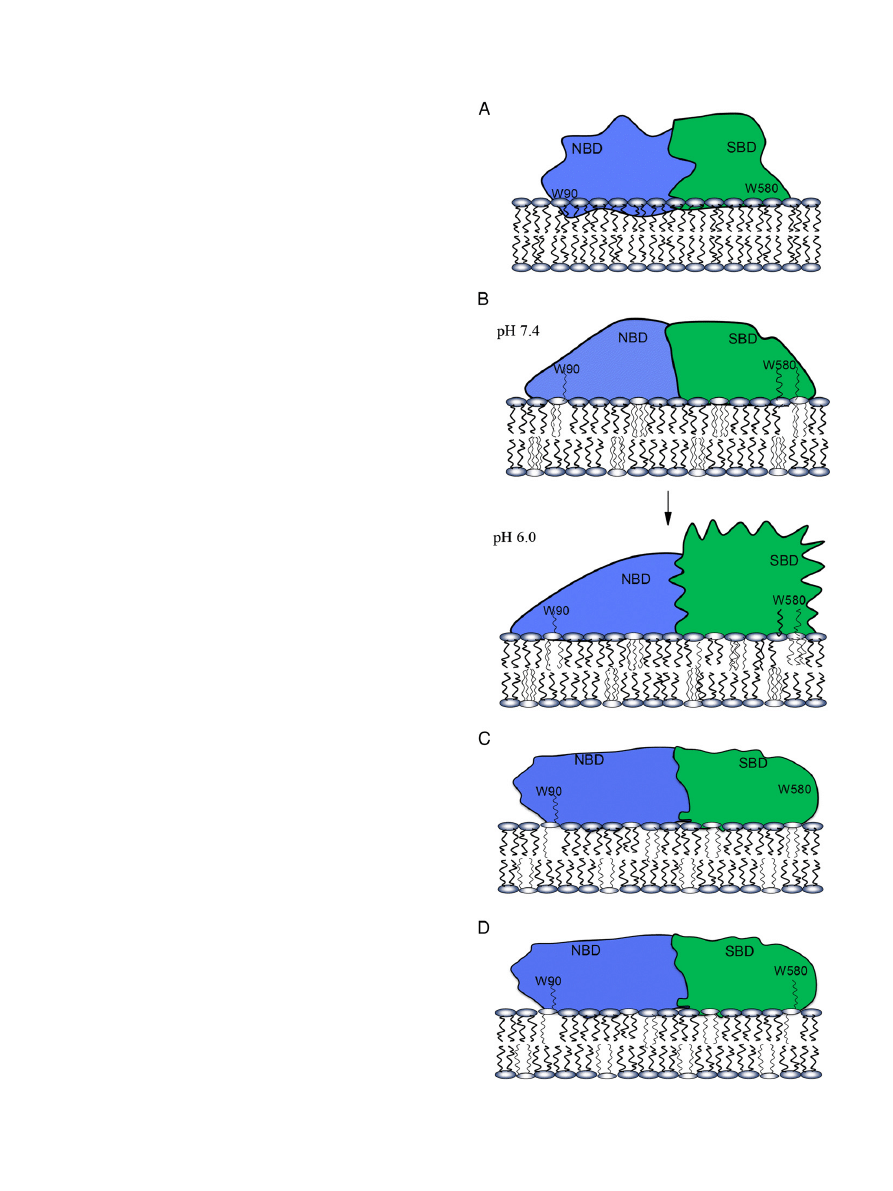
Fig. 15. Schematic illustration of the association of Hsp70 with POPC (panel A), CL/POPC
(panel B), BMP/POPC (panel C), and POPS/POPC (panel D) membranes.
1357
A.K. Mahalka et al. / Biochimica et Biophysica Acta 1838 (2014) 1344
–1361

quenching by Br
2
-PCs, suggest that Hsp70 penetrates into POPC LUV, in
a manner (i) shielding access of AcrA to the Trps at pH 7.4, (ii) bringing
W90 into a hydrophobic environment, and (iii) making W90 accessible
to quenching by brominated PCs, 6,7Br
2
-PC in particular. From parallax
analysis the distance of the Trps quenched is approx. 11.4 Å from the
bilayer center. The quenching of Hsp70-
ΔNBD and Hsp70-ΔSBD Trps
by Br
2
-6,7PC (Fig. S4, panels A & B) together with Langmuir-
film
experiments (
) comply with Hsp70 penetrating into POPC LUV.
The interaction of Hsp70 with POPC membranes concluded on the
basis of these data is schematically illustrated in
(panel A), in
order to provide a plausible scenario to explain the experimental
finding. Yet, considering the complexity of the molecular Hsp70 lipid
bilayer assembly, the depicted arrangement needs to be taken at this
time as tentative, not excluding other possible mechanisms.
Hsp70 indirectly blocks apoptotic pathways at premitochondrial
and mitochondrial level, and also at a post-mitochondrial stage
.
Hsp70 also mediates in an ATP dependent manner the translocation of
proteins across the mitochondrial membrane, by the outer and inner
membrane translocases
. Interestingly, Hsp70 protects mito-
chondria from damage by oxidative stress
. For Hsp70 acting in mi-
tochondria the negatively charged cardiolipin would be likely to attach
Hsp70 to the membranes of this organelle. The interaction of Hsp70
with CL/PC bilayers appears to be distinctly different from that with
POPC liposomes. At pH 6.0 Hsp70 binds avidly to CL/POPC (X
CL
= 0.2)
liposomes with saturation seen at L/P
≈ 50 (
, panel F). Notably,
there is a red shift in the emission of W90 under these conditions, indi-
cating a more polar environment, such as augmented exposure to water
(
). The two Trps of CL/PC bound Hsp70 are effectively quenched
by AcrA, revealing that in the presence of CL/PC LUV the Trps become ac-
cessible for AcrA in the bulk aqueous phase. Nevertheless, these Trps are
quenched also by brominated PCs, in keeping with an intercalation of
Hsp70 into the bilayer. The association of Hsp70 with CL containing
membranes is sensitive to pH, with a high af
finity interaction being ob-
served at pH 6.0 in a number of different experiments (
, panel F;
, panel D;
, panel D, and
, panel B). Our data point to
a high af
finity binding site for CL/PC membrane at acidic pH to be
contained in SBD, as demonstrated by the quenching of W580 of
Hsp70-W90F mutant by Br
2
PCs (
, panel D). Notably, because of
the high content of protein in mitochondrial membranes the local con-
tent of cardiolipin is high, causing a high local negative surface charge,
which necessarily attracts protons, generating a local low pH environ-
ment on the membrane surface
. Along these lines we previously
demonstrated the membrane surface charge to have a dramatic in
flu-
ence on the pH dependence of the lipid association of cytochrome c
.
Importantly, Hsp70 does not seem to intercalate into CL/PC
monolayers at lipid packing densities corresponding to the equilibrium
lateral surface pressures of approximately 33
–35 mN/m estimated for
biomembranes
. Intriguingly, this contradicts Trp quenching ob-
served using brominated PCs as well as brominated CL. Interestingly,
the ef
ficient quenching of Hsp70-ΔNBD and Hsp70-ΔSBD Trps by Br
2
-
6,7PC contained in CL/PC LUV, with characteristics similar as seen for
Hsp70 shows that both W90 & W580 in these constructs are contacting
the brominated phospholipid acyl chains (
, panel D). A likely
mechanism to solve this paradox is that Hsp70 binds peripherally to
the CL/PC bilayer surface, with the brominated phospholipid acyl chains
adopting the so-called extended conformation
as
first described
by us for cytochrome c
and subsequently reported for bet3
and Orf-9
. This is schematically illustrated in
(panel B),
depicting the accommodation of acyl chains into a fatty acid accommo-
dating sites contained in SBD as well NBD. To this end, two fatty acid
(FA) binding sites have been demonstrated for Hsp70, each saturating
at a 1:1 FA/protein ratio
. The structures of these fatty acid binding
sites in Hsp70
are, however not known.
Together with our data on Br
2
PCs (
), the results with Br
8
CL
imply the following mechanism. Approximately 2
–3 CL molecules
bind to Hsp70 and induce a conformational change in Hsp70, involving
opening of hydrophobic cavities, which can accommodate brominated
acyl chains of PCs. Accordingly, the Br-containing sn-2 acyl chains
would reverse their orientation extending out of the bilayer and becom-
ing accommodated into hydrophobic crevices in Hsp70. Notably, this
con
figuration results in a hydrophobic association of Hsp70 with the bi-
layers, yet without penetration of the protein into the bilayer hydrocar-
bon region
. Our data are in keeping with extended lipid acyl chains
intercalating into SBD. Notably, this acyl chain intercalation could, in ad-
dition to involving acyl chain binding cavities
, also re
flect loosening
of the tertiary structure of Hsp70 upon induction of the molten globule
(MG) state by SBD, similar to
α-lactalbumin
, in keeping with the
access of AcrA to the Trps. The far UV-CD spectra measured (
,
panel A) con
firm the above structural change upon membrane binding.
As demonstrated for cytochrome c
the above two mechanisms
are not mutually exclusive
. It seems feasible that conformational
changes induced by CL in the NBD of Hsp70, are further re
flected in
the conformations of the adjacent SBD, possibly promoting its transition
into the MG, allowing the accommodation of brominated PC acyl chains
into hydrophobic binding sites in SBD, similar to that described for
α-lactalbumin
Taken together the present results indicated SBD to adopt the MG
conformation in the presence of CL and BMP. First, red shifts in Trp emis-
sion are seen in the presence of CL/PC LUV (
). Second augmented
quenching by AcrA is evident for W580 with CL/PC LUV, in keeping with
more loose tertiary structure (
, panel D). Third, our data are in
keeping with extended lipid acyl chains intercalating into NBD and SBD.
The quenching of Trp by Br
4
BMP in BMP/PC LUV is more ef
ficient for
W90 than for W580, con
firming our previous observation that NBD con-
tains a speci
fic and pH-dependent binding site for BMP. The weak
quenching by Br
2
PCs in the presence of BMP (
, panel A) could re-
sult from BMP occupying phospholipid binding sites in Hsp70 with ex-
tension of one of its acyl chains into hydrophobic cavities within Hsp70,
similar to CL. In conclusion, Hsp70 NBD and SBD appear to bind periph-
erally onto the BMP/PC bilayer surface, analogously to a CL/PC mem-
brane. Taking into account the resemblance of the structures of BMP
and CL it is not surprising that the interactions of these lipids with
Hsp70 are similar. BMP is highly enriched in the membranes of
lysosomes and late endosomes
. The association and localization of
Hsp70 to the lysosomal membranes leads to a cytoprotective effect
and interferes with lysosomal cell death pathways
. Hsp70
–
Table 2
Values for the Stern
–Volmer quenching constants K
sv
(M
−1
) for 60 mM AcrA with 0.4
μM Hsp70 and the indicated constructs, in the presence of the indicated liposomes (95 μM total
phospholipid, corresponding to L/P
≈ 234) and at pH 7.4 and 6.0.
Hsp70
Hsp70-W90F
(W580)
Hsp70-W580F
(W90)
Hsp70-
ΔNBD
(SBD)
Hsp70-
ΔSBD
(NBD)
POPC
pH 7.4
1.85
2.52
1.34
4.16
0.13
pH 6.0
1.88
1.69
1.11
3.67
0.43
CL/PC (X
CL
= 0.2)
pH 7.4
3.75
4.26
3.79
4.72
2.21
pH 6.0
4.62
3.43
3.67
3.51
3.23
PS/PC (X
PS
= 0.2)
pH 7.4
2.90
1.78
2.3
3.64
0.79
pH 6.0
2.31
1.24
1.37
2.78
1.11
1358
A.K. Mahalka et al. / Biochimica et Biophysica Acta 1838 (2014) 1344
–1361

BMP interaction plays a crucial role in the activation of the acidic
sphingomyelinase (aSM) and the subsequent stabilization of lysosomes
by ceramide
. Direct interaction between Hsp70 and BMP is re-
quired for this activation and downstream cytoprotective properties of
Hsp70, and can be blocked by an antibody against a BMP
. Exact
molecular mechanisms of activation of the aSM by Hsp70 are unclear.
Similar to the mechanism proposed for phospholipase A2 [PLA2, 71]
Hsp70 could sustain the lifetime of the catalytically active enzyme
oligomers
. The above activation of Hsp70 can be readily assumed
to require proper orientation of Hsp70 on the lipid bilayer surfaces.
The extended phospholipid anchorage via BMP would provide a simple
mechanism (
, panel C).
Interactions between Hsp70 and PS differ from those with BMP and
CL, with similar effects by PS/PC LUV on the two domains. Hsp70 has
been shown to be enriched on the outer surface of the plasma mem-
brane of cancer cells
. While ganglioside Gb3 could contribute to
the binding of Hsp70 to the cancer cell surface
, it is of interest
that there is a loss of the asymmetric distribution of PS in the plasma
membrane of cancer cells, with exposure of this lipid in the outer sur-
face of cancer cells and vascular endothelial cell of cancerous tissues
. This expression of PS in cancer cell outer surface has been sug-
gested to provide molecular targets for cancer cell abrogating amyloid
forming host defense peptides [HDP,
].
Recent advances in our understanding of the molecular level mech-
anisms underlying protein folding/aggregation disorders emphasize the
role of membrane lipids in triggering protein aggregation and oligomer-
ization into cytotoxic intermediates
. These advances underlie
the importance of understanding of the molecular level mechanisms re-
sponsible for the membrane attachment of Hsp70. Both Hsp70 and
Hsc70 bind to phosphatidylserine
and induce liposome aggregation
, form ATP/ADP sensitive cation channels
and also larger pores
. Aggregation and oligomerization of Hsp70 in the presence of PS
causes the formation of membrane bound oligomers, arranged into
pores and as well as larger membrane permeabilizing structures, char-
acteristically to an array of amyloid type
fibrils
. The presence of
PS in the outer surface of the plasma membrane has been suggested
to be diagnostic for tumors
, and could explain the presence of
Hsp70 in the plasma membrane of cancer cells but not in normal cells
with comparable cytosolic levels of Hsp70
, thus potentially convey-
ing resistance of these cells to HDP that also bind PS
, protecting
cancer cells from apoptosis/necrosis induced by HDP.
In contrast to PS, CL does not appear to compete with Br
2
PCs for pro-
tein binding. The above
findings for PS/PC LUV could be explained by the
following mechanism. Accordingly, PS could attach Hsp70 to the bilayer
surface (
, panel D) by adopting the extended conformation
Additionally, the lack of quenching by Br
2
PC in the presence of PS
, panels E & F) is likely to result from POPS occupying phos-
pholipid binding sites in Hsp70 with extension of the PS sn-2 acyl chains
into hydrophobic cavities within Hsp70. The reason may be electrostat-
ically driven preferred interactions between the anionic PS head group
and cationic residues in Hsp70.
Our
findings suggest that the organelle specific distribution of Hsp70
is determined by speci
fic Hsp70–phospholipid interactions with BMP,
CL, and PS, localizing this chaperone in lysosomes, mitochondria, and
on the outer surface of cancer cells, respectively. Along these lines we
could also demonstrate Hsp70 to bind to POPC LUV containing N-acyl-
phosphatidylethanolamine (X = 0.2, Fig. S6,
). It seems feasible to
assume that further lipid ligands for Hsp70 may remain to be discov-
ered, the formation of which in organelles potentially causing redistri-
bution of Hsp70 within cells.
The possible functional signi
ficance of the present results is perhaps
best understood within the framework of the current views on protein
aggregation and misfolding leading to cytotoxic, lytic intermediates
and amyloid formation
and reversal of this process by
Hsp70. Recent studies have revealed apoptosis to be triggered by
partially unfolded polypeptides aggregating into toxic oligomers on
membrane surfaces and causing the permeabilization of lipid bilayers
in cellular membranes
. Several lines of evidence obtained from a
number of laboratories and dealing with different cytotoxic peptides
have merged to suggest a common underlying mechanism, in which
membranes containing either negatively charged and/or oxidized phos-
pholipid species cause the accumulation of the cytotoxic peptides onto
the membrane surface and induce their aggregation and subsequent
conversion to amyloid-like aggregates, with intermediate cytotoxic
oligomers being responsible for killing the targeted cells and causing
loss of tissue function in conditions such as Alzheimer's and Parkinson's
disease, Huntington disease, prion disease, age related macular degen-
eration, and type 2 diabetes
. Importantly, the same underly-
ing mechanisms involving lipids has been recently concluded to be
responsible for the targeting and cell death induced by host defense
peptides
, the latter thus providing an example of a novel type
of functional amyloid. In keeping with amyloid formation by HDP as
well as by endostatin
, Congo red staining deposits have been ob-
served in tumors irrespective of their anatomic location
. Hsp70
has been shown to counteract amyloid formation in vitro by Alzheimer
A
β peptide
and
α-synuclein in a Parkinson disease model
. In
addition to counteracting the aggregation and
fibril formation by amylin
, A
β-peptide
, and
α-synuclein in vitro
Hsp70 also sup-
presses neurodegeneration in several animal models of protein aggre-
gation and folding diseases
. In Huntington disease (HD)
Hsp70 localizes in the inclusion bodies formed by the mutant huntingtin
. Hsp70 may also reduce the toxic impact of amyloid forming HDP
in cancer cells. Accordingly, the overexpression of Hsp70 in
malignant tumors and its localization on the outer surface of cancer
cells suggest that an ef
ficient elimination of cytotoxic HDP aggregates
by Hsp70 could be responsible for the promotion of cancer by this chap-
erone, in keeping with the poor prognosis and malignancy of highly
Hsp70 expressing tumors
. The above mechanism would also com-
ply with the apparent protective effect from cancer by neurodegenera-
tive disorders, and vice versa, in addition to the role of oxidized lipids in
promoting aggregation of cytotoxic peptides
. The above correlation
emphasize the importance of understanding the structural characteris-
tics and membrane association of Hsp70, relating to its chaperone
function reversing the unfolding and formation of cytotoxic protein
oligomers on cell membranes
.
We recently showed Hsp70 to activate aSM in cultured cells
. We
have suggested a novel type of functional amyloid formation in control
of the activity of PLA2, with low activity enzyme monomers converting
into highly active oligomers and subsequently to inactive amyloid, this
process acting as a thermodynamic on
–off switch for PLA2 activity
, upon processing of the enzyme in the protein folding/aggregation
free energy landscape. Along these lines we also showed Hsp70 to acti-
vate PLA2 in vitro in ATP-dependent manner and suggested this activa-
tion to result from Hsp70 prolonging the lifetime of the high activity
oligomers, counteracting its inhibition by conversion into amyloid-like
aggregates
.
From a mechanistic point of view, with the formation and action of
toxic peptide oligomers in lipid membrane surfaces, the localization of
Hsp70 on these surfaces would allow its interactions with the aggregat-
ing substrate proteins, with electrostatic interactions and extended
phospholipid anchorage between the membrane and Hsp70 retaining
and orienting this enzyme strictly on the lipid membrane interface, and
allowing the binding of the substrate proteins to SBD. Adoption of the
molten globule conformation by SBD could be of functional signi
ficance
allowing SBD to become transiently immersed into a lipid bilayer, in
keeping with the observed transfer of the subunit CCT1, the substrate
binding apical domain of the CCT/TRiC chaperone through cellular mem-
branes
. Although the latter studies were conducted with a construct
containing His-tag, the hydrophobic surface exposure of molten globule
state complies with its partitioning with bilayer interior. For SBD this
would mean that it would be able to attach also to substrate sequences
which localize within the hydrophobic region of the lipid bilayer.
1359
A.K. Mahalka et al. / Biochimica et Biophysica Acta 1838 (2014) 1344
–1361

Acknowledgements
This study was supported by the European Science Foundation
EuroMEMBRANE CRP OXPL, Finnish Academy, the Sigrid Jusélius
Foundation (PKJK), Association for International Cancer Research and
Danish Medical Research Council (MJ). The authors wish to thank Prof.
Galyna Gorbenko for helpful discussions.
Appendix A. Supplementary data
Sequence for the Hsp70 and shifts in the spectral center of mass (
Δλ)
of Trp
fluorescence. Relative fluorescence intensities, and Trp quenching
by AcrA and brominated lipids of Hsp70, Hsp70-
ΔNBD (SBD), and
Hsp70-
ΔSBD. Supplementary data to this article can be found online
at
http://dx.doi.org/10.1016/j.bbamem.2014.01.022
.
References
[1]
[2]
[3]
[4]
S. Lindquist, E.A. Craig, The heat-shock proteins, Annu. Rev. Genet. 22 (1988) 631
[5]
fibroblasts. Evidence for increased cell viability and suppression of
cytokine release, J. Clin. Invest. 95 (1995) 926
[6]
B.C. Freeman, M.P. Myers, R. Schumacher, R.I. Morimoto, Identi
[7]
[8]
[9]
[10]
T.K. Nemoto, Y. Fukuma, H. Itoh, T. Takagi, T. Ono, A disul
cysteine 574 is formed in the dimer of the 70-kDa heat shock protein, J. Biochem.
139 (2006) 677
[11]
[12]
[13]
[14]
M. Jäättelä, Escaping cell death: survival proteins in cancer, Exp. Cell Res. 248
(1999) 30
–43.
[15]
M. Jäättelä, Over-expression of hsp70 confers tumorigenicity to mouse
ma cells, Int. J. Cancer 60 (1995) 689
[16]
[17]
K. Nanbu, I. Konishi, M. Mandai, H. Kuroda, A.A. Hamid, T. Komatsu, T. Mori,
Prognostic signi
ficance of heat shock proteins HSP70 and HSP90 in endometrial
carcinomas, Cancer Detect. Prev. 22 (1998) 549
[18]
B. Farkas, M. Hantschel, M. Magyarlaki, B. Becker, K. Scherer, M. Landthaler, K.
P
fister, M. Gehrmann, C. Gross, A. Mackensen, G. Multhoff, Heat shock protein 70
[19]
[20]
[21]
[22]
fister, A. Jordan, R. Scholz, R. Andreesen, G. Schmitz, H.
Schmetzer, W. Hiddemann, G. Multhoff, Hsp70 plasma membrane expression on
[23]
binding and uptake of granzyme B, J. Biol. Chem. 278 (2003) 41173
[24]
[25]
[26]
D. Mamelak, C. Lingwood, Expression and sulfogalactolipid binding speci
fic cognate heat shock protein 70, Glycoconj. J. 14
[27]
D. Mamelak, C. Lingwood, The ATPase domain of hsp70 possesses a unique binding
speci
ficity for 3′-sulfogalactolipids, J. Biol. Chem. 276 (2001) 449–456.
[28]
[29]
[30]
–Pick disease-associated lysosomal pathol-
[31]
[32]
[33]
[34]
–protein interactions, J. Biol. Chem. 270 (1995)
[35]
fibers, Biochemistry 44 (2005) 2857–2863.
[36]
[37]
[38]
[39]
fluorescence quenching by a brominated
phospholipid, Biochemistry 21 (1982) 4144
[40]
fluorescence quenching by spin-labeled
phospholipids, Biochemistry 26 (1987) 39
[41]
[42]
[43]
[44]
cence spectroscopy, Protein Sci. 9 (2000) 598
–609.
[45]
J.R. Lakowicz, Principles of Fluorescence Spectroscopy, Plenum, New York, 1983.
[46]
–42.
[47]
[48]
[49]
[50]
[51]
[52]
1360
A.K. Mahalka et al. / Biochimica et Biophysica Acta 1838 (2014) 1344
–1361

[53]
[54]
–255.
[55]
P.K.J. Kinnunen, Amyloid formation on lipid membrane surfaces, Open Biol. J. 2
(2009) 163
[56]
[57]
[58]
P.K.J. Kinnunen, On the molecular-level mechanisms of peripheral protein
[59]
[60]
[61]
[62]
P.T. Guidon Jr., L.E. Hightower, Puri
fication and initial characterization of the
[63]
–207.
[64]
A.K. Lala, P. Kaul, P.B. Ratnam, Membrane
–protein interaction and the molten glob-
ule state: interaction of alpha-lactalbumin with membranes, J. Protein Chem. 14
(1995) 601
[65]
[66]
[67]
[68]
[69]
[70]
[71]
[72]
C. Code, Y. Domanov, A. Jutila, P.K.J. Kinnunen, Amyloid-type
control of enzyme action: interfacial activation of phospholipase A2, Biophys. J.
95 (2008) 215
[73]
G. Kroemer, M. Jäättelä, Lysosomes and autophagy in cell death control, Nat. Rev.
Cancer 5 (2005) 886
[74]
fic Hsp70 plasma membrane localization is
enabled by the glycosphingolipid Gb3, PLoS One 3 (2008) e1925.
[75]
[76]
[77]
G.P. Gorbenko, P.K.J. Kinnunen, The role of lipid
fibril formation, Chem. Phys. Lipids 141 (2006) 72–82.
[78]
[79]
flet of human tumor cells and rec-
ognition by activated human blood monocytes, Cancer Res. 51 (1991) 3062
[80]
[81]
[82]
–762.
[83]
–43.
[84]
C.M. Dobson, Protein folding and misfolding, Nature 426 (2003) 884
[85]
–42) aggregation in vitro, J. Biol. Chem. 281 (2006)
[86]
[87]
[88]
[89]
[90]
[91]
fibril formation via interactions
with diverse intermediates, J. Mol. Biol. 364 (2006) 323
[92]
[93]
fibril formation via preferential binding to prefibrillar spe-
cies, J. Biol. Chem. 280 (2005) 14733
[94]
[95]
[96]
[97]
[98]
[99]
[100]
1361
A.K. Mahalka et al. / Biochimica et Biophysica Acta 1838 (2014) 1344
–1361
Document Outline
- Human heat shock protein 70 (Hsp70) as a peripheral membrane protein
Wyszukiwarka
Podobne podstrony:
1 s2 0 S0020025512001946 main
1 s2 0 S0378382002000085 main
1 s2 0 S0304397599001000 main
1 s2 0 S0006291X05021595 main
1 s2 0 S0040603111000104 main 2
1 s2 0 S0944501312001358 main
1 s2 0 S0166218X96000583 main
1 s2 0 S0304397502001342 main
1 s2 0 S0377221798003622 main (1)
1 s2 0 S0022169496031423 main
1 s2 0 S1046592814002101 main
1 s2 0 0166218X93E0153P main
1 s2 0 S0022000006001474 main
1 s2 0 S000925099800520X main
1 s2 0 S0022283610008843 main
1 s2 0 S0006291X07005785 main
1 s2 0 S0960852409006385 main
1 s2 0 S1074552103002849 main
więcej podobnych podstron