
Terms
• Cranium
The cranium comprises all of the bones of the skull except for
the mandible.
• Skull
The skull refers to all of the bones that comprise the head.
• Calvaria
The calvaria refers to the cranium without the facial bones
attached.
• Splanchocranium
The splanchocranium refers to the facial bones of the skull.
• Neurocranium
The neurocranium refers only to the braincase of the skull.
• Suture
The saw-like edge of a cranial bone that serves as joint between
bones of the skull.
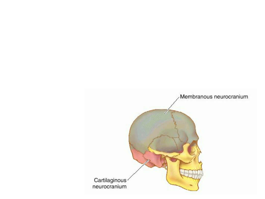
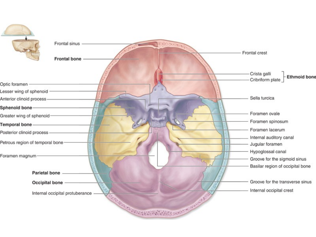
Neurocranium
Paired Cranial Bones:
Parietal
Temporal
Unpaired Cranial Bones:
Frontal
Occipital
Sphenoid
Ethmoid
Neurocranium, comprising membranous and cartilaginous
portions; the former, the calvaria, includes the frontal and
parietal and portions of the temporal, occipital, and
sphenoid bones; the latter, the chondrocranium, includes the
ethmoid and portions of the occipital, temporal, and
sphenoid bones.

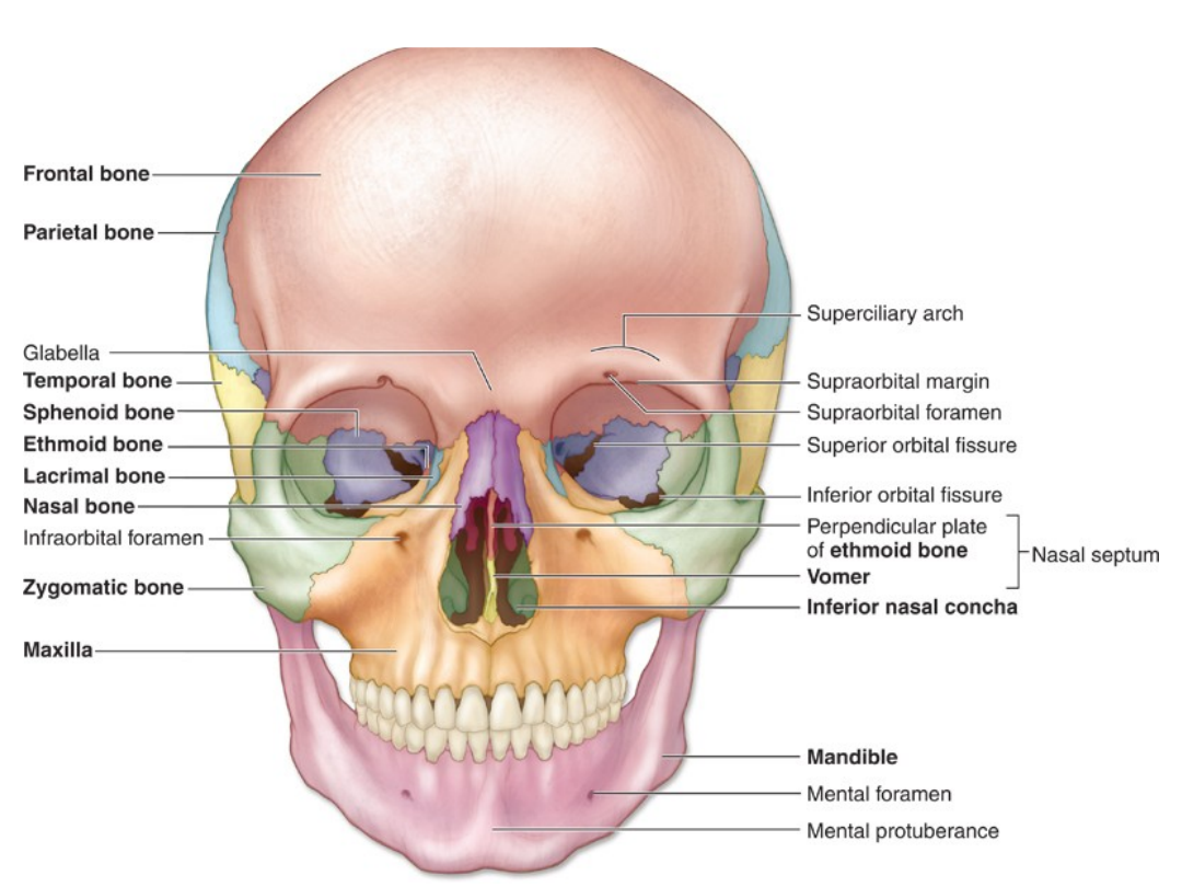
Splanchnocranium
Paired Facial
Bones:
Lacrimal
Nasal
Zygomatic
Maxilla
Palatine
Inferior Nasal
Concha
Unpaired Facial
Bones:
Vomer
Mandible
Hyoid
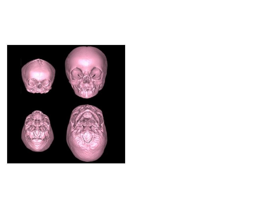
Growth of the neurocranium is
directly related to expansion
of the brain.
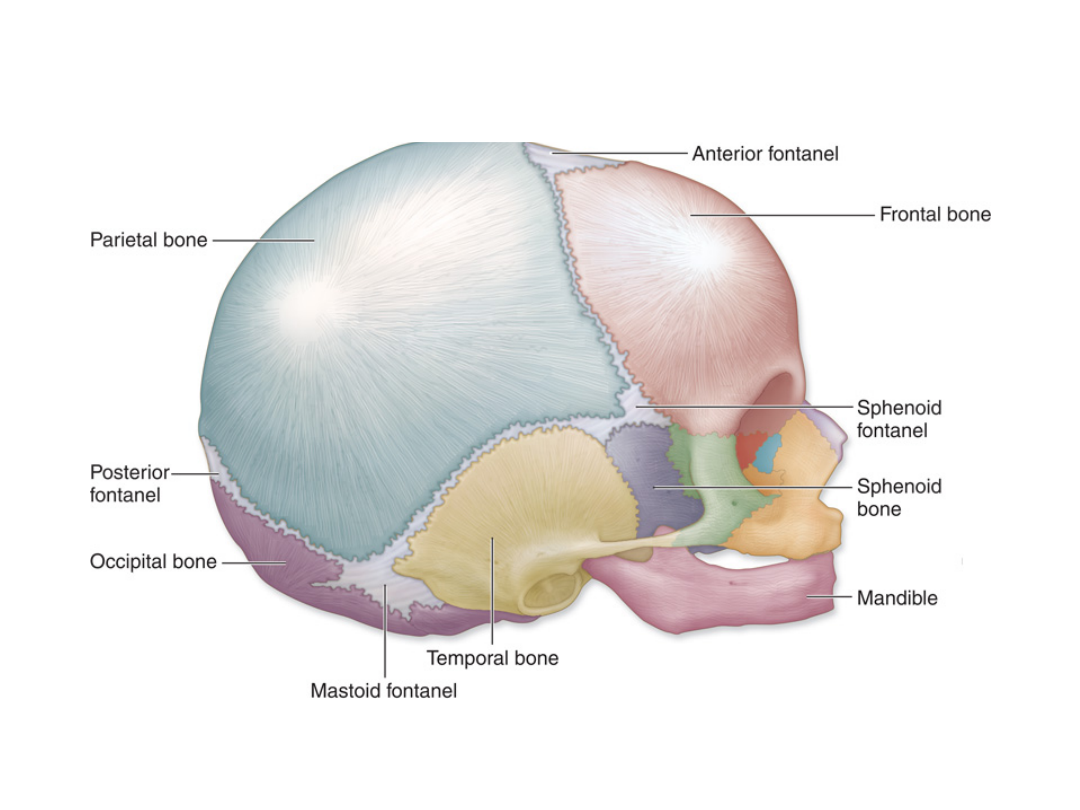
The bones of the
neurocranium are joined by
fibrous sutures, which permit
growth at the edges of the
bone. The sutures are
relatively wide at birth, with
large gaps where two or more
sutures meet. These areas are
called fontanelles, and include
the anterior fontanelle, often
refered to as a baby’s "soft
spot." Even after visible gaps
between the neurocranial
bones have closed, the
fibrous suture permits
additional growth to occur.
Eventually, the bones at that
suture fuse together. The
metopic suture normally fuses
relatively early (anywhere
from 3 to 7 years), although
the other neurocranial sutures
remain patent into adulthood.

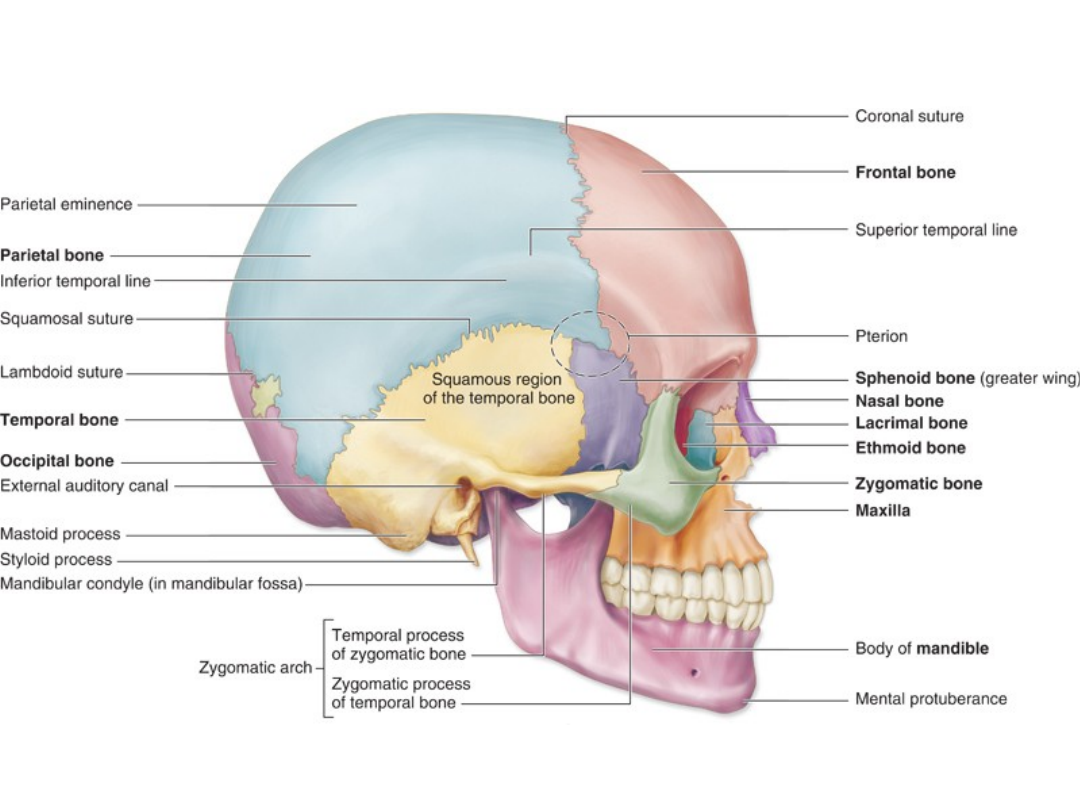
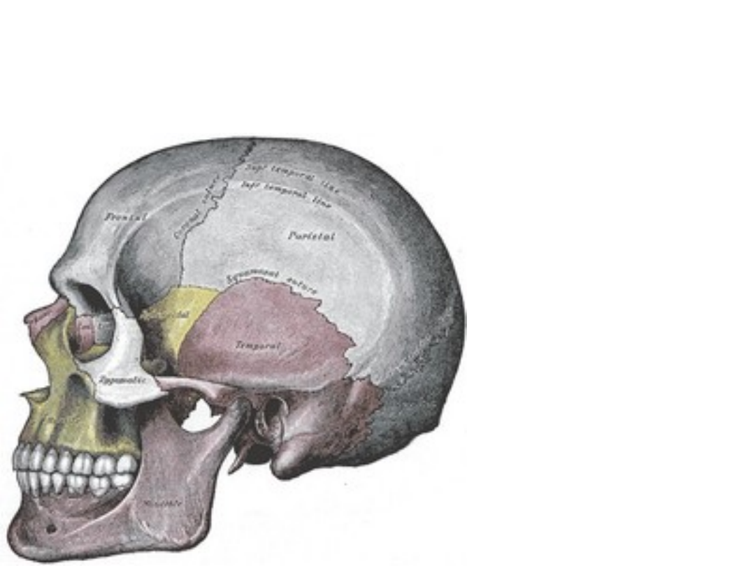
SUTURES
1.Coronal- between parietal & frontal
2.Sagittal- between right and left parietal
bones
3.Lambdoid- between parietal & occipital
4.Squamous – between parietal &
temporal
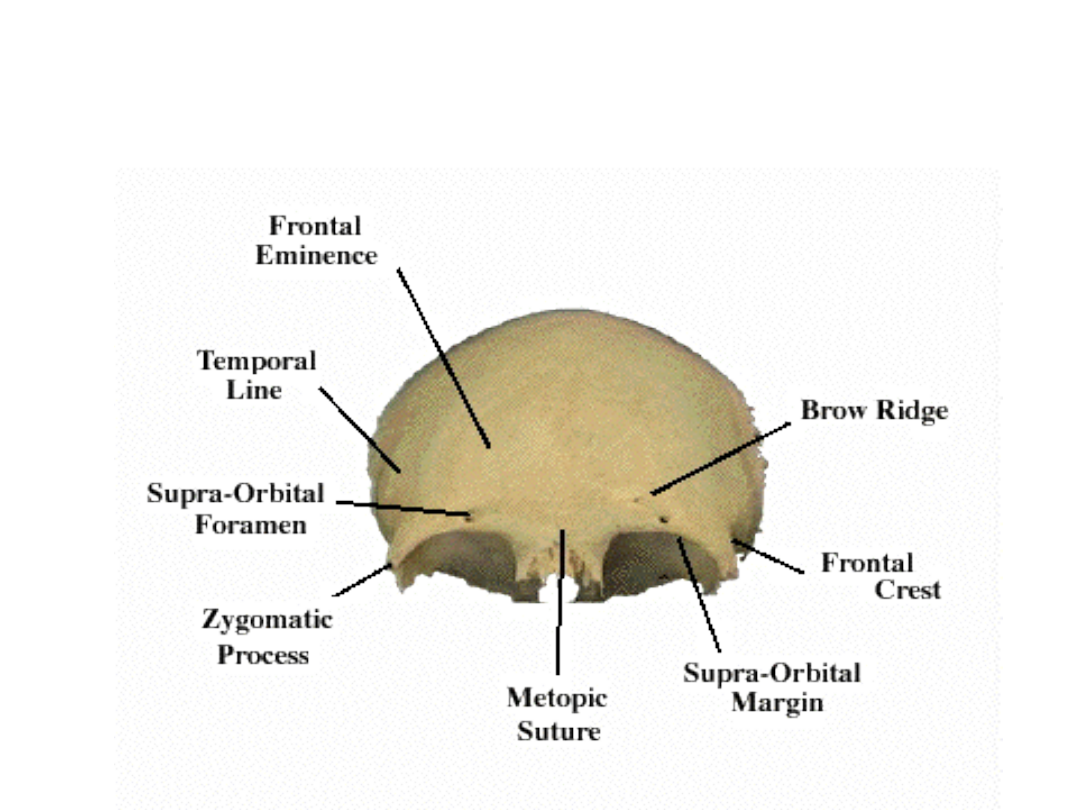
Frontal bone
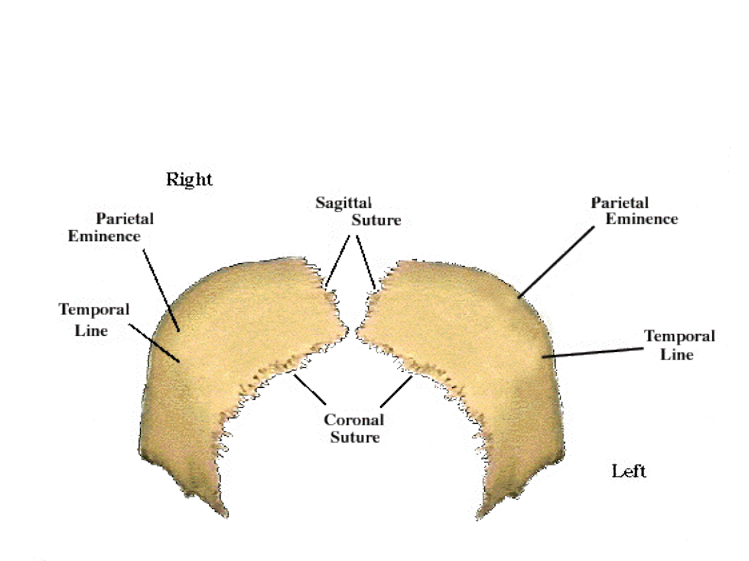
Parietal bone
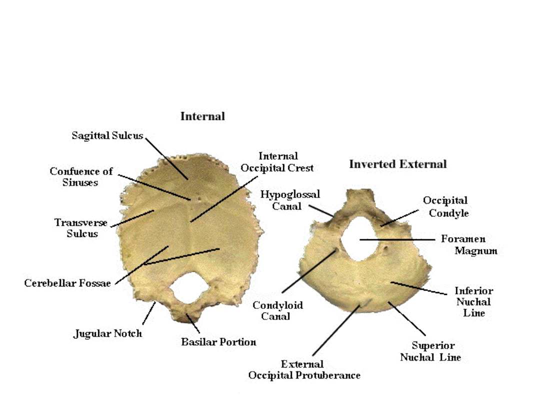
Occipital bone
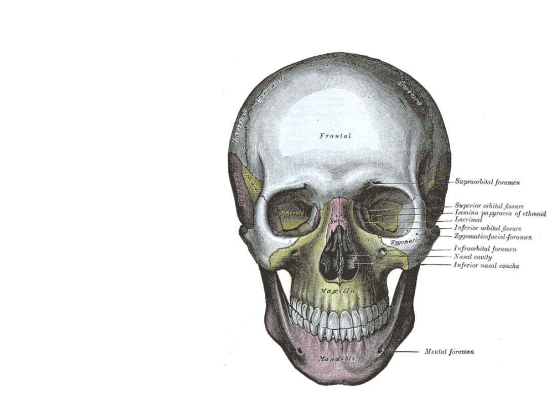
Orbit
• Provides
protection
to the
globe
• A pyramid
in shape
with apex
at foramen
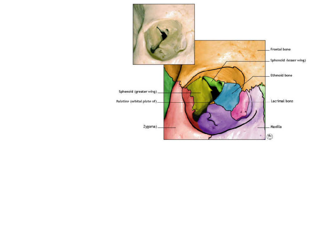
ORBIT
• Boundaries
– Superior : orbital plate
of Frontal bone and
small part of Lesser
Wing of the sphenoid
– Lateral : Greater wing
of the sphenoid and
frontal process of the
Zygomatic bone
– Medial : Orbital lamina
of the ethmoid bone
and lacrimal bones.
– Inferior : Maxillary
bone
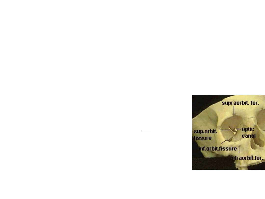
FORAMINA
INFERIOR ORBITAL FISSURE:
Carries the Maxillary Nerve (V2)
INFRAORBITAL FORAMEN
: in the orbital part of
the maxillary bone.
• It transmits the Infraorbital Nerve (V2) out of the orbit.
• It transmits the Infraorbital Artery -- an anastomotic
branch between the Angular and Maxillary Aa. (both of
which are off the External Carotid).
ETHMOID FORAMEN
: Anterior and posterior
foramina in the medial wall, transmitting structures
that are
going from orbit to the ethmoid air sinuses and nose:
• Anterior and Posterior Ethmoid Arteries, from
Opthalmic Artery
• Anterior and Posterior Ethmoid Nerves, from
Nasociliary Nerve (V1)
SUPERIOR ORBITAL FISSURE:
Between the lesser and greater
wings of the sphenoid bone.
It transmits the Superior Ophthalmic Vein.
It transmits: Oculomotor (III), Trochlear (IV), Opthalmic (V1), and
Abducens (VI)
OPTIC CANAL
: In the Lesser Wing of Sphenoid, superomedial to the
superior orbital fissure.
It transmits the Optic Nerve (II)
It transmits the Ophthalmic Artery, a branch from the Internal
Carotid.
TEMPORAL FOSSA
Boundaries:
• Temporal lines
• Zygomatic arch
Connections:
• Infratemporal
fossa
Contents:
• Temporalis
muscle
• Deep temporal
vessels and
nerves
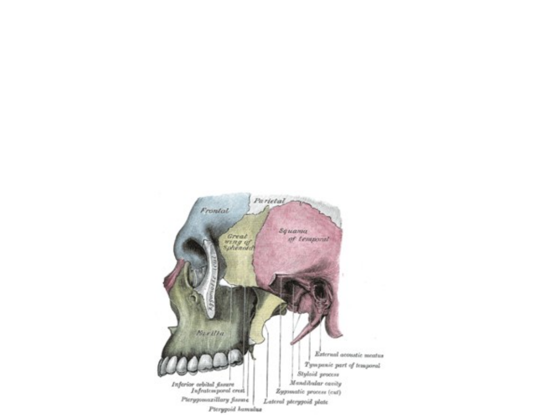
INFRATEMPORAL FOSSA
BOUNDARIES
• RAMUS OF THE MANDIBLE laterally,
• LATERAL PTERYGOID PLATE medially,
• TUBEROSITY OF THE MAXILLA anteriorly,
• CRANIAL BASE (greater wing of the sphenoid bone) superiorly.
• STYLOID PROCESS posteriorly
• The MEDIAL PTERYGOID MUSCLE bounds the fossa inferiorly

Infratemporal fossa-
connections
• The foramen ovale and foramen
spinosum open on its roof, and the
alveolar canals on its anterior wall.
• At its upper and medial part are two
fissures, which together form a T-
shaped fissure, the horizontal limb
being named the inferior orbital, and
the vertical one the pterygomaxillary.

Infratemporal fossa-
contents:
MEDIAL PTERYGOID MUSCLE
MAXILLARY ARTERY AND VEIN,
PTERYGOID PLEXUS OF VEINS,
MANDIBULAR DIVISION OF CN-V
OTIC GANGLION.
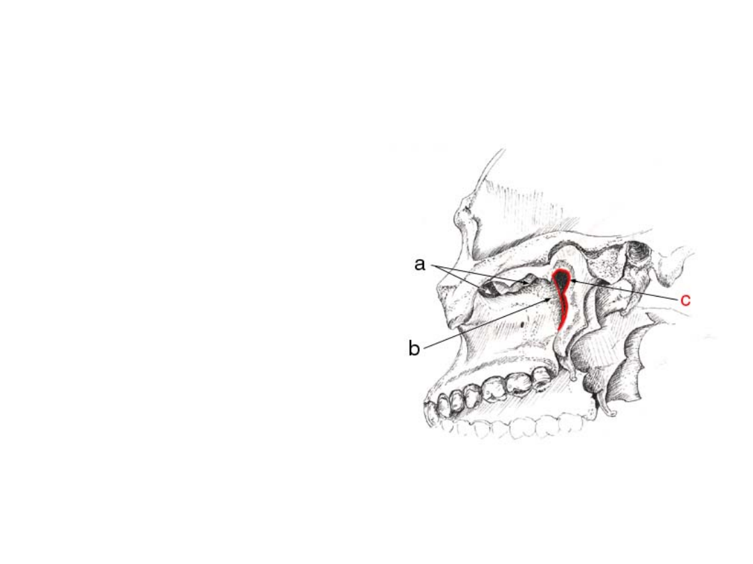
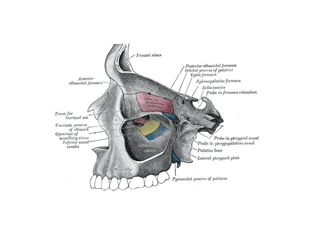
PTERYGOPALATINE FOSSA
Boundaries
Posterior:
• - root of the pterygoid plates
• - inferior surface of the greater
wing of the sphenoid bone
Anterior:
• - posterior surface of the maxilla
Superior:
• - posterior part of the inferior
orbital fissure
• - orbital process of the palatine
bone
• -body of the sphenoid bone
Inferior:
• -apex leading in to the
pterygopalatine canal
Medial:
• - perpendicular plate of the
palatine bone
Lateral:
• - location of the pterygomaxillary
fissure
a-inferior orbital fissure
b-posterior surface of the maxilla
c-pterygo-maxillary fissure (leads
to the pterygo-palatine fossa)
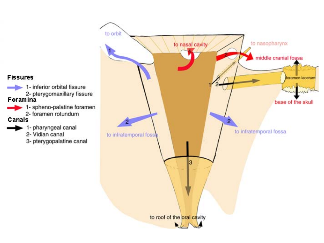

Contents:
Maxillary nerve V2 (second division of
the Trigeminal nerve),
Pterygopalatine ganglion
The third part of the Maxillary artery
• .
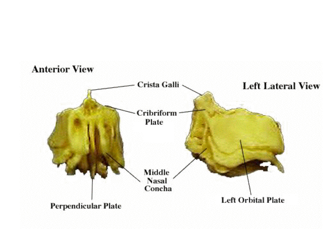
Ethmoid bone
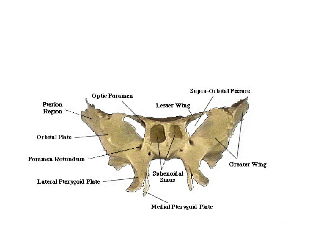
Sphenoid bone
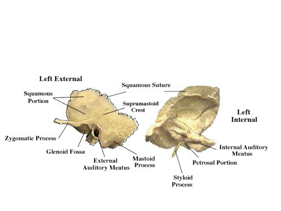
Temporal bone
Document Outline
- Slide 1
- Slide 2
- Slide 3
- Slide 4
- Slide 5
- Slide 6
- Slide 7
- Slide 8
- Slide 9
- Slide 10
- Slide 11
- Slide 12
- Slide 13
- Slide 14
- Slide 15
- Slide 16
- Slide 17
- Slide 18
- Slide 19
- Slide 20
- Slide 21
- Slide 22
- Slide 23
- Slide 24
- Slide 25
- Slide 26
- Slide 27
- Slide 28
- Slide 29
- Slide 30
Wyszukiwarka
Podobne podstrony:
Drawing the Skull Part 1
Golden Skull
Drawing the Skull Part 2
1988 Decrease of human skull size
Drawing the Skull Part 3 Teeth
A trephined skull from Iran
MEXICAN SKULL
pixelblox skull
skull QFXRT22EO74KAGUOHPCMXFHCGJX6B7WOHBWV27I
Howard, Robert E Kull The Skull of Silence
Pirate Skull
Elin Holmerin För hennes skull
A trephined skull from Iran
Robert E Howard Horror 1928 Skull Face
Kuttner, Henry & Moore, CL The Prisoner in the Skull
Rat in the Skull Rog Phillips(1)
więcej podobnych podstron