
Long-term sequential receptor activator of NF-jB
ligand (RANKL) and osteoprotegrin (OPG) expression
in lipopolysaccharide-induced rat periapical lesions
Fu-Hsiung Chuang
1,2,3
, Chi-Cheng Tsai
4
, Jeng-Huey Chen
1,2
, Ker-Kong Chen
1,2
, Yuk-Kwan Chen
1,5
,
Ying-Chu Lin
6
1
School of Dentistry, College of Dental Medicine, Kaohsiung Medical University, Kaohsiung, Taiwan;
2
Division of Conservative
Dentistry, Department of Dentistry, Kaohsiung Medical University Chung-Ho Memorial Hospital, Kaohsiung, Taiwan;
3
PhD
Program, School of Dentistry, College of Dental Medicine, Kaohsiung Medical University, Kaohsiung, Taiwan;
4
College of Dental
Medicine, Chung-Shan Medical University, Taichung, Taiwan;
5
Division of Oral Pathology and Diagnosis, Department of Dentistry,
Kaohsiung Medical University Chung-Ho Memorial Hospital, Kaohsiung, Taiwan;
6
Department of Oral Hygiene, College of Dental
Medicine, Kaohsiung Medical University, Kaohsiung, Taiwan
BACKGROUND: Long-term sequential expression of
receptor activator of NF-jB ligand (RANKL) and osteo-
protegrin (OPG) in lipopolysaccharide (LPS)-induced rat
periapical lesions has not been studied.
MATERIALS: Seventy-two 4-week-old Wistar rats were
divided into eight experimental groups and one control
group (eight animals in each).
METHODS: Lipopolysaccharide-induced periapical lesions
were produced in rats by occlusal exposure of the pulp of
their lower first molars in all experimental groups but not
the control group. The extent of periapical destruction
was measured by radiographic imaging. RANKL and OPG
mRNA were measured in all tissue sections containing
the periapical lesions as well as the control group every
week from week 1 to week 8 by real-time quantitative
reverse transcription polymerase chain reaction. RANKL
and OPG protein were determined by immunohisto-
chemistry. Osteoclasts were identified by enzyme histo-
chemistry.
RESULTS: The sequential changes in the mRNA and
protein expression of RANKL and OPG were largely
compatible with the occurrence of osteoclasts histologi-
cally and enzymes histochemically, as well as the mean
areas of the periapical lesions radiographically during
long-term observation of the LPS-induced rat periapical
lesions.
CONCLUSION: This study may be the first to demon-
strate the long-term RANKL and OPG expression every
week from week 1 to week 8 using LPS to produce
periapical infection in a Wistar rat model. The long-term
findings of high expressions of RANKL and OPG further
extend the potential application of the Wistar rat model
for future experimental trials using RANKL inhibitor to
evaluate the treatment outcome for LPS-induced rat
periapical lesions.
J Oral Pathol Med (2012) 41: 186–193
Keywords: lipopolysaccharide; OPG; osteoclast; periapical
lesion; RANKL; rat
Introduction
Periapical lesions, which are formed as a result of root
canal infection, are accompanied by an immune
response to the invading microbes and periapical bone
destruction (1–3). Bony destruction is the distinctive
function of osteoclasts. Members of the tumor necrosis
factor superfamily including receptor activator of
NF-jB ligand (RANKL) and its receptors, receptor
activator of NF-jB (RANK) and osteoprotegrin (OPG)
are involved in the formation of osteoclasts (i.e. osteo-
clastogenesis) (4, 5). Osteoclast activation and hence
bony destruction occurs as a result of RANKL binding
to the RANK receptor on the cell surface of preoste-
oclasts and mature osteoclasts (6). OPG is a decoy
receptor for RANKL (7) and hence competes with
RANK, which can attenuate bony destruction (8, 9).
Osteoclastogenesis is thus mediated via a balance
between RANKL and OPG (10).
Short-term studies of RANKL and OPG expression
have examined periapical lesions induced in a Wistar rat
model (11–13), but only short-term observation of
experimentally induced rat periapical lesions limits the
usage and further application of this rat model. To our
Correspondence:
Dr. Yuk-Kwan Chen, School of Dentistry and
Ying-Chu Lin, Department of Oral Hygiene, College of Dental
Medicine, Kaohsiung Medical University, 100 Shih-Chuan 1st Road,
Kaohsiung, Taiwan. Tel: +886-3121101
2755, Fax: +886-3210637,
E-mail: k0285@ms22.hinet.net; chulin@kmu.edu.tw
Accepted for publication June 22, 2011
J Oral Pathol Med (2012) 41: 186–193
ª 2011 John Wiley & Sons A/S Æ All rights reserved
wileyonlinelibrary.com/journal/jop
doi: 10.1111/j.1600-0714.2011.01065.x
knowledge, long-term observation of the expression of
these two cytokines has not been completed in a Wistar
rat model.
Lipopolysaccharide (LPS) is an endotoxin and is a
major component of the outer membrane of gram-
negative bacteria (14). Reviewing the English-language
literature, experimental application of LPS to the dental
pulp has been found to be able to initiate and sustain
apical periodontitis in animal models (15–17), but not in
a Wistar rat model. Previous reports have documented
that LPS is capable of directly stimulating osteoblasts to
express RANKL, resulting in osteoclast production (18)
without the involvement of RANK. Recent data have
also suggested that LPS might be directly implicated in
osteoclast differentiation via a pathway partly indepen-
dent of the aforementioned RANKL
⁄ RANK interac-
tion (19, 20). Additionally, it has been reported that
LPS-induced osteoclast formation occurs directly if
dental pulp cells are previously treated with RANKL
(21); RANKL treatment was mandatory for at least the
first 24-h, as LPS alone did not stimulate osteoclast
formation (20).
Hence, the aim of this study was to analyze the long-
term sequential expression of RANKL and OPG in
experimentally induced rat periapical lesions adminis-
tered with LPS, not only to enhance our understanding
but also to extend the potential clinical application of
this animal model. In addition, the enzyme histochem-
istry of osteoclasts and imaging analysis of periapical
bone destruction were also studied for periapical lesions
induced by LPS in a Wistar rat model in the present
study.
Materials and methods
Induction of periapical lesions
Seventy-two out-bred, 4-week-old male Wistar rats
purchased from the National Science Council Animal
Breeding Center, Taipei, Taiwan, weighing about 100 g
each at the commencement of the experiment, were
randomly divided into eight experimental groups
(groups A–H) and one control group (group I) (eight
rats in each group). The animals were housed under
constant conditions (22
C; 12-h light ⁄ dark cycle) and
fed tap water and standard Purina laboratory chow ad
libitum
. The animal-handling protocol ensured that
humane practices were adhered to throughout the
experimental process.
Subsequent to 4 weeks of acclimatization to their new
surroundings, periapical lesions were induced in all
animals of the experimental groups A–H using the
procedure documented by Stashenko et al.(22) with the
modification of using LPS to augment periapical infec-
tion. Under ketamine-HCl (5 mg
⁄ kg, intramuscular)
anesthesia, an occlusal class I cavity of the right
mandibular first molars of rats of the experimental
groups A–H was generated with magnification using a
#1
⁄ 4 round high-speed bur until the head of the bur
penetrated into the pulp chamber. With magnification, a
#8 endodontic file was subsequently introduced into the
distal root canals downwards to the root apices to
remove the pulp tissues. A paper point soaked thor-
oughly with LPS (E. Coli LPS, serotype 026: B6, Sigma,
St. Louis, MO, USA, 25 lg
⁄ 0.1 ml) was then inserted
into the distal root canal up to the root apices. Finally,
the cavity was sealed with reinforced zinc oxide eugenol
cement (Dentsply Caulk Co., Milford, DE, USA)
(23, 24).
One week after pulp exposure, all the animals of
group A were humanely sacrificed, and the mandibles
were dissected (Fig. 1A,B) and processed for image
analysis using the procedures described in the next
section.
After image analysis, the LPS-induced periapical
lesions were carefully removed. A portion of each
periapical specimen was immediately frozen in liquid
nitrogen for real-time quantitative reverse transcription
polymerase
chain
reaction
(QRT-PCR)
analysis.
Another portion was routinely processed for light
microscopy by fixation in 10% neutral-buffered forma-
lin solution, decalcification with 10% EDTA solution,
dehydration in ascending concentrations of alcohol
solution, cleaning in xylene, and finally embedding in
paraffin. Serial sections of each specimen were prepared
at a 5-lm thickness. One section was prepared for
hematoxylin–eosin staining, while the other sections
were used for enzyme histochemical staining for osteo-
clasts and immunohistochemical staining for RANKL
and OPG. Then, all animals of the other experimental
groups B–H were sacrificed, using a similar procedure,
2–8 weeks after pulp exposure, one group every week.
Finally, all rats of the unexposed pulp group (group I)
were also killed using a similar procedure.
Image analysis
Radiographs of the resected mandibles of the experi-
mental and control groups were acquired using a
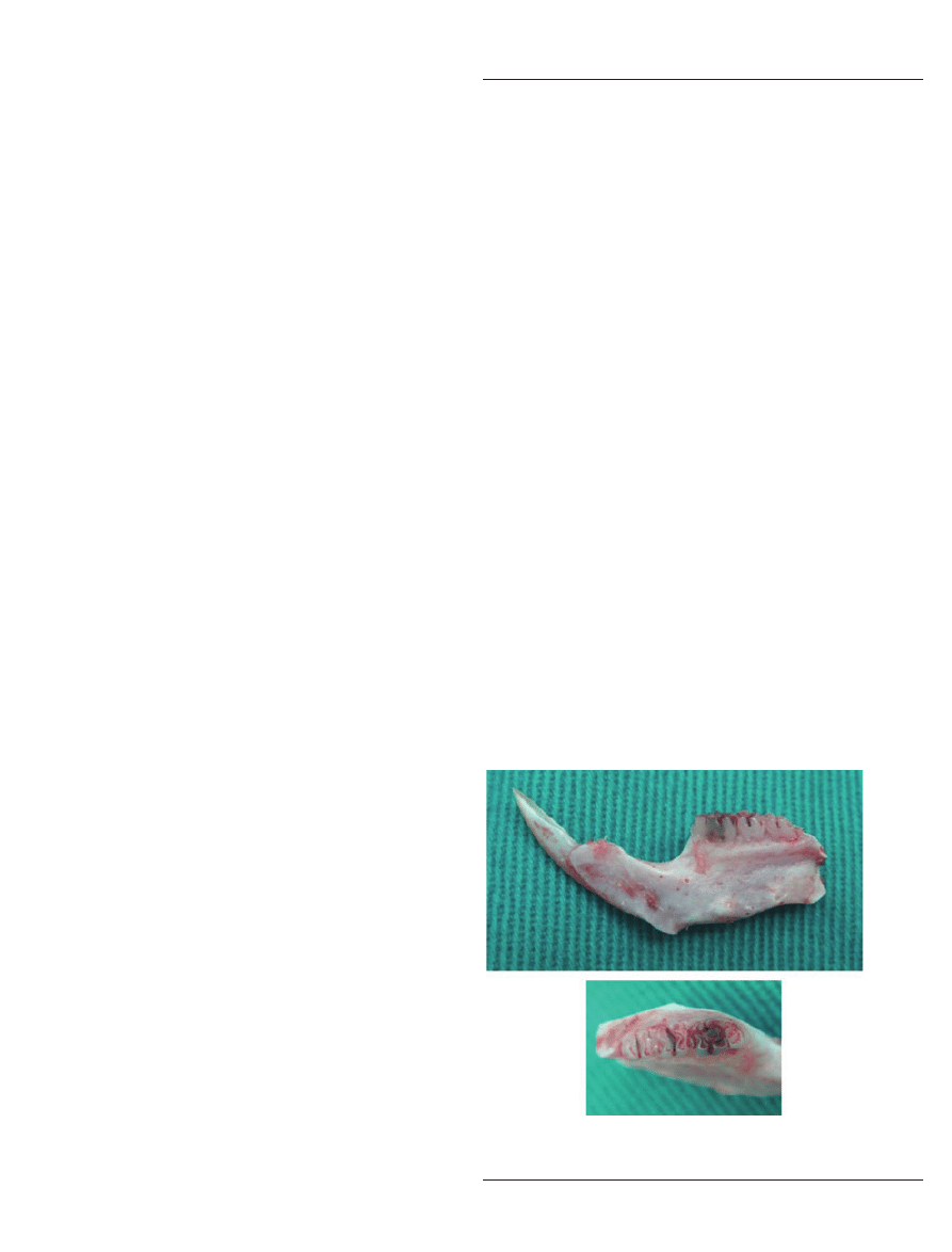
A
B
Figure 1
Lateral (A) and occlusal view (B) of a representative
dissected rat mandibular specimen.
LPS-induced rat periapical lesions
Chuang et al.
187
J Oral Pathol Med

microradiograph device and analyzed by a ScanX
image analysis system (Air Techniques, Melville, NY,
USA). Areas of the periapical lesions at the distal root
apices of the right mandibular first molars were
enumerated in pixels, which were transformed to square
millimeters using 1 mm
2
= 1000 pixels, as determined
by measuring a standard known area.
Real-time QRT-PCR
Total RNA samples were prepared from all tissues
samples of the experimental and control groups using
TRIzol Reagent (Invitrogen, Carlsbad, CA, USA). The
quality and concentration RNA was determined from
the optical density at a wavelength of 260 nm (using an
OD
260
unit equivalent to 40 lg
⁄ ml of RNA). The RNA
was resuspended in 100 ll of diethlpyrocarbonate
(DEPC)-treated water at a final concentration of
1 lg
⁄ ml. Reverse transcription was performed from
500 ng total RNA template using Taqman reverse
transcription reagent. cDNA was amplified by PCR
with oligoprimers for RANKL and OPG which were
designed with reference to the published cDNA
sequences in GenBank (Table 1). The reaction was
performed on a thermal cycler (Mx3000P
; Stratagene,
La Jolla, CA, USA) with Maxima
SYBR Green
qPCR Master Mix (2
·) added to the PCR reaction
mixture. After normalization to the expression level of
b-actin mRNA, the relative expression levels of
RANKL and OPG mRNA were presented as a
percentage change compared with the control.
Immunohistochemistry
Staining was performed using the standard avidin–
biotin peroxidase complex method (25). Rabbit poly-
clonal antibodies against mouse RANKL (Cat. no:
ab9957; Abchem Corporation, Cambridge, UK) and
OPG (Cat. no: BS1862; Bioworld Technology Inc.,
Minneapolis, MN, USA) were used. Tissue sections of
the experimental and control groups containing the
periapical areas were mounted on gelatin-chrome alum-
coated slides. Following repeated deparaffinization in
xylene and rehydration in a decreasing-concentration
ethanol series (absolute, 95%, 70%, and 30% ethanol,
and then water), tissue sections were microwave-treated
thrice (5 min each) in a citrate buffer (10 mM;
pH = 6.0) to retrieve antigenicity. Endogenous perox-
idase activity was blocked by soaking the tissue sections
in 3% hydrogen peroxide (H
2
O
2
) in methanol for
60 min. Before staining, a 10% solution of normal
rabbit serum was applied for 60 min to tissue sections to
inhibit non-specific staining. These sections were subse-
quently incubated with antibodies against RANKL and
OPG (1:100, each) overnight at 4
C. Following sub-
sequent rinsing with Tris-buffered saline (TBS, three
times, 10 min each), tissue sections were then incubated
for 60 min at room temperature with biotin-conjugated
goat anti-rabbit IgG (1:100; Vector, Burlingame, CA,
USA). Following this, all sections were washed with
TBS again (three times, 10 min each) and then incu-
bated with avidin–biotin complex conjugated with
horseradish peroxidase (Dako, Santa Barbara, CA,
USA) for a further 60 min. After washing with TBS
(three times, 10 min each), peroxidase binding was
visualized as brown reaction products via a benzidine
reaction. Each set of experiments included a human
squamous-cell carcinoma specimen known to express
RANKL and OPG (26), which served as a positive
control and ensured the reproducibility of the staining
process. A negative control, in which the primary
antibody step was omitted, was also included in each
set of experiments. In each specimen, RANKL- and
OPG-positive cells in the LPS-induced rat periapical
tissue sections were observed microscopically.
Enzyme histochemistry
Tartrate-resistant acid phosphatase (TRAP) is regarded
as a biochemical marker relatively specific for osteo-
clasts (27). The TRAP activity for all tissue sections
containing the periapical regions of the experimental
and control groups was assessed using a TRAP kit
(Sigma) in accordance with the manufacturer’s instruc-
tions.
Statistical analysis
Statistical analyses (one way ANOVA and Tukey–
Kramer tests) were performed using JUMP 7.0 software
(SAS, Cary, NC, USA). P < 0.05 was considered
significant.
Results
Histological observation
For the unexposed pulp group (group I), the periapical
area was normal and no obvious bony resorption could
be observed. Mild inflammation and small areas of
periapical alveolar bone resorption were observed for
specimens at week 1 (group A) and week 2 (group B).
The degree of inflammatory infiltration and alveolar
bone resorption became more prominent for specimens
observed at week 3 (group C) to week 8 (group H).
Furthermore, giant cells were noted for specimens
observed at week 1 (group A) and week 2 (group B)
Table 1
Oligoprimers used for real-time quantitative reverse transcriptase polymerase chain reaction in the current study
Oligoprimers
Sense
Antisense
RANKL
5
¢-ACCATCAATGCTGCCGACAT-3¢
5
¢-CTTGGCCCAGCCTCGAT-3¢
OPG
5
¢-ATATTGCCCCCAACGTTCAAC-3¢
5
¢-AGAGGGCGCATAGTCAGTAGACA-3¢
b-actin
5
¢-CTGCCCTGGCTCCTAGCA-3¢
5
¢-TAGAGCCACCAATCCACACAGA-3¢
OPG, osteoprotegrin; RANKL, receptor activator of NF-jB ligand.
LPS-induced rat periapical lesions
Chuang et al.
188
J Oral Pathol Med
(Fig. 2A), and thereafter became more prominent for
specimens observed at week 3 (group C) to week 8
(group H) (Fig. 2B,C).
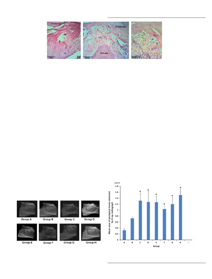
Mean area of the induced rat periapical lesions on X-ray
radiograph (mAREA
periapical
)
Representative X-ray radiographs of the experimentally
induced periapical lesions in the current experiment were
depicted in Fig. 3, and the sequential mArea
periapical
was
shown in Fig. 4.
The lowest mAREA
periapical
was observed at week 1
(group A) (0.32 ± 0.66 mm
2
), whereas the highest
mAREA
periapical
was noted at week 8 (group H)
(1.51 ± 0.19 mm
2
). Furthermore, the mAREA
periapical
significantly increased gradually from week 1 to week 3
(groups A–C), then remained more or less the same
from week 3 to week 8 (groups C–H). Starting from
week 3, the mAREA
periapical
observed each from week 3
to week 8 increased significantly as compared with the
mAREA
periapical
observed from week 1 to week 2
respectively.
Mean relative mRNA expression of RANKL
(mrRANKL
mRNA
) and OPG (mrOPG
mRNA
) for the
induced rat periapical lesions
The sequential variation of mrRANKL
mRNA
and
mrOPG
mRNA
for the current experiment was summa-
rized respectively in Fig. 5A,B. The statistical analyses
for mrRANKL
mRNA
and mrOPG
mRNA
among groups
A–I were shown in Table 2.
The mrRANKL
mRNA
observed at week 1 (group A)
and week 2 (group B) was 27% and 7%, respectively,
which were both significantly lower than that of the
unexposed
pulp
group
(group
I).
Then,
the
mrRANKL
mRNA
observed at week 3 (group C) was
elevated to 113%, which was slightly higher than
the unexposed pulp group (group I) but significantly
higher than the mrRANKL
mRNA
observed at week 1
(group A) and week 2 (group B). Subsequently, the
mrRANKL
mRNA
observed at week 4 (group D), week 5
(group E) and week 6 (group F) was decreased similarly
to 65%, 65% and 61%, respectively, which were all
significantly lower than the mrRANKL
mRNA
observed
at week 3 (group C) and that of the unexposed pulp
group (group I) but significantly higher than the
mrRANKL
mRNA
observed at week 1 (group A) and
week 2 (group B). Finally, the mrRANKL
mRNA
observed at week 7 (group G) and week 8 (group H)
was dramatically elevated to 185% and 208%, respec-
tively, both of which were significantly higher than the
mrRANKL
mRNA
observed from week 1 (group A) to
week 6 (group F), as well as that for the unexposed pulp
group (group I).
A
B
C
Figure 2
Some giant (osteoclastic-like) cells (arrows) were noted in representative lipopolysaccharide-induced rat periapical lesions at week 1
(group A) [A: hematoxylin & eosin (H–E) staining]; these became more prominent in representative rat periapical lesions observed at week 8 (group
H) (B; C: H–E staining).
Figure 3
Representative X-ray radiographs of lipopolysaccharide-
induced rat periapical lesions observed from week 1 to week 8 (groups
A–H).
Figure 4
Sequential changes in the mean area of the lipopolysaccha-
ride-induced rat periapical lesions detected by X-ray radiograph.
*P < 0.05 as compared with groups A & B respectively.
LPS-induced rat periapical lesions
Chuang et al.
189
J Oral Pathol Med
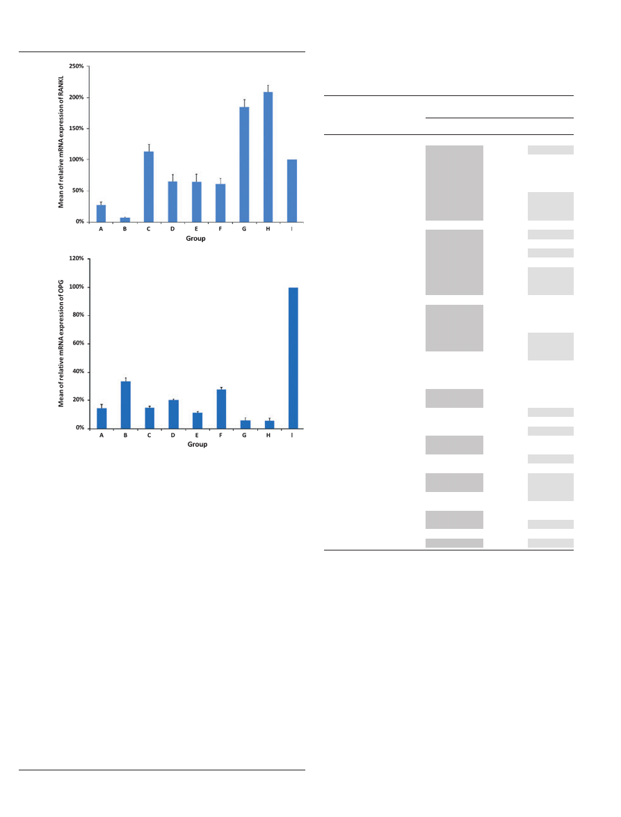
The mrOPG
mRNA
observed from week 1 to week 6
(groups A–F) fluctuated within the range of 11–33%
and then decreased significantly by week 7 (6%, group
G) and week 8 (5%, group H). Worthy of note, with the
exception of the mrOPG
mRNA
observed at week 2
(group B), the mrOPG
mRNA
observed from week 1 to
week 8 (groups A–H) was significantly lower than the
corresponding levels of mrRANKL
mRNA
observed from
week 1 to week 8 (groups A–H). Furthermore, the
mrOPG
mRNA
observed from week 1 to week 8 (groups
A–H) was significantly lower than that of the unexposed
pulp group (group I). The sequential change of the ratio
of mrRANKL
mRNA
and mrOPG
mRNA
was as shown in
Fig. 6.
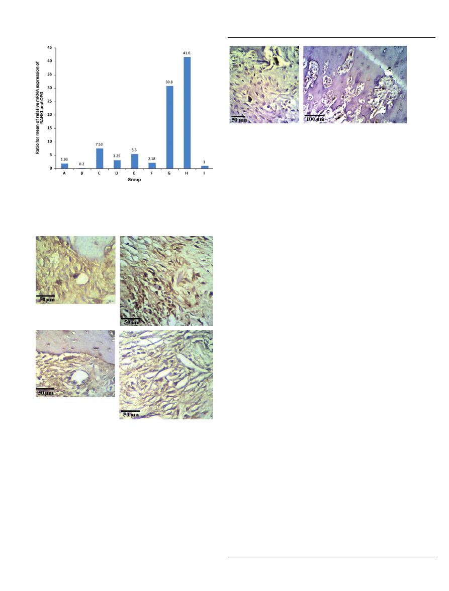
Immunohistochemical observation
Only a few RANKL- and OPG-positive cells were noted
in the tissue specimens of normal periapical areas (group
I) and the specimens containing the periapical lesions
observed at week 1 (group A) and week 2 (group B)
(Fig. 7A,B). The amount of RANKL- and OPG-
positive cells then increased in the tissue specimens
containing the periapical lesions from week 3 (group C)
to week 8 (group H) (Fig. 7C,D). Omission of primary
antisera in control sections disclosed negative findings
for RANKL and OPG activities in all sections, while the
positive control sections showed positive reactions for
RANKL and OPG activities.
Enzyme histochemical observation
Only a few osteoclast-like cells were found in the tissue
sections of normal periapical areas (group I) as well as in
the tissue sections containing the periapical lesions
A
B
Figure 5
Sequential changes in the mean of the relative mRNA
expression of receptor activator of NF-jB ligand (A) and osteopro-
tegrin (B) for the lipopolysaccharide-induced rat periapical lesions.
Table 2
Statistical analyses of the mean of relative mRNA expression
of RANKL (mrRANKL
mRNA
) and OPG (mrOPG
mRNA
) for the
induced rat periapical lesions among groups (gps) A–I
Comparisons
P values
mrRANKL
mRNA
mrOPG
mRNA
Group A
1
Gp A vs. Gp B
S
S
Gp A vs. Gp C
S
NS
Gp A vs. Gp D
S
NS
Gp A vs. Gp E
S
NS
Gp A vs. Gp F
S
NS
Gp A vs. Gp G
S
S
Gp A vs. Gp H
S
S
Gp A vs. Gp I
S
S
Group B
Gp B vs. Gp C
S
S
Gp B vs. Gp D
S
NS
Gp B vs. Gp E
S
S
Gp B vs. Gp F
S
NS
Gp B vs. Gp G
S
S
Gp B vs. Gp H
S
S
Gp B vs. Gp I
S
S
Group C
Gp C vs. Gp D
S
NS
Gp C vs. Gp E
S
NS
Gp C vs. Gp F
S
NS
Gp C vs. Gp G
S
S
Gp C vs. Gp H
S
S
Gp C vs. Gp I
NS
S
Group D
Gp D vs. Gp E
NS
NS
Gp D vs. Gp F
NS
NS
Gp D vs. Gp G
S
NS
Gp D vs. Gp H
S
NS
Gp D vs. Gp I
NS
S
Group E
Gp E vs. Gp F
NS
S
Gp E vs. Gp G
S
NS
Gp E vs. Gp H
S
NS
Gp E vs. Gp I
NS
S
Group F
Gp F vs. Gp G
S
S
Gp F vs. Gp H
S
S
Gp F vs. Gp I
NS
S
Group G
Gp G vs. Gp H
S
NS
Gp G vs. Gp I
S
S
Group H
Gp H vs. Gp I
S
S
OPG, osteoprotegrin; RANKL, receptor activator of NF-jB ligand.
1
mrRANKL
mRNA
⁄ mrOPG
mRNA
of one group (e.g. Group A) was
respectively compared each with other groups (e.g. Groups B–I) using
one-way ANOVA and Tukey–Kramer tests.
S, statistical significance (P < 0.05) (shaded areas); NS, non-statistical
significance (P > 0.05) (non-shaded areas).
LPS-induced rat periapical lesions
Chuang et al.
190
J Oral Pathol Med
observed at week 1 (group A) and week 2 (group B)
(Fig. 8A). The amount of osteoclast-like cells increased
in the tissue sections observed from week 3 (group C) to
week 8 (group H) (Fig. 8B).
Discussion
As mentioned earlier, most previous studies on RANKL
and OPG expression in experimentally induced rat
periapical lesions have only reported short-term data
(observed from week 1 to 4) (11–13), only one study
(12), to our knowledge, providing related data for week
8, but still lacking relevant information for weeks 5–7.
In this study, we documented the long-term sequential
expression of RANKL and OPG from week 1 to week 8
every week in tissue samples obtained from the
LPS-induced rat periapical lesions.
In the current study, low protein expressions of
RANKL and OPG were found using immunochemistry
in periapical tissue specimens observed at week 1 and
week 2, and the amount became more prominent in
specimens observed at week 3–8. Nevertheless, immu-
nochemistry would not provide the most specific quan-
titative changes of RANKL and OPG.
Worthy of note, by using the more specific and
advanced method of real-time QRT-PCR, delicate
quantitative alterations in RANKL and OPG mRNA
were successfully documented for LPS-induced periapi-
cal rat lesions in the present study. We found that the
mrRANKL
mRNA
observed at week 1 (group A) and
week 2 (group B) was significantly lower than in the
unexposed pulp group (group I). This is perhaps due to
the fact that, at the commencement of the experiment, a
potential unspecified mechanism has been triggered
intending to inhibit the production of RANKL so as
to suppress the growth of the experimentally induced
periapical lesions. However, with the progress of the
experiment (observed at week 3, group C), this mech-
anism is retarded, as reflected by a rebound of the
mrRANKL
mRNA,
approximating the normal value of
unexposed pulp group (group I). Therefore, the findings
in the initial period of our study were consistent with
those of previous studies (11–13). Then, on further
observation, the mrRANKL
mRNA
again declined but
became almost constant at the 4th week (group D), 5th
week (group E) and 6th week (group F), which may
imply a partial recovery of the potential mechanism of
the animals.
Nonetheless, in the final period of observation [7th
week (group G) and 8th week (group H)], we speculate
that the potential mechanism had almost completely
deteriorated, as indicated by a more or less two-fold
increase of the mrRANKL
mRNA
as compared with the
level observed in the unexposed pulp group (group I).
This finding is also supported by the highest number of
Figure 6
Sequential changes in the ratio of the mean of the relative
mRNA expression of receptor activator of NF-jB ligand and
osteoprotegrin for the lipopolysaccharide-induced rat periapical
lesions.
A
B
C
D
Figure 7
Representative photomicrograph of receptor activator of
NF-jB ligand- and osteoprotegrin-positive cells in the lipopolysac-
charide-induced rat periapical tissue specimens observed at week 1
(group A) (A and B) and week 8 (group H) (C and D).
A
B
Figure 8
Representative
photomicrograph
of
tartrate-resistant
acid phosphatase in the lipopolysaccharide-induced rat periapical
tissue specimens observed at week 1 (group A) (A) and week 8
(group H) (B).
LPS-induced rat periapical lesions
Chuang et al.
191
J Oral Pathol Med

giant (osteoclast-like) cells as well as the greatest mean
areas of the rat periapical lesions measured on X-ray
radiographs at week 7 (group G) and week 8 (group H).
This interesting finding differs from the results of
Kawashima et al.,(12) who reported a more or less
same level observed at week 8 as compared with week 4.
This discrepancy may be due to the fact that LPS, with
the capacity to upgrade the periapical infection, was
used in the present study. On the other hand, the
sequential change of the ratio of mrRANKL
mRNA
and
mrOPG
mRNA
was principally similar to the alteration in
mrRANKL
mRNA
described earlier—an initial increase
in
the
mrRANKL
mRNA
⁄ mrOPG
mRNA
ratio
was
observed at the 3rd week, which then decreased at the
4th–6th weeks but drastically increased at the 7th and
8th weeks. Consequently, the current study is, to our
knowledge, the first to report on the long-term altera-
tions of mRNA expression of RANKL and OPG in
LPS-induced periapical lesions in a rat model.
In the present study, not only alterations in
RANKL and OPG expression, but also the expression
of giant (osteoclast-like) cells histologically and en-
zymes histochemically, as well as the mean areas of the
periapical lesions radiographically, were documented
during long-term observation of LPS-induced rat
periapical lesions. We found that the trend of RANKL
and OPG in the current study has been compatible
with the results of histological, and enzyme histochem-
ical observations and imaging analyses: The later
the observation, the greater the bony destruction and
the higher the level of giant (osteoclast-like) cells.
Furthermore, the protein expression of RANKL was
consistent with TRAP activity. Hence, supported by
the histological, enzyme histochemical and imaging
examinations, our results, in line with previous animal
(11–13) and human (28–30) reports, indicated that
RANKL and OPG, as well as the RANKL
⁄ OPG
ratio, played a central role in the initiation of
periapical bone destruction.
Finally, two critical aspects for our study need further
attention. First, as the effect of LPS is concentration
dependent, a nearly equal quantity of LPS would be
expected to be available to periapical tissue of each rat
in the current study. In contrast to most in vitro studies
in which a fixed concentration of LPS would easily be
measured and applied to the culture system (20, 21, 23),
for in vivo study, this objective may not easily be
attained, especially for small animals such as rat. We
claimed that this goal would potentially be attained in
the current study, provided the paper point is saturated
with a fixed amount of LPS, and additionally, the paper
point is ascertained to be inserted correctly into the root
canal orifices; otherwise, there may be a caution of
variable concentrations of LPS to be available to the
periapical tissue of each rat.
Second, as reviewed by Suzuki et al. (31), more than
90% of the RNA transcription studies appeared in high-
impact journals employed only one housekeeping gene.
Furthermore, to our knowledge, most analyses on the
role of RANKL and OPG in the rat periapical lesion
development using real time QRT-PCR also used only
one housekeeping gene [either glyceraldehydes 3-phos-
phate dehydrogenase (GAPDH)
⁄ b-actin] (11–13, 22).
However, some authors have claimed that it is impor-
tant to use more than one housekeeping genes to obtain
the most reliable results in RNA transcription studies
(32, 33). In the current study, only b-actin was
employed as the reference gene. Hence, it is worthwhile
for future experiments to use more than one house-
keeping gene for normalization, not only to further
confirm but also to acquire the most reliable data of the
current study.
In conclusion, using LPS to produce periapical
infection in a Wistar rat model, the present study
demonstrates the long-term sequential expression of
RANKL and OPG every week from week 1 to week 8.
The long-term findings of a high RANKL level and
RANKL
⁄ OPG ratio further extend the potential appli-
cation of the Wistar rat model for future experimental
trials using RANKL inhibitor (34, 35) to evaluate the
treatment outcome for LPS-induced rat periapical
lesions by monitoring the change in RANKL expression
or the RANKL
⁄ OPG ratio in the samples.
References
1. Stashenko P, Teles R, D’Souza R. Periapical inflamma-
tory responses and their modulation. Crit Rev Oral Biol
Med
1998; 9: 498–521.
2. Takahashi K. Microbiological, pathological, inflamma-
tory, immunological and molecular biological aspects of
periradicular disease. Int Endod J 1998; 31: 311–25.
3. Kawashima N, Stashenko P. Expression of bone-resorp-
tive and regulatory cytokines in murine periapical inflam-
mation. Arch Oral Biol 1999; 44: 55–66.
4. Yasuda H, Shima N, Nakagawa N, et al. Osteoclast
differentiation factor is a ligand for osteoprotegerin
⁄
osteoclastogenesis-inhibitory factor and is identical to
TRANCE
⁄ RANKL. Proc Natl Acad Sci USA 1998; 95:
3597–602.
5. Hsu H, Lacey DL, Dunstan CR, et al. Tumor necrosis
factor receptor family member RANK mediates osteoclast
differentiation and activation induced by osteoprotegerin
ligand. Proc Natl Acad Sci USA 1999; 96: 3540–5.
6. Horowitz MC, Xi Y, Wilson K, Kacema MA. Control of
osteoclastogenesis and bone resorption by members of the
TNF family of receptors and ligands. Cytokine Growth
Factor Rev
2001; 12: 9–18.
7. Simonet WS, Lacey DL, Dunstan CR, et al. Osteoproteg-
erin: a novel secreted protein involved in the regulation of
bone density. Cell 1997; 89: 309–19.
8. Zhang H, Yano S, Miki T, et al. A novel bisphosphona-
teminodronate (YM529) specifically inhibits osteolytic
bone metastasis produced by human small-cell lung cancer
cells in NK-cell depleted SCID mice. Clin Exp Metastasis
2003; 20: 153–9.
9. Yano S, Zhang H, Hanibuchi M, et al. Combined therapy
with a new bisphosphonate, minodronate (YM529), and
chemotherapy for multiple organ metastasis of small cell
lung cancer cells in severe combined immunodeficient
mice. Clin Cancer Res 2003; 9: 5380–5.
10. Hofbauer LC, Khosla S, Dunstan CR, Lacey DL, Boyle
WJ, Riggs BL. The roles of osteoprotegerin and osteo-
protegerin ligand in the paracrine regulation of bone
resorption. J Bone Miner Res 2000; 15: 2–12.
LPS-induced rat periapical lesions
Chuang et al.
192
J Oral Pathol Med

11. Zhang X, Peng B. Immunolocalization of receptor activa-
tor of NF kappa B ligand in rat periapical lesions. J Endod
2005; 31: 574–7.
12. Kawashima N, Suzuki N, Yang G, et al. Kinetics of
RANKL, RANK and OPG expressions in experimentally
induced rat periapical lesions. Oral Surg Oral Med Oral
Pathol Oral Radiol Endod
2007; 103: 707–11.
13. Zhang X, Peng B, Fan M, Bian Z, Chen Z. The effect
of estrogen deficiency on receptor activator of nuclear
factor kappa B ligand and osteoprotegerin synthesis in
periapical lesions induced in rats. J Endod 2007; 33:
1053–6.
14. Raetz CR. Biochemistry of endotoxins. Annu Rev Biochem
1990; 59: 129–70.
15. Dahlen G, Magnusson BC, Moller A. Histological and
histochemical study of the influence of lipopolysaccharide
extracted from Fusobacterium nucleatum on the periapical
tissues in the monkey Macaca fascicularis. Arch Oral Biol
1981; 26: 591–8.
16. Mattison GD, Haddix JE, Kehoe JC, Progulske-Fox A.
The effect of Eikenella corrodens endotoxin on periapical
bone. J Endod 1987; 13: 559–65.
17. Nelson-Filho P, Leonardo MR, Silva LA, Assed S.
Radiographic evaluation of the effect of endotoxin (LPS)
plus calcium hydroxide on apical and periapical tissues of
dogs. J Endod 2002; 28: 694–6.
18. Kikuchi T, Matsuguchi T, Tsuboi N, et al. Gene expres-
sion of osteoclast differentiation factor is induced by
lipopolysaccharide in mouse osteoblasts via Toll-like
receptors. J Immunol 2001; 166: 3574–9.
19. Suda K, Woo JT, Takami M, Sexton PM, Nagai K.
Lipopolysaccharide supports survival and fusion of oste-
oclasts independent of TNF-a, IL-1 and RANKL. J Cell
Physiol
2002; 190: 101.
20. Jiang J, Li H, Fahid FS, et al. Quantitative analysis of
osteoclast-specific gene markers stimulated by lipopoly-
saccharide. J Endod 2006; 32: 742–6.
21. Jiang J, Zuo J, Chen SH, Holliday LS. Calcium hydroxide
reduces lipopolysaccharide stimulated osteoclast forma-
tion. Oral Surg Oral Med Oral Pathol Oral Radiol Endod
2003; 95: 348–54.
22. Stashenko P, Wang CY, Tani-Ishii N, Yu SM. Pathogen-
esis of induced rat periapical lesions. Oral Surg Oral Med
Oral Pathol Oral Radiol Endod
1994; 78: 494–502.
23. Hong CY, Lin SK, Kok SH, et al. The role of lipopoly-
saccharide in infectious bone resorption of periapical
lesion. J Oral Pathol Med 2004; 33: 162–9.
24. Wang QS, Gao Y, Rong L, Wang AQ. Radiological and
histopathological changes of the periapical lesions induced
by lipopolysaccharide in rats. Shanghai J Stomatology
2008; 17: 416–9.
25. Hsu SM, Raine L, Fanger H. Use of avidin-biotin-
peroxidase complex (ABC) in immunoperoxidase tech-
niques: a comparison between ABC and unlabelled
antibody (PAP) procedures. J Histochem Cytochem 1981;
29: 577–80.
26. Chuang FH, Hsue SS, Wu CW, Chen YK. Immuno-
histochemical expression of RANKL, RANK and OPG in
human oral squamous cell carcinoma. J Oral Pathol Med
2009; 38: 753–8.
27. Minkin C. Bone acid phosphatase: tartrate-resistant acid
phosphatase as a marker of osteoclast function. Calcif
Tissue Int
1982; 34: 285–90.
28. Menezes R, Bramante CM, da Silva Paiva KB, et al.
Receptor activator NFjB-ligand and osteoprotegerin
protein expression in human periapical cysts and granu-
lomas. Oral Surg Oral Med Oral Pathol Oral Radiol Endod
2006; 102: 404–9.
29. Vernal R, Dezerega A, Dutzan N, et al. RANKL in
human periapical granuloma: possible involvement in
periapical bone destruction. Oral Dis 2006; 12: 283–9.
30. Menezes R, Garlet TP, Letra A, et al. Differential patterns
of receptor activator of nuclear factor kappa B ligand
⁄
osteoprotegerin expression in human periapical granulo-
mas: possible association with progressive or stable nature
of the lesions. J Endod 2008; 34: 932–8.
31. Suzuki T, Higgins PJ, Crawford DR. Control selection for
RNA quantitation. BioTechniques 2000; 29: 332–7.
32. Bustin SA. Quantification of mRNA using real-time
reverse transcription PCR (RT-PCR): trends and prob-
lems. J Mol Endocrinol 2002; 29: 23–9.
33. Vandesompele J, De Preter K, Pattyn F, et al. Accurate
normalization of real-time quantitative RT-PCR data by
geometric averaging of multiple internal control genes.
Genome Biol
2002; 3: research0034.1–0034.11.
34. He X, Andersson G, Lindgren U, Li Y. Resveratrol
prevents RANKL-induced osteoclast differentiation of
murine osteoclast progenitor RAW 264.7 cells through
inhibition of ROS production. Biochem Biophys Res
Commun
2010; 401: 356–62.
35. Ferrari-Lacraz S, Ferrari S. Do RANKL inhibitors
(denosumab) affect inflammation and immunity? Osteo-
poros Int
2011; 22: 435–46.
LPS-induced rat periapical lesions
Chuang et al.
193
J Oral Pathol Med
Wyszukiwarka
Podobne podstrony:
Intertrochanteric osteotomy in young adults for sequelae of Legg Calvé Perthes’ disease—a long term
How effective are energy efficiency and renewable energy in curbing CO2 emissions in the long run A
Simultaneous determination of rutin and ascorbic acid in a sequential injection lab at valve system
Quality of life and disparities among long term cervical cancer suvarviors
Alexander, Smales Intelligence, Learning and Long Term Memory
Functional Origins of Religious Concepts Ontological and Strategic Selection in Evolved Minds
2008 4 JUL Emerging and Reemerging Viruses in Dogs and Cats
Angielski tematy Performance appraisal and its role in business 1
Kissoudi P Sport, Politics and International Relations in Twentieth Century
Greenshit go home Greenpeace, Greenland and green colonialism in the Arctic
Ionic liquids as solvents for polymerization processes Progress and challenges Progress in Polymer
Civil Society and Political Theory in the Work of Luhmann
RATIONALITY AND SITUATIONAL LOGIC IN POPPER
Ouellette J Science and Art Converge in Concert Hall Acoustics
Extracellular NAD and ATP Partners in immune
2008 5 SEP Practical Applications and New Perspectives in Veterinary Behavior
Mutations in the CgPDR1 and CgERG11 genes in azole resistant C glabrata
więcej podobnych podstron