
Colloids and Surfaces B: Biointerfaces 57 (2007) 250–255
Morphology and characterization of 3D micro-porous structured
chitosan scaffolds for tissue engineering
Wen-Chuan Hsieh
, Chih-Pong Chang
, Shang-Ming Lin
a
Department of Biological Science and Technology, I-Shou University, No. 1, Sec. 1, Syuecheng Road, Dashu,
Kaohsiung 84001, Taiwan, ROC
b
Department of Textile Engineering, Chinese Culture University, Taipei 11192, Taiwan, ROC
c
Department of Materials and Textiles, Oriental Institute of Technology, Pan-Chiao 220, Taiwan, ROC
Received 10 October 2006; received in revised form 1 February 2007; accepted 6 February 2007
Available online 12 February 2007
Abstract
This research studies the morphology and characterization of three-dimensional (3D) micro-porous structures produced from biodegradable
chitosan for use as scaffolds for cells culture. The chitosan 3D micro-porous structures were produced by a simple liquid hardening method,
which includes the processes of foaming by mechanical stirring without any chemical foaming agent added, and hardening by NaOH cross
linking. The pore size and porosity were controlled with mechanical stirring strength. This study includes the morphology of chitosan scaffolds,
the characterization of mechanical properties, water absorption properties and in vitro enzymatic degradation of the 3D micro-porous structures.
The results show that chitosan 3D micro-porous structures were successfully produced. Better formation samples were obtained when chitosan
concentration is at 1–3%, and concentration of NaOH is at 5%. Faster stirring rate would produce samples of smaller pore diameter, but when
rotation speed reaches 4000 rpm and higher the changes in pore size is minimal. Water absorption would reduce along with the decrease of chitosan
scaffolds’ pore diameter. From stress–strain analysis, chitosan scaffolds’ mechanical properties are improved when it has smaller pore diameter.
From in vitro enzymatic degradation results, it shows that the disintegration rate of chitosan scaffolds would increase along with the processing
time increase, but approaching equilibrium when the disintegration rate reaches about 20%.
© 2007 Elsevier B.V. All rights reserved.
Keywords: Chitosan scaffold; Biodegradable polymer; Lysozyme degradation; Tissue engineering
1. Introduction
Biomedical polymers were widely used as medical ser-
vice materials such as surgical sutures, wound dressing,
braces/fixation materials, etc., because of its biocompatibil-
ity, bioabsorbability, that it will not induce antibody from the
immune system, hypoallergenic and that it does not cause
inflammation to human tissue
. These biomedical poly-
mers used include natural polymers such as collagen, hyaluronic
acid, chitosan and alginate; microbial synthesized poly-
mers such as poly(3-hydroxybutyrate) [P(3HB)], poly(3-hydro-
xybutyrate-co-4-hydroxybutyrate) [P(3HB-co-4HB)], poly(3-
hydroxybutyrate-co-3-hydroxyvalerate) [P(3HB-co-3HV)]; and
∗
Corresponding author. Tel.: +886 7 657 7711x3469; fax: +886 7 657 9746.
E-mail address:
(W.-C. Hsieh).
artificial synthesized polymers such as poly(lactic acid),
poly(lactic-co-glycolic acid)
. Since about 20 years ago
in the 1980s, many researchers have utilized these biomedi-
cal polymers to manufacture human body tissue in organ shape
for implant, growing new tissues by molding in the shapes of
organs for use in repairing body tissue imperfection or dam-
ages
. Therefore, biomedical polymers have become the
extremely popular material in this new research domain of tis-
sue engineering. In order to effectively and massively cultivate
the tissue cells, these biomedical polymers usually designed as
three-dimensional structure (scaffolds) of interconnected pores
with high porosities for easily implant into the human body.
Hence, this kind of scaffolds must have good biocompatibil-
ity, biodegradability, high porosity, and possess appropriate
mechanical properties
Chitosan is extracted and purified from the shells of shrimp,
crab and other crustaceans, and from some of the fungi cell walls.
0927-7765/$ – see front matter © 2007 Elsevier B.V. All rights reserved.
doi:

W.-C. Hsieh et al. / Colloids and Surfaces B: Biointerfaces 57 (2007) 250–255
251
It is an abundant deposit of natural cationic polysaccharides
second only to cellulose on earth’s reserves. The amino and
hydroxyl groups in its molecular structure can be easily modified
chemically to produce other derivatives. Chitosan can be made
into gelatin, orb, fiber and membrane shapes for various uses.
Moreover, because chitosan can be easily obtained and confirms
to tissue engineering application requirements; can be implanted
into human body and causes no harm; it is a notably suitable
material for use in tissue engineering
. At present time, there
are not many related research for chitosan direct application in
3D scaffolds. Furthermore, most of these researches are mainly
on 3D composite scaffolds that mixed with other materials for
permanent implant in human body
This research uses chitosan as the only crude materials,
utilize mechanical agitation to produce foaming bubbles, and
through a simple liquid hardening method to produce the chi-
tosan scaffolds. The purpose is to avoid any excessive chemical
additives in the manufacturing processes that could possibly
cause harmful effects to the human body. We observed the
produced sample’s formation status through SEM, and inves-
tigated the influence of various concentrations of chitosan and
hardening agent, stirring rate of homogenizer and other vari-
ables to the pore sizes production and pore distribution of the
chitosan scaffolds samples. In addition, we measure water con-
tent/uptake ratio and use compression tensile test to investigate
the influence of structural variation to chitosan scaffolds’ physi-
cal properties. Lastly, we conduct in vitro enzymatic degradation
to observe the condition while chitosan scaffolds being dis-
integrated by enzyme so we could assess the feasibility of
chitosan scaffolds’ use for cell culture in tissue engineering
application.
2. Experimental
2.1. Preparation of chitosan scaffolds
Chitosan [poly(
-(1-4-2-amino-2-deoxy-d-glucopyranase)]
was purchased from Wako Pure Chemical Industries Co. (80
◦
of
deacetylation). Place 1–3% (C-1%, C-2%, C-3%) chitosan and
evenly dissolved in 1N acetic acid solution to form thick chitosan
solution. Use the Homogenizer (HG-300D + K12S, Shuang-Tai,
Taiwan) set at 2000–6000 rpm speed to mechanically agitate and
stir to foam bubbles in the thick chitosan solution. Then pour
into a regulate container having 5% aqueous sodium hydroxide
solution (NaOH, First Chemical, Taiwan) to conduct liquid hard-
ening processes for chitosan porous scaffolds to slowly harden
into formation. Wash with distilled water at least three times to
remove remaining traces of alkali, and cure with Freeze Dryer
(FD-3a2d, Fuyuan Co., Taiwan), then stored in desiccator as the
samples for this research.
C-1%, C-2% and C-3% represent the chitosan scaffolds sam-
ples produced under 1%, 2% and 3% of chitosan concentration,
4000 rpm, 5% NaOH. The liquid hardening method we used is
the above processes of producing the 3D micro-porous chitosan
scaffolds, including the foaming by mechanical stirring without
adding any chemical foaming agent, and the hardening process
of cross-linked by NaOH.
2.2. Analytical methodology
2.2.1. Pore size and porosity
By using a scanning electron microscope (SEM) apparatus,
the pore sizes of the cross section and the average pore diame-
ter of samples were observed, we could derive the mean value
and standard deviation (N = 20) of the pore sizes of the sam-
ples. However, porosity (P) is obtained from calculation formula
below. It is calculated from the density of chitosan and the den-
sity of actual porous scaffolds sample produced. The density
of chitosan (
ρ) is 1.342 g/cm
3
. So the calculation formula of
porosity (P) is defined as follows:
porosity (%)
=
V
m
− V
p
V
m
× 100 =
V
m
− (W
m
/ρ)
V
m
× 100 (1)
V
m
is the total volume of chitosan scaffolds (cm
3
), V
p
the actual
volume of chitosan (cm
3
) and W
m
is the mass of scaffold (g).
2.2.2. Water absorption
The water absorption of the chitosan samples were obtained
by following these steps: the chitosan scaffolds samples were
prepared into round shape specimens with diameter about 2 cm.
The absolute-dry weight (
w
0
) of the samples were measured
and then were placed into 37
◦
C PBS buffer solution to satu-
rate with liquid. The samples were then taken out quickly after
24 h and placed into the Automatic Water Content Measuring
System (AWCMS)
. The measurements of the samples’ sat-
urated weight (
w
t
) were not taken until the weight displayed on
AWCMS no longer changes. The water absorption (W) of these
samples was calculated using the following equation:
water absorption (%)
=
w
t
− w
0
w
0
× 100
(2)
2.2.3. Compressive strength and strain
The compression strength and strain data of the chitosan
scaffolds samples were obtained from the following processes.
First place 5 cm (L)
× 1 cm (H) × 2 cm (W) (L, H, W denoted the
length, height and width, respectively) size chitosan scaffolds
samples into a 25
± 2
◦
C, 65
± 2% RH Programmable Tem-
perature & Humidity Chamber (THS-A, KSON) for 24 h, then
use Micro Hardness Tester (HMV-2T, SHIMADZU) to measure
samples’ compressive strength and strain. The compression rate
was set at 10 mm/min.
2.2.4. In vitro enzymatic degradation
First the absolute-dry weight (
w
0
) of the chitosan scaffolds
samples were measured, and the samples were placed into 37
◦
C
PBS buffer solution, simultaneously add in 5
g/ml of Lysozyme
(PSTi Co., Taiwan). Then the samples were placed in an Orbital
Shakers (SO-701, GMB Co.) with temperature set at constant
37
◦
C to do the time course weight-change experiment of enzy-
matic degradation. After the samples were washed with distilled
water and freeze-dried, they were placed into a 25
± 2
◦
C,
65
± 2% RH Programmable Temperature & Humidity Cham-
ber for 24 h and the weight loss ratio was then calculated. The
weight loss ratio of these samples was calculated using the
252
W.-C. Hsieh et al. / Colloids and Surfaces B: Biointerfaces 57 (2007) 250–255
following equation:
weight loss (%)
=
w
0
− w
t
w
0
× 100
(3)
3. Results and discussion
3.1. Morphology of the scaffolds structure
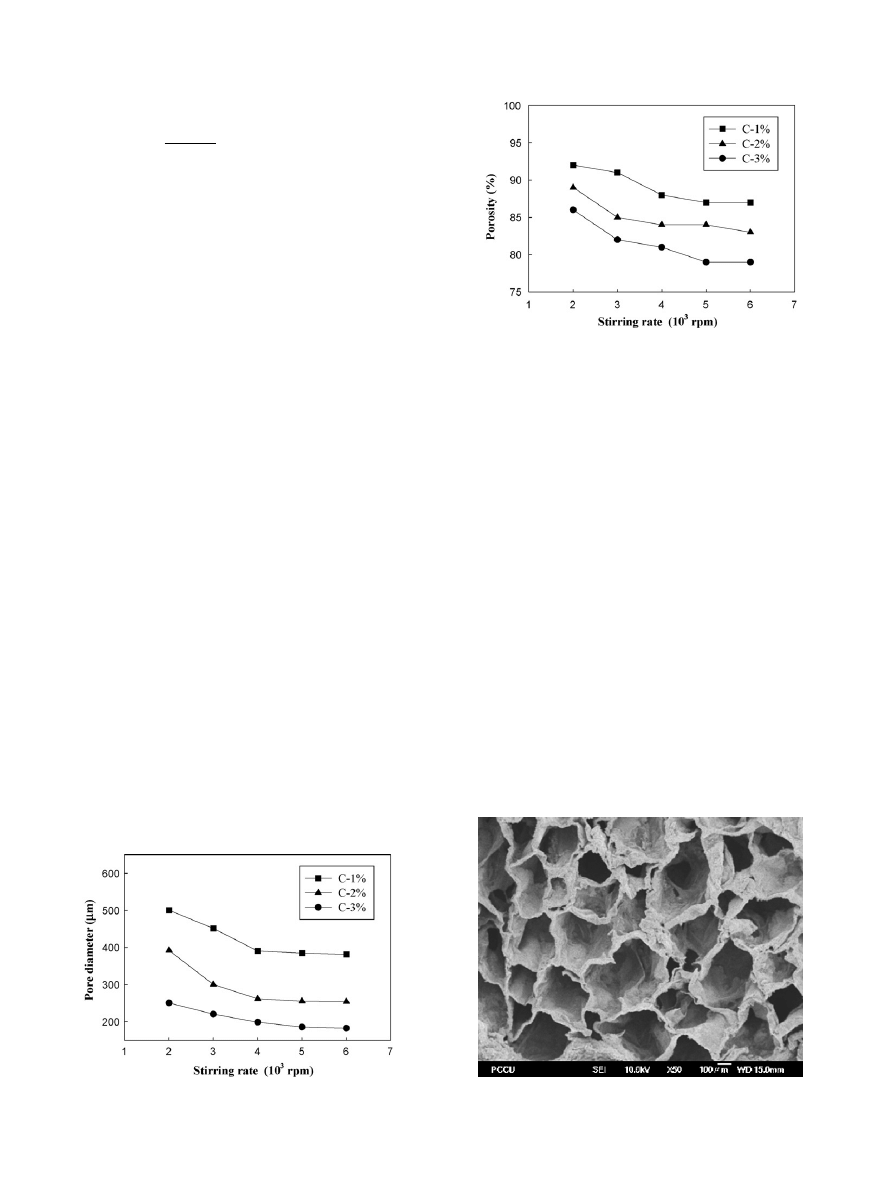
shows different pore sizes of 3D porous scaffolds pro-
duced under various concentration of chitosan and stirring rate of
homogenizer. Testing samples were produced by using 1, 2, and
3% concentration of chitosan solution, foaming using homog-
enizer with 2000, 4000, and 6000 rpm stirring rate, then add
5% NaOH to harden into shape. From the figure we could see
that the samples’ pore diameter decrease while the stirring rate
increase. Take C-1% chitosan scaffolds for example, when stir-
ring rate increases from 2000 rpm to 4000 rpm, its pore diameter
rapidly decreases from 500
m to about 400 m. The reason is
that for homogenizer the higher the stirring rate the greater the
shear force of the cutting head. Nevertheless, when the stirring
rate reaches 4000 rpm and higher, the change in porous diame-
ter of the chitosan scaffolds is minimal. Both C-2% and C-3%
chitosan scaffolds also have similar results.
also shows that the porous diameter decreases along
with the concentration of chitosan solution decreases. Higher
chitosan solution concentration also easier to obtain smaller
diameter foam bubbles. In our laboratory preparation experi-
ments we discovered that when chitosan solution concentration
is lower than 1%, the sample’s viscosity is too low for foaming,
on the other hand, when the chitosan solution concentration is
higher than 3%, the sample’s viscosity is too high thus the sam-
ple would stick to the homogenizer’s cutting heads and were
unable to mix homogeneously.
The pore diameters of C-3% chitosan scaffolds are smaller
than those of C-2% and C-3%. The reason is that C-3% has
higher viscosity therefore it is easier to maintain the smaller
pore diameter after foaming. To sum it up, we are certain that
chitosan solution concentration and homogenizer stirring rate
both variables have great influence to the pores’ formation in
scaffolds.
Fig. 1. The pore sizes of chitosan scaffolds at various concentration of chitosan
and various stirring rate (5% NaOH).
Fig. 2. The porosity of chitosan scaffolds at various concentration of chitosan
and various stirring rate (5% NaOH).
is the porosities for chitosan scaffolds of various chi-
tosan solution concentrations. Porosity is calculated from Eq.
in Sections
. As shown in the figure, porosity would
decrease along with the increase of both homogenizer stirring
rate and chitosan solution concentration. In addition, that under
these set conditions, no matter how high the concentration or
stirring rate is, the porosity of chitosan scaffolds constantly
maintains in high level above 80%. The formation of chitosan
scaffolds samples produced were always inspected and observed
with SEM.
is the SEM photo taken from a 2% chitosan scaffolds
after the 5% NaOH liquid hardening processes. From
can be seen that although no chemistry foaming agent was added
in the manufacture process, yet the 3D scaffolds samples with
pore sizes of 200–500
m
−1
, and 80% porosity could still be
obtained by controlling the concentration of chitosan and the
stirring rate of homogenizer.
3.2. Water absorption
The water absorption property of the raw materials influ-
ences not only the maintaining of the chitosan scaffolds’ shape
Fig. 3. Morphology of chitosan scaffold produced by liquid hardening method
(2% chitosan, 4000 rpm, 5% NaOH).
W.-C. Hsieh et al. / Colloids and Surfaces B: Biointerfaces 57 (2007) 250–255
253
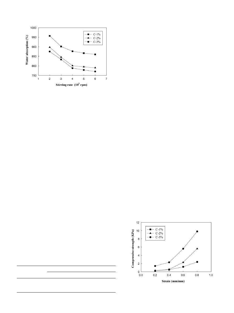
Fig. 4. The water absorption diagram of chitosan 3D porous scaffolds at various
concentrations and various stirring rates.
and form, but also affects the cells’ growth. In the cell cultiva-
tion processes of tissue engineering, if long culture time were
required and high water absorption materials were used, the scaf-
folds might saturated with water and expand, causing deform and
affecting the proliferation and division of cells.
shows
the diagram of water absorption for 1–3% chitosan scaffolds
under 2000–6000 rpm stirring rates. The change ratio of water
absorption is the water absorption in relation to the homogenizer
stirring rates, which would show the influence of stirring rates to
the amount of water absorbed. As the diagram illustrated, along
with the increase of both chitosan solution concentration and
homogenizer stirring rate, the change ratio of water absorption
reveals a reducing trend. Regardless of the chitosan scaffolds
sample is C-1%, C-2% or C-3%, if the homogenizer stirring
rate is between 2000 and 4000 rpm, the change ratio of water
absorption decrease rapidly, as shown in
as the variation
are between 8.5 and 10.8%. If the stirring rate reaches above
4000 rpm the decrease of the change ratio of water absorption
tend to slow down and not as rapidly, also shown in
the variation are between 1.4 and 2.2%. When compared these
experiment results with
, we could find that under the
manufacturing condition of 2000–4000 rpm stirring rate, pore
sizes would quickly become smaller and porosity lower, result
in the change ratio of water absorption rapidly reduced. On the
other hand, when samples are manufactured under the condition
of 4000–6000 rpm stirring rate, the above-mentioned shrink in
pore sizes and porosity tend to slow down, thus reducing the
change ratio of water absorption. Therefore, we believe that the
samples’ ratio of water absorption can be changed not only by
the samples’ material properties, but also by varying the pore
Table 1
The water absorption (changing ratio) for chitosan scaffolds at the manufacturing
conditions of 2000–6000 rpm stirring rate
Samples
Stirring rate
2000–4000 rpm
4000–6000 rpm
C-1%
957–876 (
−8.46%)
876–860 (
−1.83%)
C-2%
898–801 (
−10.80%)
801–790 (
−1.39%)
C-3%
875–788 (
−9.94%)
788–771 (
−2.16%)
sizes and porosity of the samples as well as change the structure
of chitosan scaffolds and control the samples’ water absorption
property.
As far as the influence of chitosan concentration changes
to the chitosan scaffolds samples’ ratio of water absorption,
shown in
, apparently we could see that along with the
concentration of chitosan solution increase the water absorp-
tion tends to decrease. Relatively to 6.2–8.1% between C-1%
and C-2%, the difference between C-2% and C-3% is minimal
(about 1.6–2.6%). From
we could find that
the ratio of water absorption of chitosan scaffolds has very high
relevance to pore sizes, porosity and water absorption. When chi-
tosan solution concentration is within the range of 1–3%, larger
pore sizes having higher porosity thus would have higher ratio
of water absorption. Obviously the scaffolds has higher ratio
of water absorption as it has more water storage space (than
surface area). While mechanical stirring rate of homogenizer
is under 4000 rpm, along with the increase of stirring rate the
pore size and porosity decrease. The decrease of pore size may
causes capillary phenomenon and increases some water absorp-
tion
. However, we believe the main reason for the water
absorption to decrease should be the decrease of the whole sam-
ple’s porosity that reduces the sample’s specific surface area for
water absorption. We could also see similar results from other
correlated literatures
3.3. Mechanical properties
During cell cultivation, 3D porous scaffolds need to have
appropriate strength for cells attachment.
shows the chi-
tosan scaffolds’ compressive strength–strain curves of C-1%,
C-2% and C-3%. In the initial compression phase (strain < 0.4),
chitosan scaffolds exhibits poor resistance to tension stress, but
demonstrates better resistance to tension stress when strain is
greater than 0.4. Moreover, C-3% has stronger compressive
strength than C-2% and C-3%. Chitosan is essentially a non-
water-soluble material, even soaked in water for a long period
(such as in PBS for cell culture) should have minimal influence
to the compressive strength of chitosan scaffolds. Therefore,
the deciding factor of chitosan scaffolds’ mechanical properties
Fig. 5. The compressive strength and strain diagram of chitosan 3D porous
scaffolds at concentration of chitosan 1–3% (4000 rpm, 5% NaOH).
254
W.-C. Hsieh et al. / Colloids and Surfaces B: Biointerfaces 57 (2007) 250–255
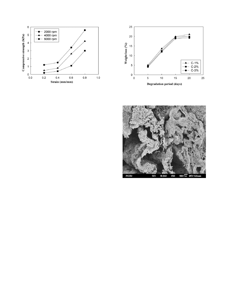
Fig. 6. The compressive strength and strain diagram of chitosan scaffolds at
manufacturing conditions of stirring rate 2000–6000 rpm (2% chitosan, 5%
NaOH).
is the concentration of chitosan added in the solution.
is the compressive strength–strain diagram for chitosan scaf-
folds manufactured under various homogenizer stirring rates.
From the diagram, we could see that chitosan scaffolds would
have stronger mechanical strength along with the increase of
homogenizer stirring rate. The reason is that higher homogenizer
stirring rate produces smaller pore diameter in the scaffolds, and
smaller pore sizes could gain larger surface area, which could
withstand stronger compression force. In summary, we found
that chitosan scaffolds manufactured under the conditions of
higher chitosan concentration and higher homogenizer stirring
rate would possess better mechanical properties.
3.4. In vitro degradation
The ultimate goal for the application of chitosan 3D porous
scaffolds in tissue engineering were the hope that it could dis-
integrated naturally along the cells growth. As a result, the
time to degradation would affect the condition of the cells
growth. Lysozyme could hydrolyze the bindings between N-
acetylmuramic acid and N-acetylglucosamine in some bacteria’s
cell wall. Therefore, this research uses lysozyme as degradation
enzyme to investigate through the time course of degradation
conditions of chitosan 3D porous scaffolds.
shows the
enzymatic degradation experiments of scaffolds with various
chitosan concentrations. From the diagram, the degradation rates
of chitosan C-1%, C-2% and C-3% all increase as time increases;
moreover, degradation would decelerate slightly as chitosan con-
centration increases. Take chitosan scaffolds C-2% as example,
first 15 days presents fast degradation rate nearly in 45
◦
angle,
but the degradation decelerate after 15 days, and almost horizon-
tal when degradation rate reaches 20%. Possible explanation for
enzyme degradation rate of chitosan scaffolds were only about
20% are related to the function active sites on lysozyme. Its func-
tion active sites are primarily
-(1–4)-glycosidic bonds between
polysaccharide. The chitosan used in this research has about
80% degree of deacetylation. Because it only has about 20%
-
(1–4)-glycosidic bond, the chitosan scaffolds reach equilibrium
when it has 20% degradation rate.
shows SEM micrograph
of chitosan scaffold (2% Chitosan, 4000 rpm, 5% NaOH) after
Fig. 7. The weight loss curves of chitosan scaffolds samples under the time
course of degradation (4000 rpm, 5% NaOH).
Fig. 8. SEM micrograph of chitosan scaffold after 20 days enzymatic degrada-
tion at 37
◦
C (2% chitosan, 4000 rpm, 5% NaOH).
20 days enzymatic degradation at 37
◦
C. As you can see from
the picture, after enzyme degradation for 20 days the chitosan
scaffolds obviously has partially disintegrated on the surface.
4. Conclusion
In this research we create a simple liquid hardening method
to manufacture 3D chitosan porous materials. The main purpose
is to investigate the feasibility of chitosan scaffolds application
in cell culture of tissue engineering. The following conclusions
were reached.
(1) With chitosan concentration in 1–3% range, chitosan porous
scaffolds could obtain good formability. When chitosan con-
centration is smaller than 1%, there’s insufficient viscosity
thus scaffolds forming is not good, on the other hand, chi-
tosan concentration higher than 3% the viscosity too high
to cause difficulty for formation.
(2) The pore size and porosity will reduce along with the
increase of chitosan concentration. Higher chitosan con-

W.-C. Hsieh et al. / Colloids and Surfaces B: Biointerfaces 57 (2007) 250–255
255
centration is easier to obtain smaller pore diameter foaming
bubbles. The pore size and porosity also will reduce along
with the increase of homogenizer’s stirring rate. But when
the stirring rate is greater than 4000 rpm, the pore size
change of chitosan scaffolds is minimal.
(3) The higher the chitosan concentration, the lower the water
absorption of chitosan scaffolds; similarly, increase the
homogenizer’s stirring rate also reduce the water absorption
of the chitosan scaffolds. By changing the concentration of
chitosan and homogenizer’s stirring rate, the water absorp-
tion of chitosan scaffolds samples can be controlled.
(4) From the mechanical property test of the chitosan scaffolds,
we could see that the compressive strength of chitosan scaf-
folds increases along with chitosan concentration increases;
in addition to that, the compressive strength of chitosan
scaffolds also increases along with homogenizer’s stirring
rate.
(5) In the enzymatic degradation tests, weight loss of chitosan
scaffolds increases along with time of degradation, but stop
further weight loss when the loss reaches 20% of chitosan
scaffolds weight.
Acknowledgement
The work was supported in part by the National Science
Council Taiwan, ROC, under Grant No. 94-2622-E-034-001-
CC3.
References
[1] L.E. Niklason, Engineering of bone grafts, Nat. Biotechnol. 18 (2000)
929–930.
[2] N. Saito, T. Okada, H. Horiuchi, N. Murakami, J. Takahashi, M. Nawata,
H. Ota, K. Nozaka, K. Takaoka, A biodegradable polymer as a cytokine
delivery system for inducing bone formation, Nat. Biotechnol. 19 (2001)
332–335.
[3] Z. Li, H.R. Ramay, K.D. Hauch, D. Xiao, M. Zhang, Chitosan-alginate
hybrid scaffolds for bone tissue engineering, Biomaterials 26 (2005)
3919–3928.
[4] H. Mitomo, W.C. Hsieh, K. Nishiwaki, K. Kasuya, Y. Doi, Poly(3-
hydroxybutyrate-co-4-hydroxybutyrate) produced by Comamonas aci-
dovorans, Polymer 42 (2001) 3455–3461.
[5] W.C. Hsieh, K. Kasuya, H. Mitomo, Crystallization and evalua-
tion of physical properties of microbial poly(3-hydroxybutyrate-co-4-
hydroxybutyrate), Sen-i Gakkaishi (Japan) 58 (2002) 428–431.
[6] W.C. Hsieh, H. Mitomo, K. Kasuya, T. Komoto, Enzymatic degradation and
aminolysis of microbial poly(3-hydroxybutyrate-co-4-hydroxybutyrate)
single crystals, J. Polym. Environ. 14 (2006) 78–97.
[7] Y.W. Wang, Q. Wu, G.Q. Chen, Attachment, proliferation and differentia-
tion of osteoblasts on random biopolyester poly(3-hydroxybutyrate-co-3-
hydroxyhexanoate) scaffolds, Biomaterials 25 (2004) 669–675.
[8] J. Jiang, N. Kojima, T. Kinoshita, A. Miyajima, W. Yan, Y. Sakai, Cultiva-
tion and induction of fetal liver cells in poly-l-lactic acid scaffold, Mater.
Sci. Eng. C 24 (2004) 361–363.
[9] S. Levenberg, N.F. Huang, E. Lavik, A.B. Rogers, J. Itskovitz-Eldor,
R. Langer, Differentiation of human embryonic stem cells on three-
dimensional polymer scaffolds, PNAS 100 (2003) 12741–12746.
[10] D.W. Hutmacher, Scaffolds in tissue engineering bone and cartilage, Bio-
materials 24 (2000) 2529–2543.
[11] R.L. Price, M.C. Waid, K.M. Haberstroh, T.J. Webster, Selective bone cell
adhesion on formulations containing carbon nanofibers, Biomaterials 11
(2003) 1877–1887.
[12] H. Petite, V. Viateau, W. Bensaid, A. Meunier, C. de Pollak, M. Bour-
guignon, K. Oudina, L. Sedel, G. Guillemin, Tissue-engineered bone
regeneration, Nat. Biotechnol. 18 (2000) 959–963.
[13] S. Yang, K.F. Leong, Z. Du, C.K. Chua, The design of scaffolds for use
in tissue engineering. Part I. Traditional factors, Tissue Eng. 7 (2001)
679–689.
[14] G. Chen, T. Ushida, T. Tateshi, A biodegradable hybrid sponge nested with
collagen microsponges, J. Biomed. Mater. Res. 51 (2000) 273–279.
[15] P.R. Austin, C.J. Brine, J.E. Castle, J.P. Zikakis, Chitin new facets of
research, Science 212 (1981) 749–753.
[16] A. Chenite, C. Chapat, D. Wang, C. Combes, M.D.C. Buschmann, D. Hoe-
mann, J.C. Leroux, B.L. Atkinson, F. Binette, A. Selmani, Novel injectable
neutral solutions of chitosan form biodegradable gels in situ, Biomaterials
21 (2000) 2155–2161.
[17] K.D. Harpreet, A.R. Ray, A.K. Panda, Three-dimensional chitosan
scaffold-based MCF-7 cell culture for the determination of the cytotoxicity
of tamoxifen, Biomaterials 26 (2005) 979–986.
[18] K. Rezwana, Q.Z. Chena, J.J. Blaker, A.R. Boccaccini, Biodegradable and
bioactive porous polymer/inorganic composite scaffolds for bone tissue
engineering, Biomaterials 27 (2006) 3413–3431.
[19] C.P. Chang, K.B. Cheng, The study of automatic measuring method applied
on nonwoven fabric, J. Chin. Textile Eng. 15 (1997) 1–15.
[20] H. Markus, T. Miller, Pore-morphology-based simulation of drainage in
totally wetting porous media, Adv. Water Resourc. 24 (2001) 243–255.
[21] S. Gerhard, Electrochemistry of capillary systems with narrow pores. I.
Overview, J. Membr. Sci. 150 (1998) 151–157.
[22] C.P. Chang, C.C. Hsu, The formation and water content of synthetic fiber
growing media, Mater. Sci. Eng. A 433 (2006) 100–103.
Document Outline
- Morphology and characterization of 3D micro-porous structured chitosan scaffolds for tissue engineering
Wyszukiwarka
Podobne podstrony:
Production and Characterisation of extracts
#1038 Types and Characteristics of Apartments
Rewicz, Tomasz i inni Isolation and characterization of 8 microsatellite loci for the ‘‘killer shri
Drying, shrinkage and rehydration characteristics of kiwifruits during hot air and microwave drying
Improved Characterization of Nitromethane, Nitromethane Mixtures, and Shaped Charge Jet
Fit sphere unwrapping and performance analysis of 3D fingerprints
Characteristics, treatment and utilization of residues from MSW
GL Morphology The analysis of word structure
Detection and Molecular Characterization of 9000 Year Old Mycobacterium tuberculosis from a Neolithi
Characterization of microwave vacuum drying and hot air drying of mint leaves (Mentha cordifolia Opi
Eurocode 6 Part 2 1996 2006 Design of Masonry Structures Design Considerations, Selection of Mat
Corrosion behavior and surface characterization of titanium
2001 In vitro fermentation characteristics of native and processed cereal grains and potato
Structure and properties of Ti
Microwave drying characteristics of potato and the effect of different microwave powers on the dried
Bell locality and the nonlocal character of nature
DESIGN AND DEVELOPMENT OF MICRO TURBINE
więcej podobnych podstron